Urinary System
Kidney & the Nephron
Slice a fresh kidney in half along its long axis and the first thing you see is two zones. The outer rim is a pale, granular cortex studded with tiny red dots — the glomeruli. The inner zone is the medulla, arranged into 8–18 conical renal pyramids whose striated, parallel-lined appearance comes from straight tubules and vasa recta all running in the same direction. The apex of each pyramid — the papilla — pokes into a minor calyx, the funnel that catches finished urine.
Look closer at the cortex and you will see vertical stripes running from medulla up into the cortex like rays of sunlight. These are the medullary rays — bundles of straight tubules (proximal straight, thick ascending, collecting duct) that belong to the medulla but invade cortical territory. Between rays sits the cortical labyrinth — a tangle of renal corpuscles and convoluted tubules. This cortex-vs-medulla, labyrinth-vs-ray geography is the single most useful map you can carry into a histology exam.
The functional unit doing all the work is the nephron. Follow a drop of filtrate from start to finish: it begins in the renal corpuscle (glomerulus + Bowman capsule), runs into the proximal convoluted tubule (PCT), dives down as the thin descending limb, hairpins through the thin ascending limb, climbs back up the thick ascending limb, brushes past its own glomerulus at the distal convoluted tubule (DCT), slips through a short connecting tubule and finally joins a collecting duct. The collecting duct is technically not part of the nephron — it is shared real estate where several nephrons drain together — but you must always think of nephron + duct as one drainage tree.
Think of the cortex as the filtering floor (where blood is sieved) and the medulla as the concentration floor (where water is pulled back). The medullary rays are the elevators connecting them, and the calyces are loading docks at the bottom. About 85% of nephrons are cortical with short loops (light work). The remaining 15% are juxtamedullary — their corpuscles sit deep on the corticomedullary border and their loops plunge far into the medulla. These long-loop nephrons are the only ones that can crank up the medullary osmotic gradient enough to produce concentrated urine. Lose them (in chronic kidney disease) and you lose the ability to concentrate.
A condensed map of the components:
| Component | Parts |
|---|---|
| Renal corpuscle | Glomerulus + Bowman's capsule |
| Renal tubule | Proximal tubule (convoluted + straight) → thin segment (loop of Henle) → distal tubule (straight + convoluted) |
Collecting ducts receive the DCT but are not part of the nephron — they are a separate embryologic lineage (ureteric bud, not metanephric blastema). Renal corpuscles + convoluted tubules lie in the cortical labyrinth; straight tubules run in the medullary rays and medulla.
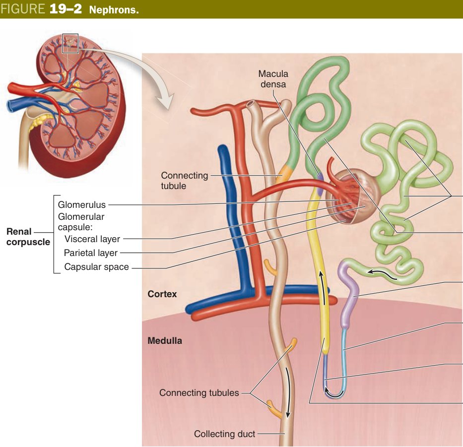
Renal Corpuscle
The renal corpuscle is the kidney's filtration head. Picture a fist (the glomerulus) pushed into a balloon (Bowman capsule) — the fist invaginates the balloon so deeply that two layers of balloon wall cling to the fist. The outer wall you can still see is the parietal layer (simple squamous); the inner wall now plastered onto the capillary loops is the visceral layer, made of bizarre octopus-shaped cells called podocytes. Between the two layers is the capsular (urinary) space where filtrate collects.
Every corpuscle has two poles. At the vascular pole, the afferent arteriole enters and the efferent arteriole leaves — two arterioles in series, a kidney peculiarity that lets the glomerulus run at high pressure. At the urinary pole, opposite the vascular pole, the capsule funnels filtrate into the proximal convoluted tubule. Examiners love to swap these — remember: distal tubule touches the vascular pole (macula densa); proximal tubule leaves the urinary pole.
Most capillary beds drain into venules. The glomerulus drains into another arteriole, and that arteriole is muscular and contractile. By tuning afferent vs efferent tone independently the kidney can hold glomerular pressure stable across a huge range of systemic blood pressure — this is autoregulation. ACE inhibitors dilate the efferent arteriole preferentially, dropping glomerular pressure (good for diabetics' GBM, bad in renal artery stenosis).
The cellular cast:
- Glomerulus: a tuft of fenestrated capillaries (large 70–90 nm pores, no diaphragm) between an afferent (enters) and efferent (leaves) arteriole; supported internally by intraglomerular mesangial cells.
- Bowman's capsule: parietal layer = simple squamous; visceral layer = podocytes wrapping the capillaries with foot processes (pedicels).
- Vascular pole = where arterioles enter/leave; urinary pole = where the capsule becomes the proximal convoluted tubule.
Sitting in the angles between the capillary loops, never on the urinary side of the basement membrane, are the mesangial cells. These are unusual cells with three day-jobs: (1) contractile — they squeeze and relax to change the filtering surface area; (2) phagocytic — they clean trapped immune complexes off the basement membrane (which is why mesangial expansion is a hallmark of immune-complex nephritis); and (3) structural — they secrete the matrix that holds the tuft together. Their cousins outside the glomerulus, the extraglomerular mesangial cells (lacis cells), sit at the vascular pole and form part of the juxtaglomerular apparatus (see 13.4).
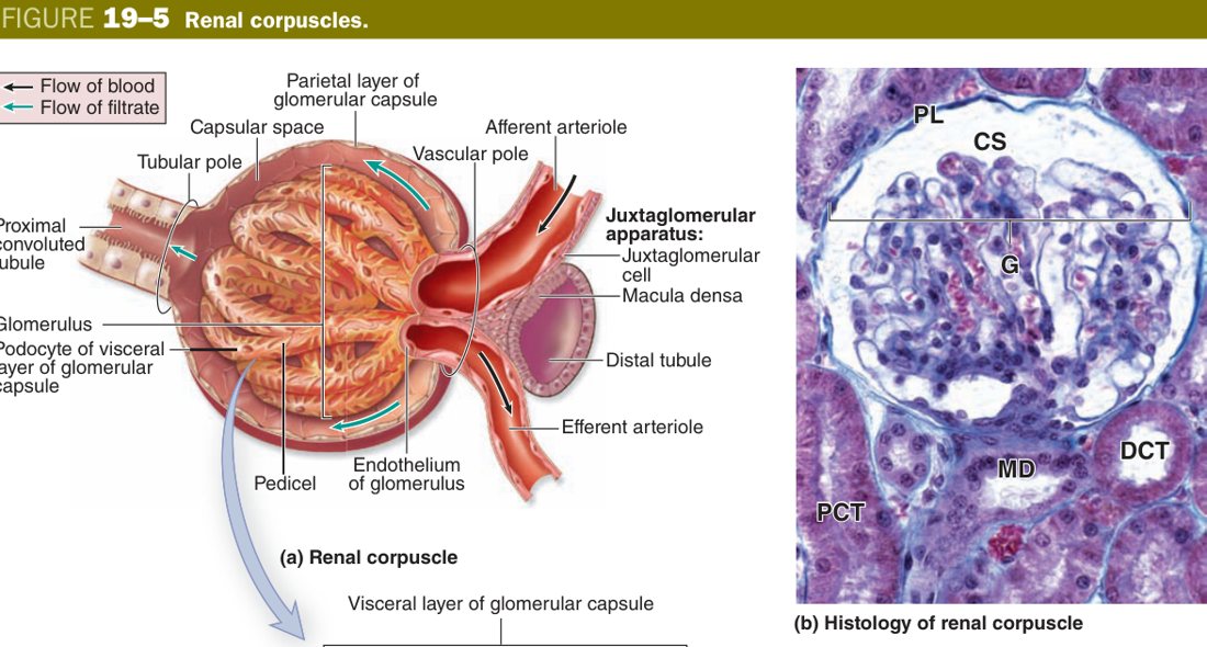
Plasma cannot just leak into Bowman space — if it did, you would lose every protein in your blood every minute. Between blood and filtrate sit three molecular sieves stacked on top of each other, collectively the filtration barrier. Each layer rejects something slightly different, and each layer has a signature disease when it breaks.
Filtration barrier: the three layers plasma crosses to form the filtrate — (1) fenestrated endothelium of the glomerular capillary; (2) the fused glomerular basement membrane (a selective macromolecular filter, 4–8 nm); (3) the filtration slits between podocyte foot processes, bridged by a slit diaphragm. Function: filters blood plasma into Bowman's space.
Layer one is the fenestrated endothelium. The pores are wide and open (no diaphragm), so this layer mostly stops blood cells — it is a coarse screen, not a fine one. Layer two is the glomerular basement membrane (GBM), the only place in the body where two basement membranes (endothelial + podocyte) fuse into one thick sandwich. On EM you see three sublayers: lamina rara interna, lamina densa in the middle, and lamina rara externa. The middle lamina densa is a meshwork of type IV collagen (sets the size cutoff) plus laminin, while the rare layers are studded with heparan sulphate proteoglycan — intensely negatively charged. Albumin is also negatively charged at physiological pH, so it is repelled before it even reaches the slits. Layer three is the podocyte slit diaphragm: between pedicels of adjacent podocytes a zipper-like membrane made of the protein nephrin stretches across a ~25-nm slit. Nephrin is the final, finest sieve.
Lose the charge of the GBM (T-cell cytokines neutralise heparan sulphate) → albumin pours through → minimal change disease — on EM the podocyte foot processes are also flattened ("effacement") but the membrane looks normal on LM. Thicken the GBM with immune deposits and you get membranous nephropathy (anti-PLA2R antibody, "spike-and-dome" silver stain). Mutate nephrin (congenital Finnish nephrotic syndrome) and the zipper itself is broken. The filtration barrier is one of the rare structures where you can read a histology lecture and a pathology lecture as a single story.
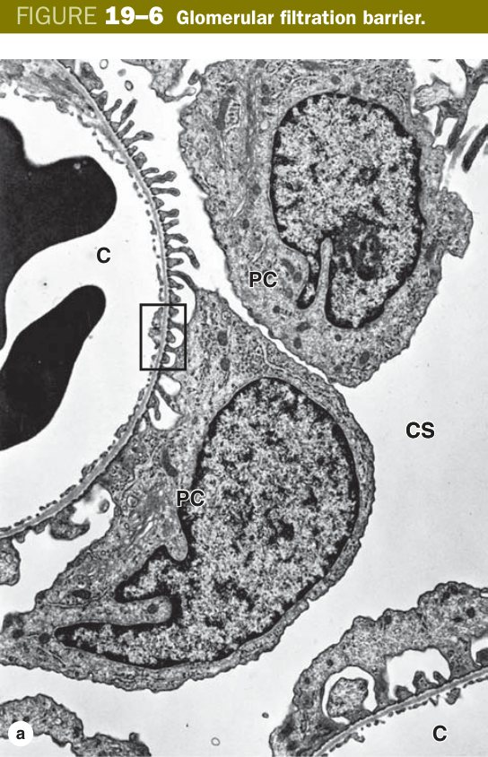
Alport syndrome — X-linked mutation in the α5 chain of type IV collagen; the GBM splits and laminates ("basket-weave" on EM) → haematuria, deafness, lens defects. Goodpasture syndrome — IgG antibodies against the α3 chain of type IV collagen attack GBM and alveolar basement membrane → crescentic glomerulonephritis + lung haemorrhage. Diabetic nephropathy — non-enzymatic glycation thickens the GBM and expands the mesangium (Kimmelstiel–Wilson nodules) — histological correlate of microalbuminuria.
Proximal & Distal Convoluted Tubules
After filtrate leaves Bowman space at the urinary pole, the very first thing that happens is bulk reabsorption — not because the kidney can't filter less, but because evolution chose to filter generously (~180 L/day) and reclaim almost everything downstream. That reclamation begins in the proximal convoluted tubule (PCT), where ~65% of filtered sodium and water, all filtered glucose, all filtered amino acids, and the bulk of bicarbonate go back into the blood.
To do this work the PCT cell is built like a tiny absorption factory. Look at the apical surface and you will see a tall brush border of microvilli — massive surface-area amplification for transporters. Look at the cytoplasm and it is packed with mitochondria (because reabsorption against an electrochemical gradient costs ATP), making the cytoplasm stain a deep eosinophilic pink on H&E. Look at the basolateral surface and you will see deep infoldings with lateral interdigitation — more surface area, more Na/K-ATPase. The lumen looks narrow and fuzzy because microvilli fill it. There are fewer visible nuclei per ring of tubule because each cell is so large.
The loop of Henle is a hairpin diving into the medulla. The thin descending and thin ascending limbs are lined by a flat simple squamous epithelium — so thin and so pale they are easy to mistake for capillaries until you look for red cells inside. Their job is passive: water leaves the descending limb (it is permeable to water but not solute) while solutes leave the ascending limb (the reverse). The thick ascending limb (TAL) is back to simple cuboidal and runs the famous Na/K/2Cl cotransporter — the target of furosemide. The TAL is impermeable to water but pumps salt out, leaving dilute filtrate and a salty medulla. This is the engine of the countercurrent multiplier.
Think of the PCT as the workhorse cell — tall, dark-pink, fuzzy-edged, fewer nuclei in section because the cells are huge. Think of the DCT as the fine-tuning cell — shorter, paler, with a clear wide lumen and lots of small nuclei crowded together because the cells are smaller. PCT fuzz = brush border; DCT clean lumen = no brush border. If you see a tubule on a slide and can count the nuclei easily, it's a DCT.
The distal convoluted tubule (DCT) is structurally the reverse of the PCT — simple cuboidal but smaller cells, NO brush border, fewer mitochondria, less eosinophilic, wider clearer lumen, more nuclei visible per ring. Functionally the DCT runs the Na/Cl cotransporter (target of thiazide diuretics) and is the site where the wall is modified into the macula densa where it grazes its own afferent arteriole.
| Feature | PCT | DCT |
|---|---|---|
| Epithelium | Simple cuboidal/low columnar, large cells | Simple cuboidal, smaller cells |
| Cytoplasm | Strongly acidophilic | Less acidophilic (paler) |
| Brush border | Yes (abundant long microvilli) | No |
| Lumen | Narrow, irregular ("fuzzy") | Wider, clearer |
| Nuclei / section | Fewer (cells larger), borders indistinct | More nuclei seen |
| EM | Microvilli + basal infoldings + mitochondria + lateral interdigitation | More basal infoldings (Na⁺/K⁺-ATPase), fewer/short microvilli |
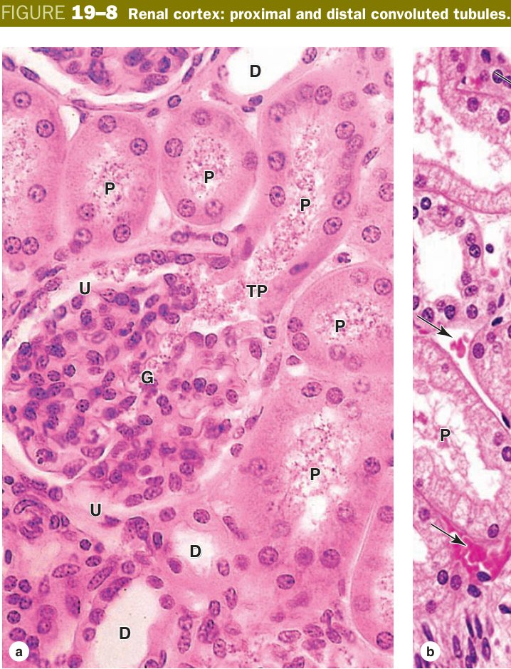
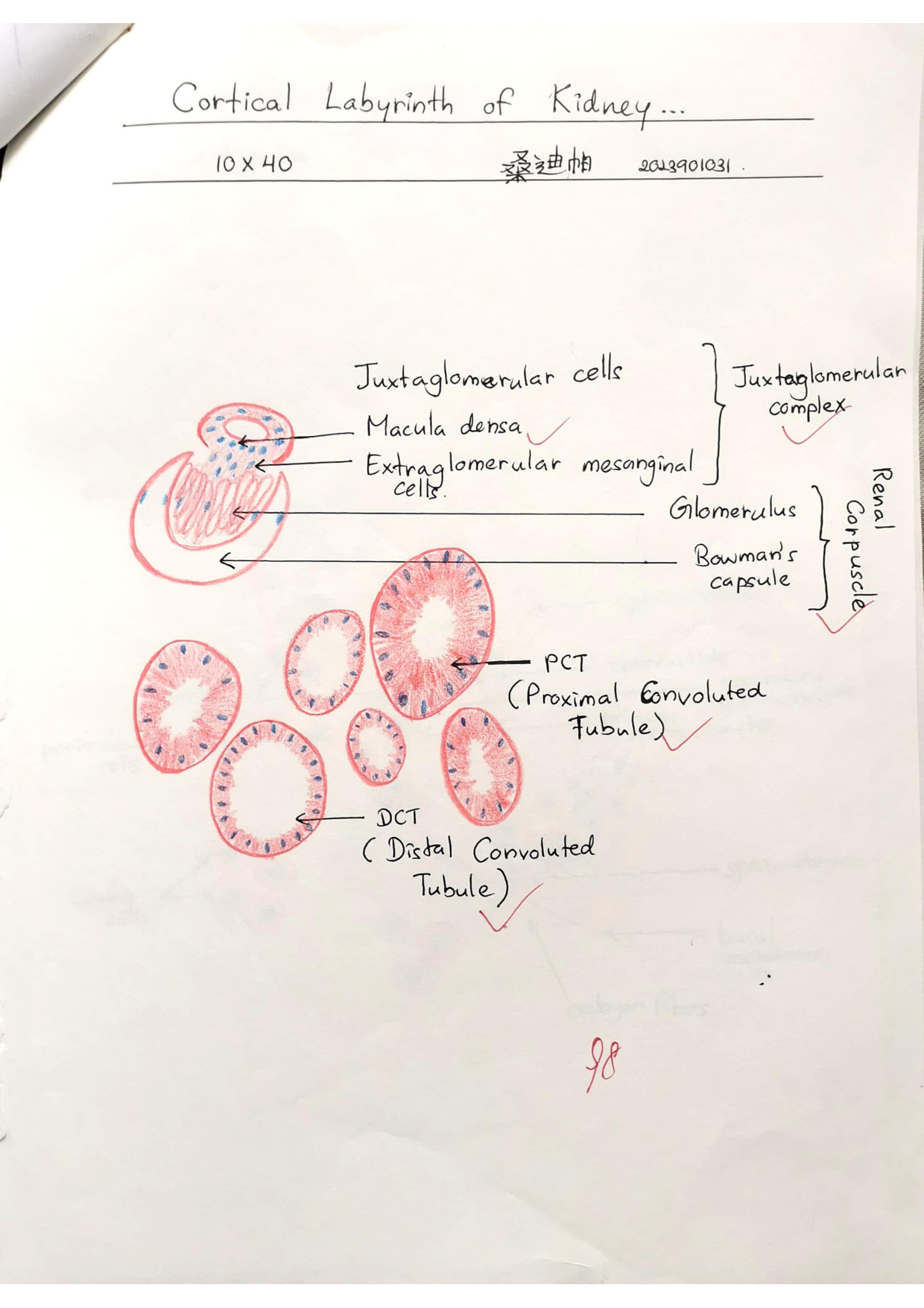
PCT = Pink, Packed, brush-border, fuzzy narrow lumen, fewer nuclei. DCT = paler, clear wide lumen, more nuclei, no brush border. PCT cells are bigger → fewer per ring.
Acute tubular necrosis (ATN) kills PCT cells first because they are mitochondria-rich and most ATP-dependent — their swollen, sloughed brush borders pack the lumen as the classic "muddy brown" granular casts in urine. Renal cell carcinoma (clear cell type) arises from PCT epithelium — the clear cytoplasm comes from dissolved-out lipid and glycogen. Loop diuretics (furosemide) act on the TAL Na/K/2Cl; thiazides act on the DCT Na/Cl.
Juxtaglomerular Apparatus (JGA)
After the TAL climbs back up to the cortex, it does something extraordinary: the rising tubule swings around and touches the same glomerulus that spawned it, brushing against its own afferent arteriole at the vascular pole. Why? So that one nephron can feel what its own filtration is doing and feed back on it. This anatomical handshake is the juxtaglomerular apparatus (JGA), and it has three cellular characters.
The first is the macula densa — literally "dense spot" — a patch of DCT epithelium where the cells become tall, narrow and crowded so that their nuclei pile up tightly. Their job is to sense the NaCl concentration in the tubular fluid. If filtration is too brisk, NaCl arrives in excess; if too sluggish, NaCl is low. The macula densa reads this number and signals next door. The second character is the juxtaglomerular (JG) cell — modified smooth muscle of the afferent arteriole wall, swollen with PAS-positive granules of renin. The third is the extraglomerular mesangial cell (lacis cell), a connector cell in the angle between afferent and efferent arterioles, linked by gap junctions to both the macula densa and the JG cells.
Think of the JGA like a room thermostat wired to a boiler. The macula densa is the temperature sensor (reading tubular NaCl). The lacis cells are the wires. The JG cells are the boiler — when NaCl drops, they fire renin into the blood, kicking off the renin–angiotensin–aldosterone cascade, which raises blood pressure and pushes more salt back into the tubule. Conversely, a salty signal closes the boiler down. Beta-blockers cool this thermostat (sympathetic input on JG cells); ACE inhibitors break the cascade downstream.
At the vascular pole, three components regulate blood pressure & filtration:
| Component | Structure / role |
|---|---|
| Macula densa | Modified DCT cells — tall, narrow, closely-packed; a Na⁺ chemoreceptor |
| Juxtaglomerular (JG) cells | Modified smooth muscle of the afferent arteriole; secrete renin |
| Extraglomerular mesangial cells | Connect the two (via gap junctions) |
Macula densa: a plaque on the wall of the distal convoluted tubule at the vascular pole of its own renal corpuscle; the epithelial cells become narrower and taller (columnar) with closely-packed nuclei. It is a Na⁺ (sodium) chemoreceptor linked to the extraglomerular mesangium by gap junctions.
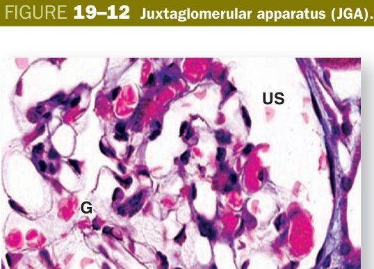
Renal artery stenosis → chronically under-perfused afferent arteriole → JG cells dump renin → secondary hypertension that responds poorly to standard antihypertensives but dramatically to ACE inhibitors (which break the angiotensin II loop). Bartter syndrome mimics chronic furosemide — defective Na/K/2Cl in TAL → macula densa "senses" low NaCl → runaway renin → hypokalaemic metabolic alkalosis with normal blood pressure.
Collecting Ducts & Excretory Passages
The collecting duct is where the kidney does its final water- and acid-base accounting. Two cell types share its wall. Principal cells are pale-staining with distinct cell borders (those crisp polygonal outlines are a giveaway); they handle Na reabsorption (under aldosterone) and water reabsorption (under ADH, via aquaporin-2 channels inserted into the apical membrane). Intercalated cells are darker and scattered between principals; the α-intercalated cell secretes H⁺ (acid handling), the β-intercalated cell secretes HCO3⁻ (alkali handling). ADH is the master switch — without it the duct is impermeable to water and you produce dilute urine; with it, water is pulled back into the salty medulla and urine concentrates.
From the papilla, urine drips into the minor calyx, pools in the major calyx and renal pelvis, then runs down the ureter. The whole tract from minor calyx to bladder is lined by transitional epithelium (urothelium) — a stretchy 3-to-6-layered epithelium whose surface (umbrella) cells carry rigid plaques of the protein uroplakin. When the bladder is empty the umbrella cells fold like an accordion; when full they stretch flat. This trick lets the epithelium go waterproof at every volume.
You learned in the gut that smooth muscle is arranged inner circular + outer longitudinal. The upper ureter does the opposite: inner longitudinal + outer circular. In the lower third it adds a third outer longitudinal layer to help drive urine into the bladder. Easy to remember: gut squeezes onwards; ureter squeezes open. Don't be tricked by the swap.
The bladder has the same urothelium plus a thick muscular wall — the detrusor — arranged in three interwoven smooth-muscle layers. Crucially, the bladder has no muscularis mucosae and no submucosa, so the urothelium sits directly on a loose lamina propria over the detrusor. The urethra changes lining as it descends: in the male the prostatic portion stays transitional, the membranous and proximal spongy portions are stratified or pseudostratified columnar, and near the glans it becomes stratified squamous (matching the skin). The much shorter female urethra is mostly stratified squamous along its length.
- Collecting ducts: simple cuboidal → columnar epithelium, pale cytoplasm with distinct cell borders; ADH-responsive (water reabsorption); run in medullary rays/pyramids to the papilla.
- Ureter & bladder: lined by transitional epithelium (urothelium) with umbrella cells, on a muscular wall (recall Unit 2).
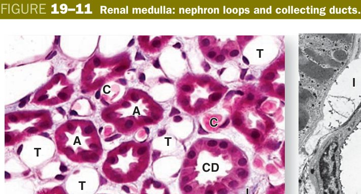
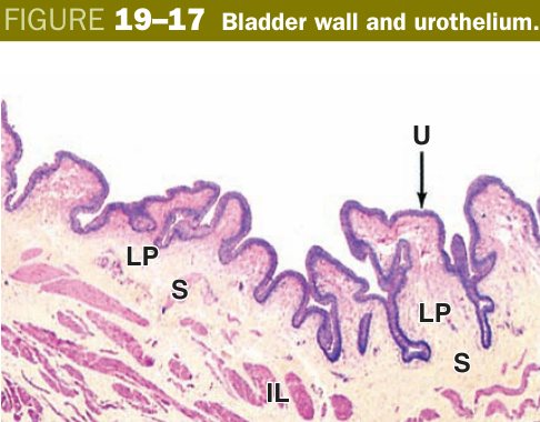
Damage to the filtration barrier → proteinuria (nephrotic syndrome); anti-GBM antibodies → Goodpasture. JG-cell renin drives the renin–angiotensin system (hypertension). Pyelonephritis — ascending bacterial infection of pelvis + tubules — classic sediment is WBC casts. Transitional cell (urothelial) carcinoma arises anywhere urothelium lies (renal pelvis, ureter, bladder) and is the dominant bladder cancer in industrialised countries (aniline dyes, smoking). BPH grows in the prostate's transition zone (around the urethra → obstruction) while prostate cancer arises peripherally.
TMU Exam Drill
📝 Open the full TMU Question Bank — 20 MCQ + 6 terms + 5 essays →
Authentic Tianjin Medical University past-paper questions (2021 Final & the multi-section Final with answer key) mapped to this unit, in the real exam format. Click Show answer to self-test.
□ Single best answer
- A. Located in cortical labyrinth & renal column.
- B. Composed of glomerulus and renal capsule.
- C. At the urinary pole it is continuous with the distal tubule.
- D. At the vascular pole the afferent arteriole enters & efferent leaves.
- E. Function is to filter blood plasma.
- A. On the wall of the distal tubule.
- B. The epithelial cells become stratified cuboidal.
- C. The cells are closely packed.
- D. Sensitive to sodium ion concentration.
- E. Connect to extraglomerular mesangial cells by gap junctions.
□ Fill in the blank
- (fill three segments)
□ True or false
□ Explain the following terms
□ Structure essay
Urinary system complete
Nephron, filtration barrier, PCT/DCT & the JGA mastered. Next: Respiratory System.