Endocrine Glands
General Features
Every organ system in the body has its own private postal service — nerves carry urgent signals on dedicated wires, but the endocrine system writes letters and drops them straight into the bloodstream. There is no duct, no tube, no addressed envelope. The hormone enters the circulation, washes past every cell in the body, and only the cells carrying the correct receptor pick the letter up. Slow compared with a nerve impulse, but with one priceless advantage: a single drop of hormone can reach a billion targets at once.
Under the microscope this strategy has a very predictable look. Endocrine cells are arranged in cords, clumps or follicles, never in the duct-lined acini you saw in the pancreas or salivary glands. Hugging them everywhere is a dense lacework of fenestrated capillaries — capillaries with pores in their endothelium, like postboxes already cut open. The cell secretes, the hormone slips through the fenestra, and within seconds it is on its way through the body.
There are two ultrastructural personalities you must learn to recognise. Protein and peptide secretors (anterior pituitary cells, parathyroid chief cells, islet cells) are packed with rough endoplasmic reticulum and obvious membrane-bound secretory granules — the same factory plan as a plasma cell. Steroid secretors (adrenal cortex, Leydig, theca) look completely different: pale, foamy cytoplasm full of smooth ER, tubular-cristae mitochondria and lipid droplets, because cholesterol is the raw material and the steroid enzymes live in the SER and mitochondria.
A neuron is a telegraph: point-to-point, one synapse, one target, milliseconds. An endocrine cell is a radio station: it broadcasts on a hormone frequency and only cells tuned to that receptor pick it up. The trade is speed for reach. The pituitary is the broadcasting tower for the whole network — small, central, and unreasonably powerful.
Walk through a protein-hormone cell and you see assembly lines: RER stacks, ribosomes, vesicles stamped with hormone and waiting at the door. Walk through a steroid cell and you see a refinery: oily lipid droplets feeding tubular mitochondria, smooth ER snaking everywhere, almost no granules. Same job, completely different architecture — because peptides are packaged but steroids diffuse straight out as soon as they are made.
Pituitary Gland (Hypophysis)
The pituitary sits in the sella turcica like a pea on a stalk, and it is really two glands fused into one. The anterior part (adenohypophysis) is glandular epithelium that grew up from the roof of the embryonic mouth as Rathke’s pouch. The posterior part (neurohypophysis) is nervous tissue that grew down from the floor of the diencephalon. They meet, they fuse, but they never become the same thing — on every slide you can still see the epithelial cords on one side and the pale nerve-fibre meshwork on the other.
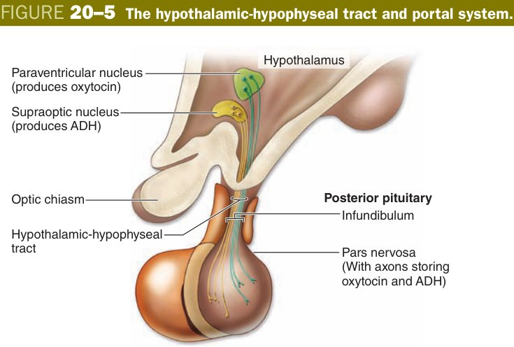
This double origin explains everything about how the gland works. The anterior lobe makes its own hormones in response to chemical messages from the hypothalamus, delivered through a tiny portal blood system. The posterior lobe makes nothing: it simply stores and releases hormones that were synthesised by hypothalamic neurons and shipped down their axons. One is a factory taking orders by mail; the other is a warehouse with a railway running into it.
Picture the embryo at six weeks. The roof of the primitive mouth pushes up a little bag of epithelium. The floor of the brain pushes down a little finger of neural tissue. They meet, kiss, and the epithelial bag wraps around the neural finger. That handshake is your pituitary — forever after, the front half thinks like skin and the back half thinks like brain.
The bulk of the anterior pituitary is the pars distalis: cords of secretory epithelial cells separated by sinusoidal fenestrated capillaries. When the pathologist stains the slide, two populations jump out — cells that take up dye (chromophils) and cells that look pale and empty (chromophobes). Within the chromophils, those that prefer acidic dyes go pink (acidophils) and those that prefer basic dyes go purple-blue (basophils). The whole exam pattern for this section is built on that one tinctorial split.
Acidophils make up roughly forty percent of the pars distalis and they secrete only two hormones — growth hormone (somatotrophs) and prolactin (lactotrophs or mammotrophs). Basophils are fewer (about ten percent) but carry the other four anterior-pituitary hormones: TSH, ACTH, FSH and LH. Chromophobes are pale because they are either resting/degranulated chromophils or undifferentiated stem cells — not a separate hormone line. Whenever an exam asks “which hormone do basophils not make?” the answer is GH, because GH is an acidophil product.
| Cell | Stain | Hormone |
|---|---|---|
| Somatotroph | Acidophil | Growth hormone (GH) |
| Mammotroph (lactotroph) | Acidophil | Prolactin |
| Thyrotroph | Basophil | TSH |
| Corticotroph | Basophil | ACTH |
| Gonadotroph | Basophil | FSH & LH |
| Chromophobe | Pale | Degranulated / stem (non-secretory) |
Behind the cell types is the wiring. The hypothalamus sits above the gland, but its releasing and inhibiting hormones never reach the systemic blood. Instead they drip from hypothalamic neurons into a primary capillary plexus in the median eminence; that plexus drains into portal venules running down the pituitary stalk; the venules open into the secondary plexus bathing the pars distalis. The whole thing is called the hypothalamo-hypophyseal portal system and it concentrates hypothalamic hormones one hundred-fold before they ever touch a pituitary cell.
A small pars intermedia sits between front and back, often a thin row of basophil-like cells around colloid-filled cysts (the residue of Rathke’s pouch lumen). A thinner pars tuberalis wraps the upper stalk. Neither is heavily tested in the TMU exam — recognise them, then move on.
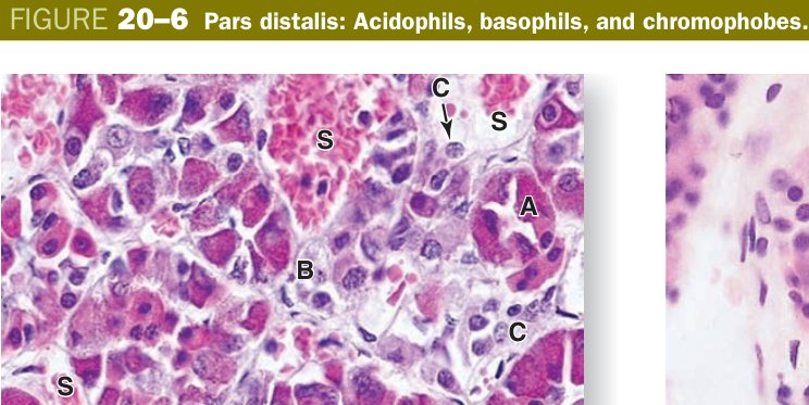
If you remember nothing else, remember the colour code. Pink cells (acidophils) build — growth hormone builds your body, prolactin builds milk. Blue cells (basophils) command the other glands — thyroid, adrenal, ovary and testis. So “blue commands, pink builds.” A pink-cell tumour gives gigantism or galactorrhoea; a blue-cell tumour gives Cushing’s or hyperthyroidism.
Acidophils = GH + Prolactin (“Acidophils Are for Growth & milk”). Basophils = “B-FLAT”: FSH, LH, ACTH, TSH. So GH is an acidophil product — basophils cannot make it.
A somatotroph adenoma in an adult gives acromegaly (jaw, hands, frontal bossing); in a child before growth-plate closure it gives gigantism. A prolactinoma is the commonest pituitary tumour — women present with amenorrhoea/galactorrhoea, men with low libido. A corticotroph adenoma drives ACTH and produces Cushing disease. After post-partum haemorrhage the anterior pituitary can infarct (Sheehan syndrome) and the mother fails to lactate — the first lost hormone is prolactin from acidophils.
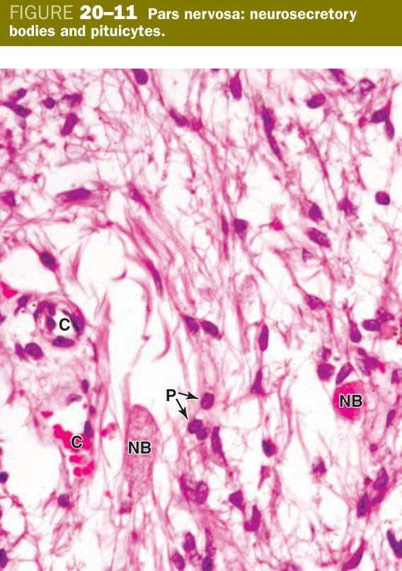
Turn the slide to the posterior lobe and the picture changes completely. There are almost no cell bodies — just a pale tangle of unmyelinated axons, scattered glial nuclei, and capillaries. The axons belong to magnocellular neurons whose cell bodies sit far above in the hypothalamus: the supraoptic nucleus (mostly antidiuretic hormone, ADH) and the paraventricular nucleus (mostly oxytocin). They synthesise their hormone in the cell body, package it with a carrier protein called neurophysin, and ship the granules down the axon by axonal transport. The pars nervosa is just where the axon terminates and the hormone is finally released into the blood.
Along the way you will see beaded swellings in the axons — these are Herring bodies. They are not separate cells; they are dilated axon terminals stuffed with neurosecretory granules, waiting for an action potential to trigger release. The other nuclei you see scattered between the axons belong to pituicytes: modified astrocyte-like glia that simply support the axons.
- Unmyelinated axons of hypothalamic neurons (supraoptic → ADH; paraventricular → oxytocin) + glial pituicytes + capillaries.
- Herring bodies = dilated axon terminals storing neurosecretory granules.
Herring body: a dilated portion of the axon terminals of hypothalamic secreting neurons in the pars nervosa, where neurosecretory granules accumulate; appears as homogeneous acidophilic masses. Contains oxytocin and ADH (transported down the axons).
Damage the hypothalamic-pituitary axis (surgery, tumour, head trauma) and the ADH supply fails — the kidney can no longer concentrate urine and the patient pours out litres of dilute urine: central diabetes insipidus. The opposite, SIADH, is inappropriate over-release of ADH (small-cell lung cancer is the classic ectopic source) — the patient retains water and goes hyponatraemic. Oxytocin deficiency is rarely symptomatic, which is why DI dominates the clinical picture of posterior pituitary failure.
Thyroid & Parathyroid
The thyroid is the only endocrine gland in the body that stores its hormone outside the cell, and that single design quirk explains everything about its histology. The structural unit is the follicle: a hollow sphere lined by one layer of simple cuboidal follicular cells, with the lumen completely filled by a pink-staining gel called colloid. The colloid is iodinated thyroglobulin — the hormone precursor — sitting safely outside the cells, weeks of hormone supply already pre-made.
When TSH from the basophils tells the thyroid to release hormone, the follicular cell pinches a drop of colloid back into itself, digests the thyroglobulin in lysosomes, and frees T4 and T3 into the basal capillary network. The taller the cell looks under the microscope, the more active it is — in hyperthyroid Graves disease the cells go columnar and the colloid gets eaten back to thin scallops; in hypothyroid Hashimoto disease the cells flatten and the colloid heaps up. The histology literally shows you the patient’s TFTs.
Wedged between the follicles — or in the basal part of the follicular epithelium — are pale, larger cells with no contact with colloid. These are the parafollicular cells (also called C cells). They are not derived from the same endoderm as the follicular cells; they migrated in from the neural crest. They secrete calcitonin, a peptide that lowers blood calcium by inhibiting osteoclasts. Calcitonin is a minor calcium hormone in adult humans, but it matters clinically because C cells give rise to medullary thyroid carcinoma.
| Cell / gland | Structure | Hormone |
|---|---|---|
| Thyroid follicular cell | Simple cuboidal epithelium around colloid-filled follicles (colloid = thyroglobulin); rich RER, Golgi | Thyroxine (T4) & triiodothyronine (T3) |
| Parafollicular (C) cell | Larger, paler cells between/within the follicular epithelium; neural crest origin | Calcitonin (lowers blood Ca²⁺) |
| Parathyroid chief (principal) cell | Small, dark, in cords | PTH (raises blood Ca²⁺) |
| Parathyroid oxyphil cell | Large, deeply eosinophilic, mitochondria-packed | Function uncertain; increase with age |
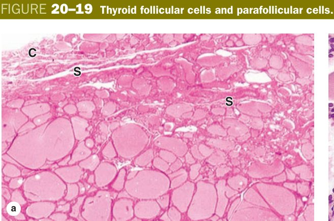
Imagine each follicle as a tiny clay pot. The potters (follicular cells) build the pot, fill it with honey (colloid), and seal it shut. When the body needs energy they reach back into the pot, scoop a spoon of honey, refine it inside themselves into pure T3/T4, and pass it out the back door into the blood. No other endocrine gland keeps weeks of hormone in storage like this — which is why patients with autoimmune thyroid disease can take months to become symptomatic; the honey pot has to empty first.
Hashimoto thyroiditis — lymphocytic infiltrate destroys follicular cells, follicles shrink, colloid disappears, patient becomes hypothyroid. Graves disease — TSH-receptor autoantibodies drive the cells columnar and scallop the colloid; hyperthyroid + ophthalmopathy. Riedel thyroiditis — gland replaced by fibrous tissue, stone-hard. Thyroglossal duct cyst — midline neck mass from incomplete obliteration of the thyroid’s embryonic descent. Cancers: papillary (most common, Orphan-Annie nuclei), follicular (haematogenous spread to bone), medullary (C cells → calcitonin, RET mutation, part of MEN-2), anaplastic (elderly, lethal).
Four pinhead-sized glands sit on the back of the thyroid, embedded in or just behind its capsule. They were discovered late in history because they were always mistaken for thyroid tissue — until surgeons realised that removing them caused fatal hypocalcaemic tetany. The parathyroid is the body’s minute-to-minute calcium thermostat, and its histology reflects how relentlessly it works.
Inside, you see two cell types in cords with a fenestrated capillary network. Chief (principal) cells are small and dark, with the typical protein-secreting machinery of RER and Golgi. They secrete parathyroid hormone (PTH), which raises blood calcium by stimulating osteoclasts, increasing renal calcium reabsorption and activating vitamin D to absorb calcium from the gut. Oxyphil cells are larger, brightly eosinophilic and crammed with mitochondria. Their function is still uncertain after a century of staring at them; they appear after puberty and increase with age. With age you also see clusters of adipocytes infiltrating the gland — a normal finding, not pathology.
Imagine a pair of taps over a bathtub of calcium. PTH (chief cells) fills the tub — pulling calcium from bone, kidney and gut. Calcitonin (C cells) drains the tub — shutting down osteoclasts. In adult humans the calcitonin tap is small; PTH does almost all the regulation. So lose your parathyroids and calcium crashes within hours; lose your C cells (e.g. total thyroidectomy) and calcium hardly moves.
Primary hyperparathyroidism — parathyroid adenoma over-secretes PTH; “stones, bones, abdominal groans, psychic moans” (kidney stones, osteitis fibrosa cystica, peptic ulcer, depression). Secondary hyperparathyroidism — chronic kidney disease lowers vitamin D and traps phosphate, low calcium drives compensatory PTH up, ending in renal osteodystrophy. Hypoparathyroidism after accidental removal at thyroid surgery → Chvostek and Trousseau signs, perioral tingling, tetany. MEN-1 = parathyroid + pituitary + pancreatic islet tumours; MEN-2 = parathyroid + medullary thyroid + phaeochromocytoma.
Adrenal (Suprarenal) Gland
The adrenal sits on top of each kidney like a yellow tricorn hat, and like the pituitary it is really two glands fused into one. The cortex grew in from mesoderm and secretes steroid hormones; the medulla migrated in from the neural crest and secretes catecholamines. On a freshly cut slice the cortex is bright yellow (lipid droplets full of cholesterol) and the medulla is brown-grey (chromaffin granules), a colour split visible to the naked eye.
Sweep through the cortex from outside in and you cross three zones in fixed order: glomerulosa, fasciculata, reticularis. Each zone has a different cell arrangement, a different cytochrome P450 enzyme set, and a different steroid output. This is the textbook’s “GFR = Salt, Sugar, Sex” pattern, and it shows up on almost every histology paper.
The zona glomerulosa is the outermost thin band — small cells in rounded clusters or arches. They are the only adrenal cells that express aldosterone synthase and they answer to angiotensin II through the renin-angiotensin system, not to ACTH. Their product is the mineralocorticoid aldosterone, which makes the kidney retain sodium and excrete potassium.
The zona fasciculata is the broadest middle zone, easily fifty to seventy percent of the cortex. Its cells run in long straight cords, two cells thick, with sinusoidal capillaries between. They look pale and foamy because their cytoplasm is packed with lipid droplets — hence the affectionate nickname spongiocytes. They make the glucocorticoid cortisol in answer to ACTH from the corticotroph basophils.
The zona reticularis is the innermost cortical band, with anastomosing cords of darker cells. They have less lipid (most has been used up making steroids) and accumulate brown lipofuscin pigment with age, which is why the zone looks dusky. Their product is weak androgens, principally DHEA, which the periphery converts to testosterone and oestrogen.
| Zone (outer→inner) | Cells | Hormone | Driven by |
|---|---|---|---|
| Zona glomerulosa | Rounded/arched clusters | Mineralocorticoids (aldosterone) | Angiotensin II / K+ |
| Zona fasciculata | Straight cords of lipid-rich “spongiocytes” | Glucocorticoids (cortisol) | ACTH |
| Zona reticularis | Anastomosing cords, lipofuscin | Androgens (DHEA) | ACTH |
| Medulla | Chromaffin cells (+ ganglion cells, central vein) | Adrenaline & noradrenaline | Preganglionic sympathetic nerves |
Cut deep enough into the adrenal and the cortical cords end abruptly at the central medulla. The cells here are large and basophilic, arranged in clumps around capillaries and central veins. They go brown when fixed in chromate salts — the reaction that earned them the name chromaffin cells. Genetically they are modified postganglionic sympathetic neurons: the embryo planned to make them into ganglion cells, then decided to drop the axon and turn them into endocrine cells instead. They are innervated directly by preganglionic sympathetic fibres, and a single sympathetic discharge dumps a flood of adrenaline (about 80%) and noradrenaline (about 20%) straight into the central vein.
Why is the medulla buried inside the cortex? Because cortisol from the fasciculata washes through the medullary sinusoids on its way out, and one of cortisol’s side jobs is to induce phenylethanolamine-N-methyltransferase, the enzyme that converts noradrenaline to adrenaline. Without high cortisol exposure, the medulla makes mostly noradrenaline. Anatomy and biochemistry beautifully reinforce each other.
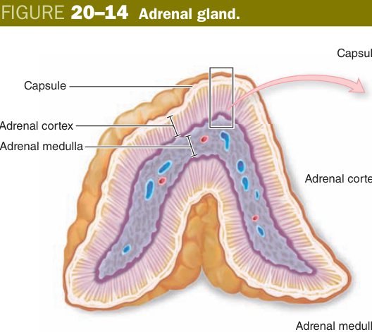
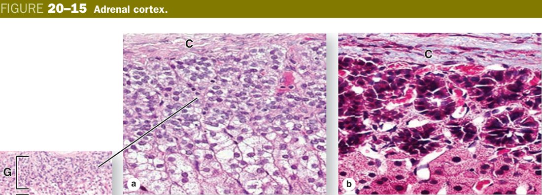
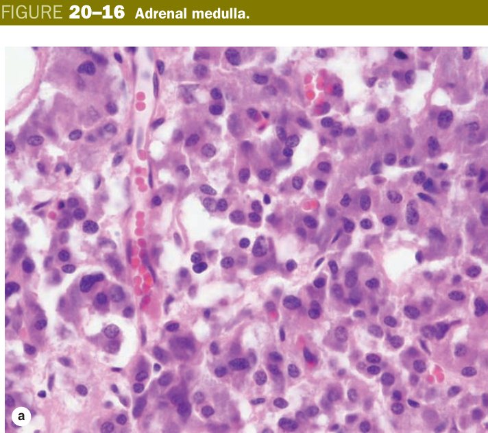
The cortex is a slow, steady hormonal shower — cortisol drips out for hours, aldosterone tunes salt over days, DHEA simmers in the background. The medulla is a wall-switch: sympathetic nerve fires, adrenaline floods the body in seconds, your pupils blow open and your heart leaps. Same organ, two different timescales — because evolution sewed a fast neural emergency line into the middle of a slow steroid factory.
“Salt, Sugar, Sex — the deeper you go, the sweeter it gets.” Glomerulosa → aldosterone (salt); Fasciculata → cortisol (sugar); Reticularis → androgens (sex). Medulla → adrenaline.
Conn syndrome — glomerulosa adenoma over-secretes aldosterone → hypertension + hypokalaemia. Cushing syndrome — excess cortisol (pituitary adenoma, ectopic ACTH, or adrenal tumour) → moon face, buffalo hump, abdominal striae, glucose intolerance. Addison disease — autoimmune destruction of the entire cortex → low cortisol + aldosterone, low Na/high K, hyperpigmentation (high ACTH drives MSH). Congenital adrenal hyperplasia — usually 21-hydroxylase deficiency, cortisol and aldosterone fall, the precursors shunt into the androgen pathway, baby girl is virilised. Phaeochromocytoma — chromaffin-cell tumour of the medulla, “rule of 10s” (10% bilateral/extra-adrenal/malignant/familial); paroxysmal hypertension, headache, sweating; part of MEN-2.
Pancreatic Islets & Pineal Gland
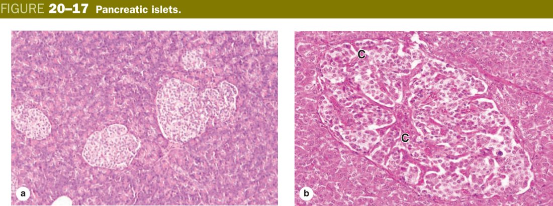
You already met the islets of Langerhans in Unit 11 with the exocrine pancreas; they reappear here because they are endocrine. Pale islands of cords sit scattered among the dark exocrine acini, each ringed by a fenestrated capillary basket. The four cell types are not distinguishable on routine H&E — you need immunohistochemistry — but the proportions are worth remembering: B (β) cells about 70% → insulin, A (α) cells about 20% → glucagon, D (δ) cells about 5% → somatostatin, and PP cells → pancreatic polypeptide. The B cells lie centrally; the A cells form a rim at the periphery.
Tucked at the back of the third ventricle, the pineal is a tiny cone of nervous tissue, no bigger than a grain of rice. Its main cells are the pinealocytes — modified neurons with long branched processes — which secrete melatonin at night and quietly in the day. Light hitting the retina is the cue: optic information runs to the suprachiasmatic nucleus, then down a sympathetic chain to the pineal, and noradrenaline switches off melatonin production. Block the light path and the pineal pumps melatonin out around the clock — which is why melatonin tablets help jet lag.
Scattered between pinealocytes are astrocyte-like interstitial cells, and clumped throughout are pale concretions of calcium phosphate called corpora arenacea or brain sand. They appear in childhood, grow with age, and have no known function — but radiologists love them: on a plain skull X-ray the calcified pineal is a midline marker, and lateral shift signals a mass.
Think of the pineal as a tiny lighthouse that only turns its lamp on when the world outside goes dark. The lamp’s glow is melatonin, and every cell with a melatonin receptor uses it to set its own clock. Block the sky (shift work, jet lag, blue-light screens) and the lighthouse gets confused; the whole body’s rhythm shifts with it.
Diabetes mellitus = islet B-cell (insulin) failure (autoimmune in type 1, insulin-resistance + B-cell exhaustion in type 2). Goitre / Graves / Hashimoto distort thyroid follicles. Cushing (excess cortisol, zona fasciculata) and Addison (whole cortex destroyed) are mirror diseases. Pituitary somatotroph adenoma → gigantism/acromegaly (recall the Unit 4 growth plate). A pineal tumour in a child can compress the tectum (Parinaud syndrome, upward-gaze palsy) and shorten the night-time melatonin signal, occasionally producing precocious puberty.
TMU Exam Drill
📝 Open the full TMU Question Bank — 20 MCQ + 6 terms + 5 essays →
Authentic Tianjin Medical University past-paper questions (2021 Final & the multi-section Final with answer key) mapped to this unit, in the real exam format. Click Show answer to self-test.
□ Single best answer
- A. glucocorticoid
- B. mineralocorticoid
- C. androgen
- D. epinephrine
- E. thyroid hormone
- A. glucocorticoid
- B. mineralocorticoid
- C. androgen
- D. epinephrine
- E. norepinephrine
- A. TSH
- B. ACTH
- C. FSH
- D. LH
- E. growth hormone (somatotropin)
□ Fill in the blank
- (fill three zones)
- (fill three basophils + the connection)
□ True or false
□ Explain the following terms
Endocrine system complete
Pituitary, thyroid, adrenal zones & islets mastered. Next: Urinary System.