Digestive Glands
Salivary Glands
The salivary glands do something deceptively complex: they take roughly a litre of plasma-derived fluid each day, dress it up with digestive enzymes, lubricating mucins, antibodies and electrolytes, and deliver it to your mouth on demand. To get all of that into one organ they use a modular plan — acini (the secretory berries on the end of branches) connected to a tree of ducts that progressively modify what the acini just produced.
You meet three pairs of major glands and dozens of minor ones scattered through your lips, palate and tongue. The big three each pick a personality. The parotid is purely serous — watery, enzyme-rich, full of zymogen granules and famously dotted with adipocytes that lift out as clear holes on H&E. The submandibular is mixed but mostly serous, throwing crescent-shaped serous demilunes onto mucous acini. The sublingual flips that around — mostly mucous with a few serous caps.
Think of the acinar cell as a tiny shop. A serous cell is a busy enzyme factory: dark basophilic basal cytoplasm packed with rough ER, an apical hoard of pink zymogen granules, a round central nucleus. A mucous cell is a warehouse for gloopy mucin: clear, bubbly, almost empty-looking cytoplasm and a flat nucleus squashed against the basement membrane like someone leaning on a wall.
Wrapping the acinus — squeezed between the secretory cells and the basement membrane — sits the myoepithelial cell, the “basket cell.” It has long branching arms full of actin and myosin and behaves like a hand cupped around a juice sac: when secretomotor nerves fire, it squeezes the acinus and helps fling its contents into the duct. Myoepithelial cells also coat the smallest intercalated ducts but disappear by the time you reach the striated duct.
Compound exocrine glands with serous, mucous or mixed acini, a duct system (intercalated → striated → excretory) and myoepithelial cells.
| Gland | Acini | Secretion |
|---|---|---|
| Parotid | Pure serous | Watery, enzyme-rich (amylase); basophilic pyramidal cells with zymogen granules |
| Submandibular | Mixed, mostly serous (serous demilunes) | Seromucous |
| Sublingual | Mixed, mostly mucous | Mainly mucous |
Saliva does not leave the acinus the same fluid it arrives at the mouth. The duct tree edits it. Intercalated ducts (simple cuboidal) collect raw secretion. Striated ducts (tall columnar with basal infoldings packed with mitochondria) actively pump Na+ back out of the saliva and dump K+ and HCO3- in, leaving the final saliva hypotonic and slightly alkaline — recall the basal membrane infoldings you saw in Unit 2 (epithelia). The excretory duct, lined initially by stratified columnar that turns into stratified squamous as it approaches the oral mucosa, just carries the finished product.
Mumps is paramyxovirus parotitis — swollen, painful parotids. Sjögren syndrome is autoimmune destruction of salivary and lacrimal glands → dry mouth and eyes. The most common salivary tumour is pleomorphic adenoma of the parotid; Warthin tumour (also parotid) is the second most common; mucoepidermoid carcinoma is the most common malignant salivary tumour. Stones (sialolithiasis) classically lodge in the submandibular (Wharton's) duct because its secretion is thicker and the duct runs uphill.
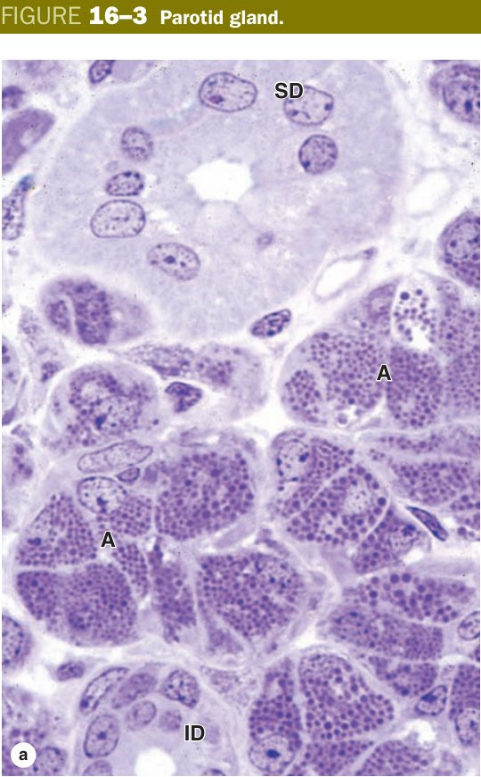
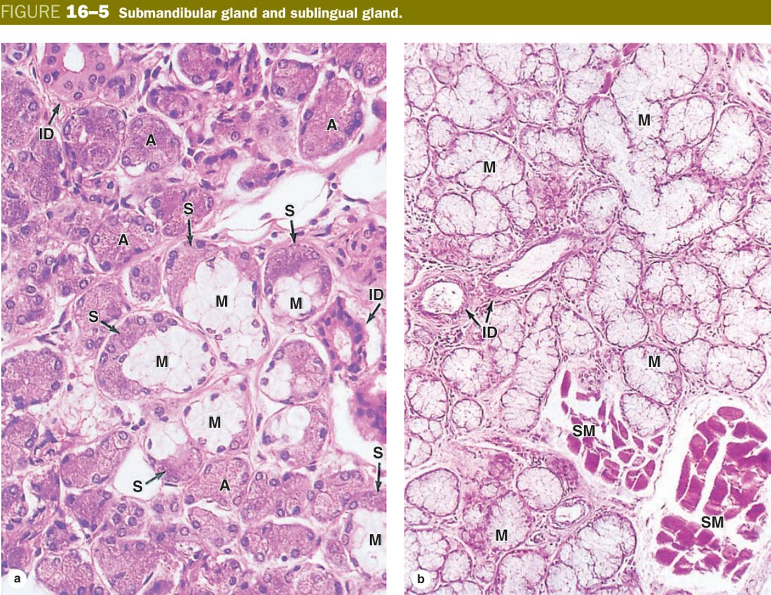
Read the duct tree as a small kidney glued to each gland: the acinus filters; the duct fine-tunes. The further you walk down the duct, the bigger the lumen, the taller the epithelium and the less ionic modification happens. The striated duct is the workhorse — if you find a duct with vertical basal lines on H&E, you are looking at mitochondria-packed columnar cells doing electrolyte work.
| Duct | Epithelium | Key Feature |
|---|---|---|
| Intercalated duct | Simple cuboidal/squamous; short narrow ducts leaving acini | In serous glands these are long; mucous glands have short/absent intercalated ducts |
| Striated duct (secretory duct) | Simple tall columnar; prominent basal striations (interdigitating plasma membrane infoldings packed with mitochondria) | Actively reabsorbs Na+, secretes K+ → hypotonic saliva; only in salivary glands |
| Excretory duct | Changes from stratified columnar → stratified squamous near the oral cavity | Drains into the oral mucosa; no ionic modification |
Star-shaped contractile cells basket cells lying between the acinar cells and the basement membrane; contain actin & myosin; squeeze the acinus like a hand to aid secretion. Present in both serous and mucous glands and in intercalated ducts. They do not line the striated duct.
Parotid = Pure Serous (both start P). Sub-Mandibular = Mostly serous (Mixed). Sub-Lingual = Mostly mucous. Demilunes = serous caps on mucous acini (best seen in submandibular).
Pancreas
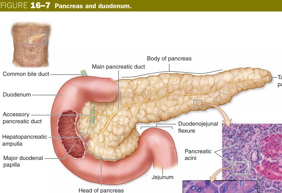
The pancreas is two organs jammed into one capsule. The bulk of the tissue is a vast serous exocrine gland that pours digestive enzymes into the duodenum; sprinkled through that exocrine sea are pale islands of endocrine cells — the islets of Langerhans — that quietly run blood-glucose homeostasis. Histologically you can usually tell which is which from across the room: exocrine acini are deep purple and grainy; islets look like pale pink ghosts because their cells secrete hormones straight into capillaries and so do not stockpile zymogen.
Each exocrine acinus is a cluster of serous cells with the same architecture as the parotid — basal RER making zymogen, apical granules waiting for cholecystokinin to release amylase, lipase, trypsinogen, chymotrypsinogen and the other proenzymes. The proenzymes part matters: the pancreas dares not activate trypsin inside itself, so it ships everything inactive and lets the duodenal brush border enzyme enterokinase fire the cascade outside the gland.
Imagine pushing a finger into a soft tennis ball. The finger does not disappear — it dimples in. The duct system does the same thing to each pancreatic acinus: the first cells of the intercalated duct (pale, cuboidal, with little cytoplasm) actually sit inside the acinus. Those intra-acinar duct cells are the centroacinar cells, and they are a histological fingerprint unique to the pancreas.
From there the duct system is intercalated → intralobular → interlobular → main pancreatic duct (of Wirsung). The duct cells — not the acinar cells — secrete the alkaline, bicarbonate-rich watery fluid that neutralises gastric acid in the duodenum. There are no striated ducts here. That is a reliable exam discriminator: striated duct = salivary; centroacinar cell = pancreas.
A mixed gland: a large exocrine part + scattered endocrine islets.
| Part | Structure / function |
|---|---|
| Exocrine | Serous acini (basophilic basal RER + apical zymogen granules) secreting digestive enzymes; centroacinar cells begin the duct; no striated ducts; duct cells add HCO3- |
| Endocrine — islets of Langerhans | Pale spherical clusters scattered among acini, rich in fenestrated capillaries; cells: β (~70%, centre) → insulin; α (~20%, periphery) → glucagon; δ (~5%) → somatostatin; PP → pancreatic polypeptide; rare ε → ghrelin |
The islets themselves are tightly organised. Beta cells, which make insulin, dominate the centre — about 70% of the islet. Around the rim you find alpha cells producing glucagon (about 20%) and delta cells producing somatostatin (about 5%). PP cells and the rare epsilon (ghrelin) cells are scattered. The islet capillary network is fenestrated and the cells release hormones straight through it.
Beta cells are the main power station (insulin = “store the glucose”). Alpha cells are the back-up generator that kicks in when fuel runs low (glucagon = “release the glucose”). Delta cells are the dispatcher with the kill-switch — somatostatin damps both sides down so neither runs away. The whole grid is wired straight into fenestrated capillaries.
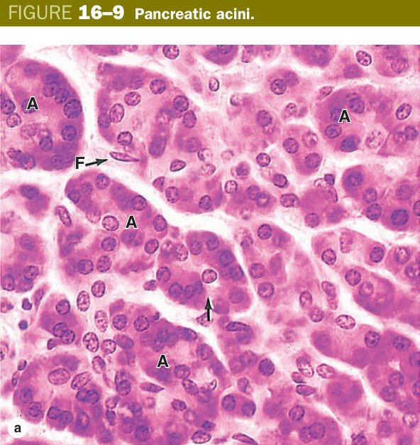
Acute pancreatitis — trypsinogen activates inside the gland (gallstone obstruction, alcohol) and the pancreas digests itself; serum amylase/lipase shoot up. Chronic pancreatitis — repeated injury → fibrosis, calcification, exocrine and eventually endocrine failure. Cystic fibrosis — CFTR mutation thickens duct secretion → ductal obstruction and exocrine deficiency. Type 1 DM — autoimmune destruction of β cells. Type 2 DM — insulin resistance with later β-cell exhaustion. Ductal adenocarcinoma arises from duct epithelium and has a grim prognosis. Insulinoma (β-cell tumour) causes fasting hypoglycaemia; gastrinoma (Zollinger–Ellison) causes intractable peptic ulcers.
Liver — the Hepatic Lobule (essay-grade)
The liver is the body's central biochemical factory — it makes plasma proteins, stores glycogen, detoxifies xenobiotics, manufactures bile and filters incoming gut blood through a resident army of macrophages. To do all of that, its 1.5 kg of tissue is built from one repeating module so simple that exam questions revisit it every year: the hepatic lobule.
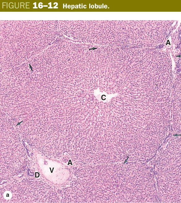
Anatomists have argued for a century about which way to draw that module. Three competing definitions exist — each emphasising a different function — and you need to know all three. The classic lobule centres on a central vein and is the easiest to see on slides. The portal lobule centres on a portal triad and follows bile drainage. The liver acinus of Rappaport centres on a terminal portal venule and explains why hepatocytes near the central vein die first in shock. We will build them up in that order.
Picture a city block. If you draw the block around the central drain (sewer), you get the classic lobule — blood flow. If you draw it around the central well (bile duct), you get the portal lobule — bile drainage. If you draw a diamond between two wells and two drains, you get the acinus — oxygen supply. Same city, different maps for different questions.
Hepatic lobule: the classic structural unit — a polygonal (hexagonal) prism with a central vein in its axis and portal triads at its corners. Plates/cords of hepatocytes radiate from the central vein, separated by hepatic sinusoids.
Now zoom into the classic lobule. From the central vein, one-cell-thick plates of polygonal hepatocytes radiate outward like spokes on a wheel, branching and anastomosing as they go. Between every two plates runs a sinusoid — a wide, leaky capillary that carries mixed portal-venous and hepatic-arterial blood centripetally toward the central vein. At the six corners of the hexagon sit the portal triads: a branch of the hepatic artery, a branch of the portal vein and a bile duct, glued together in connective tissue.
| Component | Detail |
|---|---|
| Central vein | Smallest tributary of the hepatic vein; receives sinusoidal blood |
| Hepatic plates/cords | Anastomosing single rows of hepatocytes radiating from the central vein |
| Hepatocyte | Polygonal; 1–2 round central nuclei (often polyploid); acidophilic cytoplasm with basophilic RER patches; abundant SER, mitochondria, glycogen, peroxisomes |
| Hepatic sinusoids | Wide, irregular discontinuous capillaries between plates; fenestrated, no basal lamina; lined by endothelial cells + Kupffer cells (macrophages) |
A hepatocyte is a polygonal cell about 20 µm across with one or two large central nuclei — binucleate and polyploid forms are normal here because hepatocytes routinely double their genome to keep up with biosynthetic load. On H&E the cytoplasm looks pink with scattered basophilic patches; those basophilic patches are stacks of rough ER. The unusually pink areas are smooth ER and glycogen.
Drill into the EM and the cell becomes a list of organelles you already know — just amplified. The smooth ER is colossal: this is where cytochrome P450 enzymes detoxify drugs, ethanol, steroid hormones and bilirubin, and where lipid and steroid metabolism happen. Phenobarbital and other inducers can double the SER in days. The rough ER exports plasma proteins — albumin, fibrinogen, prothrombin, the clotting cascade — which is why liver failure presents with low albumin and a high INR. Mitochondria are everywhere (around 2000 per cell, more than any other parenchymal cell). Peroxisomes oxidise long-chain fatty acids and detoxify hydrogen peroxide via catalase. Glycogen sits in the cytosol as pink PAS-positive rosettes; it builds up after a meal and is mobilised by glucagon between meals. Lipid droplets appear in small numbers normally; if they take over the cell you are looking at steatosis.
| Organelle / Inclusion | Detail & Significance |
|---|---|
| SER (smooth ER) | Extremely abundant; site of detoxification (CYP450 enzymes), steroid & lipid metabolism, glycogen synthesis. Proliferates with drug inducers (e.g. phenobarbital). |
| RER (rough ER) | Synthesises plasma proteins: albumin, fibrinogen, prothrombin, clotting factors; seen as basophilic patches. |
| Mitochondria | ~2 000 per hepatocyte — most of any cell; provide ATP for the metabolically demanding hepatocyte. |
| Peroxisomes | Contain catalase; oxidise long-chain fatty acids, detoxify H2O2. |
| Glycogen | Appears as PAS-positive pink granules; stored in the cytosol; mobilised under glucagon stimulation. |
| Golgi complex | Packages and secretes proteins into bile and sinusoidal blood. |
| Lipid droplets | Normal in small amounts; accumulate in steatosis (fatty liver). |
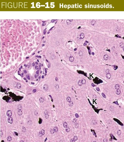
The sinusoid is the liver's signature capillary. It is deliberately leaky — discontinuous, with large fenestrations and almost no basal lamina — so that plasma can wash directly over the hepatocyte surface and exchange anything short of an erythrocyte. Between the sinusoidal endothelium and the hepatocyte microvilli sits a thin gap called the perisinusoidal space of Disse. That space is where the real metabolic traffic happens.
Three cell types share the sinusoidal wall and the space of Disse. Endothelial cells form the leaky lining. Kupffer cells, fixed macrophages of the mononuclear phagocyte system, sit inside the sinusoidal lumen and phagocytose old red cells, bacteria carried up from the gut, and immune complexes. Hepatic stellate cells of Ito live inside the space of Disse; in health they store vitamin A in lipid droplets, but in chronic injury they transform into myofibroblasts that lay down collagen — the engine of cirrhosis.
The sinusoid is a slow muddy river with deliberately broken banks. Plasma soaks out into the marketplace (space of Disse) where hepatocytes lean over their stalls and exchange goods. Kupffer cells are river police boats patrolling for invaders. Ito cells are the warehouse keepers on the bank — calm in peacetime, but they bring out the bricks and start building walls (fibrosis) when there is trouble.
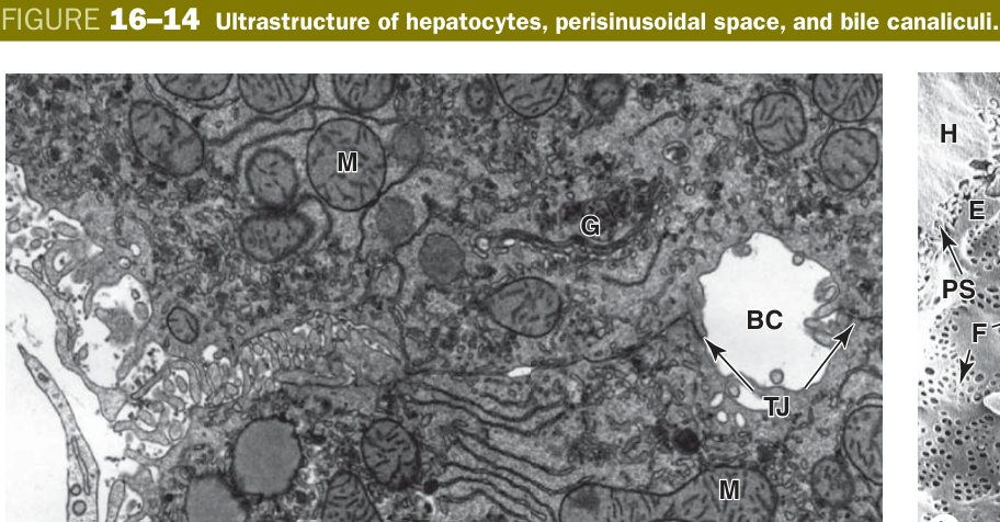
Bile takes the opposite path. Between any two adjacent hepatocytes the apposed plasma membranes form a tiny groove sealed shut by tight junctions, with microvilli sticking into the lumen — that is the bile canaliculus. It is too small to see in routine H&E and is classically demonstrated by silver impregnation. Bile flows from these canaliculi centrifugally (outward, away from the central vein) to the lobule edge, drains into a transition channel called the canal of Hering, and empties into the bile duct of the portal triad.
- Perisinusoidal space (of Disse): between the sinusoidal endothelium and the hepatocyte; contains plasma, hepatocyte microvilli and hepatic stellate (Ito) cells that store vitamin A — site of metabolic exchange.
- Bile canaliculi: tiny channels formed by the indented plasma membranes of two adjacent hepatocytes, sealed by tight junctions, with microvilli; shown by silver impregnation. Bile flows away from the central vein toward the portal triad.
- Kupffer cells: sinusoidal macrophages (MPS) that phagocytose old erythrocytes & debris.
At each corner of the hexagon, embedded in a triangle of connective tissue, sits the portal area containing the portal triad: a branch of the hepatic artery (oxygen), a branch of the portal vein (nutrients from the gut) and an interlobular bile duct (drainage), often accompanied by a small lymphatic. The hepatic artery and portal vein together pour into the sinusoids; the bile duct collects bile coming the other way.
Hold the directions of flow in your head as opposites. Blood moves centripetally (corners → centre): portal vein + hepatic artery → sinusoids → central vein → sublobular vein → hepatic vein → IVC. Bile moves centrifugally (centre → corners): bile canaliculi → canals of Hering → interlobular bile duct → hepatic ducts → common hepatic duct.
Functionally, the liver wears six hats simultaneously. It secretes bile for fat emulsification. It synthesises plasma proteins — albumin (oncotic pressure), clotting factors (II, VII, IX, X), most carrier proteins. It handles metabolism — glycogen storage and release, gluconeogenesis, lipoprotein assembly, urea synthesis. It detoxifies drugs and xenobiotics in zone-3 SER. It stores vitamins A (Ito cells), D, B12, iron and copper. And it filters portal blood through Kupffer macrophages before that blood reaches the systemic circulation.
- Portal triad (portal area): at lobule corners — a branch of the hepatic artery + branch of the portal vein + an interlobular bile duct (± lymphatic).
- Blood flow: portal vein + hepatic artery → sinusoids → central vein → sublobular → hepatic vein. Bile flow is opposite: canaliculi → bile ductule (canal of Hering) → bile duct.
- Functions: bile secretion, plasma-protein synthesis (albumin), metabolism & detoxification, glycogen/vitamin storage; Kupffer phagocytosis; Ito cells store vitamin A.
If the classic lobule answers “where does the blood drain?”, Rappaport's acinus answers “where is the oxygen?” Pick any terminal portal venule and its companion arteriole as the centre, then draw a diamond out to the two flanking central veins. That diamond is the liver acinus, and it is divided into three concentric zones based on distance from the inflow.
Zone 1 (periportal) hepatocytes are closest to the incoming oxygenated blood, so they are the best fed and the first to regenerate. They lead oxidative metabolism, gluconeogenesis and bile acid handling. They are also the first to meet anything coming up from the gut — including viruses — which is why viral hepatitis tends to start here. Zone 3 (centrilobular / perivenous) hepatocytes sit furthest from the inflow and stew in low-oxygen blood. They specialise in CYP450 detoxification (good for clearing drugs, dangerous when those drugs make toxic intermediates), glycolysis and lipogenesis. They die first in ischaemia, shock and right-heart failure, which is why “centrilobular necrosis” is the classic histology of congestive hepatopathy and paracetamol overdose. Zone 2 is the buffer in between.
Imagine swimmers holding their breath as they push from one end of a pool (portal triad, full oxygen) to the other (central vein, empty). The swimmers at the start (zone 1) are fine. The ones in the middle (zone 2) struggle. The ones at the far end (zone 3) drown first whenever the supply is cut.
| Zone | Location | Oxygenation | Vulnerability |
|---|---|---|---|
| Zone 1 (periportal) | Closest to portal triad vessels | Best oxygenated | First damaged by toxins/viral hepatitis; first to regenerate; site of oxidative metabolism & gluconeogenesis |
| Zone 2 (mid-zone) | Intermediate | Intermediate | Intermediate vulnerability; yellow fever necrosis |
| Zone 3 (perivenous/centrilobular) | Closest to central vein | Least oxygenated | First to undergo ischaemic necrosis (right heart failure, shock, paracetamol); site of fatty change; detoxification enzymes (CYP450) highest here |
Viral hepatitis (A, B, C, D, E): hepatocyte inflammation; chronic B and C lead to cirrhosis and HCC. NAFLD/NASH: obesity-driven fatty change starting in zone 3. Cirrhosis: chronic injury activates Ito cells → myofibroblasts → collagen → nodular scarring and distortion of vascular architecture. Portal hypertension follows: oesophageal varices, caput medusae, splenomegaly, ascites. Hepatocellular carcinoma arises on a cirrhotic background. Jaundice: pre-hepatic (haemolysis), hepatic (hepatocyte failure) or post-hepatic (biliary obstruction). Obstruction of bile canaliculi or ducts produces a conjugated hyperbilirubinaemia with pale stools and dark urine.
Gallbladder
The gallbladder has one job: hold the bile the liver makes between meals, concentrate it 5–10 fold, and squeeze it into the duodenum when a fatty meal arrives. Its histology is shaped entirely around that storage-and-concentration brief. Importantly, the wall is unusual: it has no muscularis mucosae and no submucosa, so the textbook gut-tube plan you learnt in Unit 10 does not apply here. Going from lumen out you meet only mucosa → muscularis → perimuscular connective tissue → adventitia/serosa.
The mucosa is folded into low villus-like ridges when the bladder is empty and flattens out when full. It is lined by a simple columnar epithelium covered in apical microvilli, supported by mitochondria-rich cytoplasm. Those mitochondria power Na+/K+-ATPase pumps along the lateral membranes; sodium is pumped out into the lateral intercellular space, water follows by osmosis, and bile is concentrated as it sits in the lumen. There are no goblet cells and no glands — the gallbladder absorbs, it does not secrete.
Bile arrives from the liver dilute, like seawater. The gallbladder epithelium is a row of solar pumps: it sucks out sodium and water, leaving behind a thick concentrate of bile salts, cholesterol and bilirubin pigments. When CCK fires from the duodenum, the smooth muscle wall contracts and squirts the concentrate down into the gut.
A peculiar feature unique to the gallbladder is the Rokitansky–Aschoff sinus: a mucosal diverticulum that punches through the lamina propria and dives into the muscle layer. A few exist normally; they multiply and deepen in chronic cholecystitis, where they become little pockets in which bile, bacteria and even stones can lodge.
A pear-shaped musculomembranous sac that stores and concentrates bile (5–10×). Wall has four layers (no muscularis mucosae, no submucosa).
| Layer | Structure |
|---|---|
| Mucosa | Simple columnar epithelium with numerous microvilli (brush border) + abundant mitochondria for active Na+/water absorption; lamina propria beneath. Mucosa forms low folds when empty, flattens when full. |
| Rokitansky–Aschoff sinuses | Epithelial invaginations (diverticula) penetrating into the muscularis — a diagnostic feature of the gallbladder; can harbour stones & infection. |
| Muscularis | Smooth muscle in interlacing bundles (oblique, longitudinal, circular — no distinct layers); no muscularis mucosae. |
| Adventitia/Serosa | Dense fibrous tissue; peritoneum (serosa) covers the free surface; liver surface = adventitia only. |
Unlike the gut tube, the gallbladder wall goes directly mucosa → muscularis → adventitia/serosa. The absence of muscularis mucosae means the Rokitansky–Aschoff sinuses extend directly into the muscle layer. This is a classic exam distinction.
Cholelithiasis (gallstones) come in two flavours: cholesterol stones (most common, yellow, the 4Fs — fat, female, fertile, forty) and pigment stones (black in haemolysis, brown in infection). Acute cholecystitis usually starts when a stone impacts the cystic duct → bile stasis → chemical and bacterial inflammation, classically eliciting a positive Murphy sign (arrest of inspiration on palpation of the RUQ). Chronic cholecystitis deepens the Rokitansky–Aschoff sinuses and can end in porcelain gallbladder — a calcified wall with raised carcinoma risk. Stone migration into the common bile duct (choledocholithiasis) causes obstructive jaundice; lodgement at the ampulla can trigger gallstone pancreatitis.
TMU Exam Drill
📝 Open the full TMU Question Bank — 20 MCQ + 6 terms + 5 essays →
Authentic Tianjin Medical University past-paper questions (2021 Final & the multi-section Final with answer key) mapped to this unit, in the real exam format. Click Show answer to self-test.
□ True or false
□ Structure essay
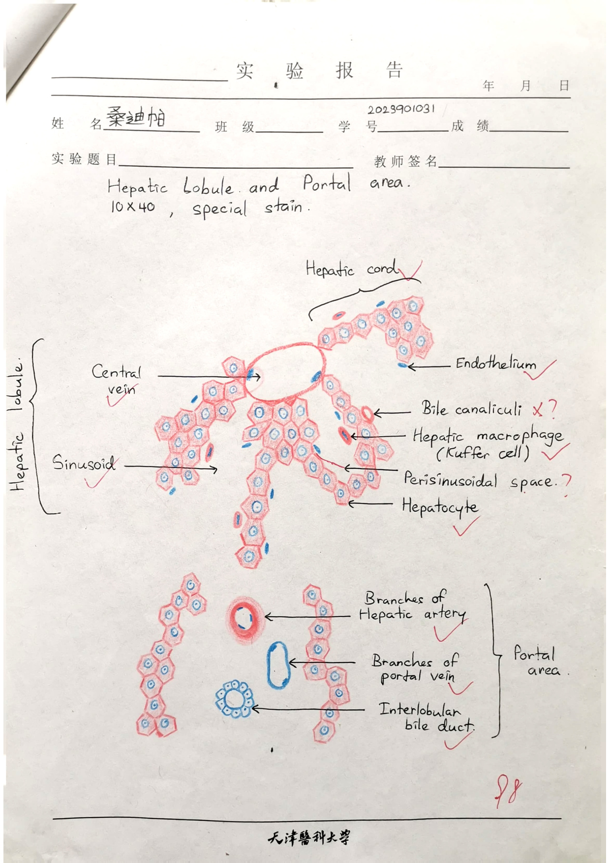
- Hepatic cords/plates — hepatocytes radiating from the central vein, anastomosing.
- Hepatic sinusoids — between the cords; lined by endothelium + Kupffer cells.
- Perisinusoidal space (of Disse) — between sinusoidal endothelium & hepatocytes.
- Bile canaliculi — tiny channels formed by indented plasmalemmas of opposing hepatocytes.
- Portal areas at corners: portal triad (hepatic artery, portal vein, bile duct).
Digestive glands complete
Salivary, pancreas (acini + islets) & the liver lobule essay mastered. Next: Endocrine System.