Digestive Tract
The General 4-Layer Plan
Before you memorise a single cell type, learn the wall. From the lower oesophagus to the anal canal, the digestive tube is built the same way: four concentric tunics, each with its own job, stacked from the lumen outward. Once you can see the four layers in your mind, every regional speciality — gastric glands, intestinal villi, colonic crypts — is just a local variation on this single body plan.
The innermost is the mucosa, and it has three sub-layers of its own: an epithelium facing the lumen (whatever epithelium that region needs), a lamina propria of loose connective tissue underneath it carrying capillaries, glands and lymphoid tissue, and a thin slip of smooth muscle called the muscularis mucosae that gently moves the mucosa independently of the rest of the wall. Below the mucosa lies the submucosa — denser connective tissue holding the larger arteries, veins and lymphatics, plus the submucosal (Meissner) plexus of enteric neurons. In two places only — the oesophagus and the duodenum — the submucosa also contains glands.
Outside the submucosa comes the muscularis externa: an inner ring of circular smooth muscle that constricts the lumen, an outer sleeve of longitudinal smooth muscle that shortens the tube, and sandwiched between them the myenteric (Auerbach) plexus — the second enteric ganglion network, driving peristalsis. Together Meissner and Auerbach make up the enteric nervous system, the so-called "second brain" of the gut that can run motility and secretion even when the vagus is cut. The outermost layer is either a serosa (mesothelium + thin connective tissue, slippery, present wherever the gut is intraperitoneal) or an adventitia (connective tissue only, present where the gut is retroperitoneal and fixed to the body wall, e.g. most of the oesophagus and the rectum).
Picture a garden hose with four layers stitched around it. The inner liner that touches the water is the mucosa (different lining for different "fluids" — abrasive food, acid, chyme, faeces). Around that, padding with wiring inside it — the submucosa with Meissner's plexus. Around that, two muscle sleeves running at right angles to each other with a second wiring loom between them — muscularis externa with Auerbach's plexus. And finally either a slippery raincoat (serosa, free to slide) or a rough canvas wrap (adventitia, glued to the body wall). Every regional difference is just a swap of the inner liner.
| Layer | Components |
|---|---|
| Mucosa | Epithelium + lamina propria (loose CT, glands, vessels, lymphoid tissue) + muscularis mucosae (thin smooth muscle) |
| Submucosa | Dense CT, larger vessels, submucosal (Meissner's) plexus; oesophageal & duodenal glands |
| Muscularis externa | Inner circular + outer longitudinal smooth muscle; myenteric (Auerbach's) plexus between them |
| Serosa / adventitia | Serosa = mesothelium + CT (intraperitoneal); adventitia = CT only (retroperitoneal) |
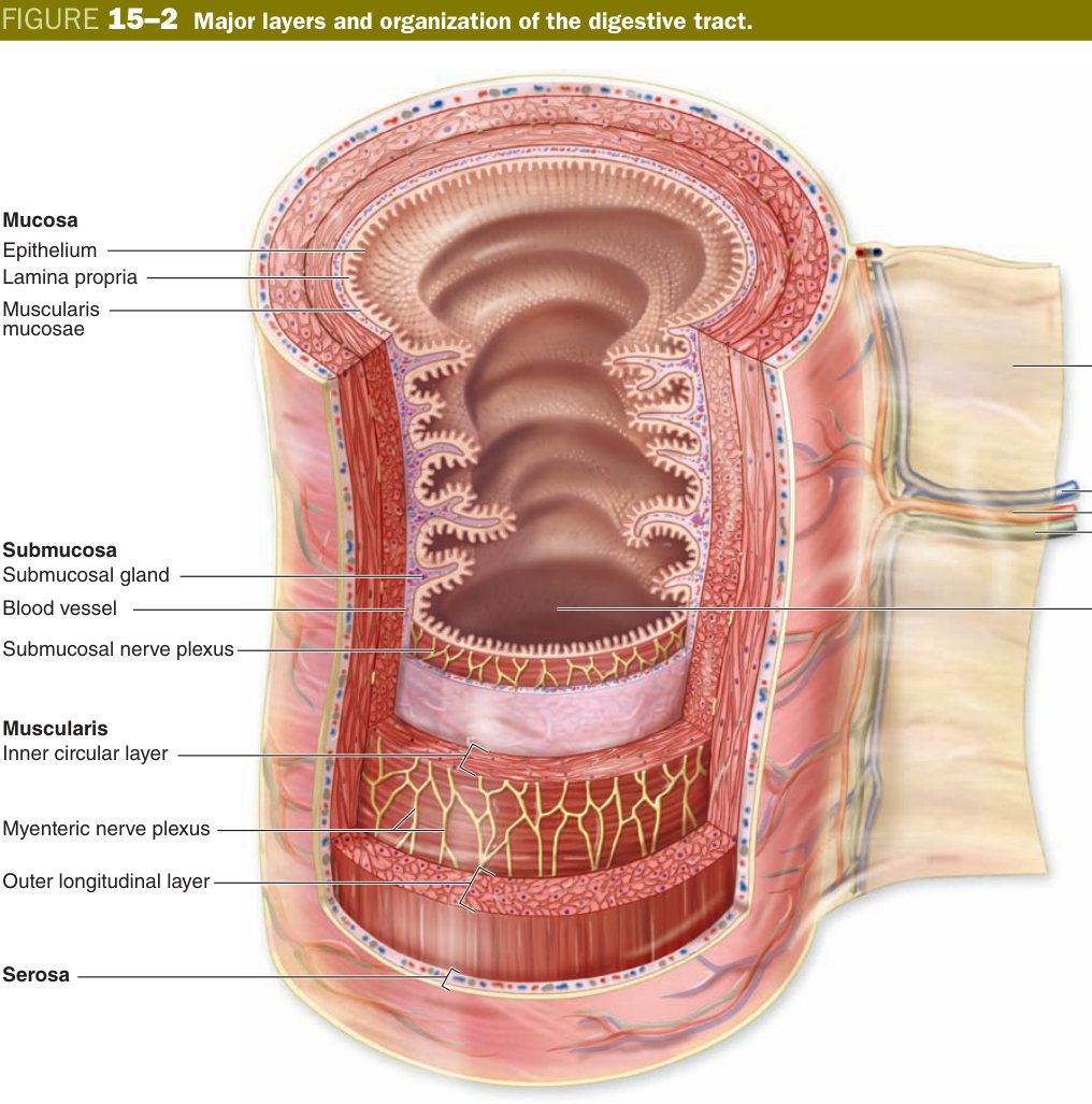
Hirschsprung's disease: failure of neural-crest cells to colonise the distal hindgut leaves a segment with no Auerbach plexus (and no Meissner). The aganglionic segment cannot relax — so stool piles up proximal to it, the upstream colon balloons into a megacolon, and the neonate fails to pass meconium in the first 48 h. The diagnosis is made histologically: a rectal biopsy showing absence of ganglion cells in both plexuses.
Oesophagus
The oesophagus has one job: deliver a bolus of dry, lumpy, scratchy food from the pharynx to the stomach in seconds, without injuring its own wall. Everything about its histology serves that brief. The lining you would expect is therefore a tough, abrasion-resistant epithelium — and that is exactly what you see down the microscope: non-keratinised stratified squamous epithelium, the same protective design used in the mouth, the vagina and the cornea. Many cell layers means losing the top few to a passing crust of toast costs the organ nothing — the basal cells just push up replacements.
Underneath, the lamina propria carries small mucous cardiac oesophageal glands near the stomach junction. The real workhorse mucus glands, however, sit one layer deeper in the submucosa — the oesophageal proper glands, scattered along the whole length, draining via short ducts onto the surface. The mucus they release coats the bolus and lubricates its slide. Remember that submucosal glands are unusual; only two regions of the gut have them, and the oesophagus is one.
The muscularis externa tells you where you are along the tube. The upper one-third is skeletal muscle (continuous with the pharynx, under voluntary control at the start of the swallow); the middle third is mixed skeletal and smooth, transitioning; the lower one-third is smooth muscle, taking over for involuntary peristalsis into the stomach. The outermost layer is mostly adventitia — the oesophagus is fixed in the posterior mediastinum and is not free to slide — only the short intra-abdominal segment below the diaphragm acquires a serosa.
Think of the oesophagus as a kitchen-grade silicone slide for rough food. You want the inside layer thick and replaceable (stratified squamous, like calloused skin), oil glands embedded in the wall (submucosal mucous glands), and at the top end you keep voluntary control to start the slide (skeletal muscle), but once gravity and peristalsis take over you switch to autopilot (smooth muscle).
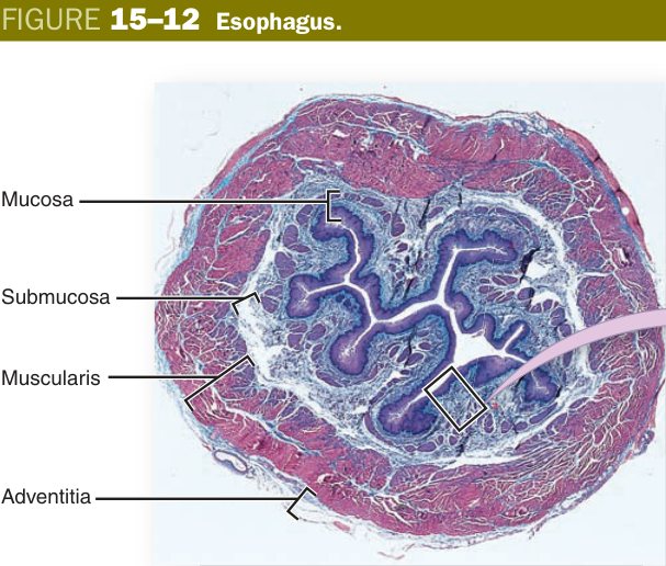
If chronic acid reflux burns the lower oesophagus, the stratified squamous lining cannot survive in that chemical environment, and stem cells switch programmes. They begin to differentiate instead into simple columnar epithelium with goblet cells — the kind of lining you would normally see in the intestine. This is Barrett's oesophagus: an example of metaplasia, a reversible change of one mature epithelium into another. The metaplastic columnar mucosa is more resistant to acid but is also a known precursor to oesophageal adenocarcinoma. So the histological diagnosis is biopsy-confirmed intestinal-type columnar epithelium in the distal oesophagus.
Stomach Mucosa (essay-grade)
The stomach is the only organ in the body that deliberately makes its own lumen as corrosive as a battery — pH 1–2 hydrochloric acid mixed with the protease pepsin. That single fact controls the histology of every layer. The lining must produce the acid, secrete the enzyme, hire endocrine staff to regulate the operation, and at the same time defend itself from being digested. So the mucosa is a five-cell-type chemical factory wrapped in a protective mucus blanket.
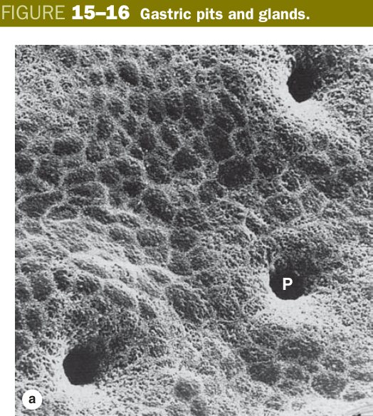
The luminal surface, and the lining of the shallow pits that dip down into it, is a single layer of simple columnar surface mucous cells. Every one of these cells secretes a thick, viscid, bicarbonate-rich mucus that pastes itself over the epithelium as an unstirred mucus-bicarbonate layer. Across this layer there is a remarkable pH gradient — pH 1 at the luminal surface, pH 7 against the epithelial apex — so the cells themselves never see the acid they help to manufacture. This is the gastric mucosal barrier, and breaking it is the first step in every peptic ulcer.
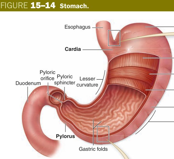
Each shallow surface depression is a gastric pit; deep to the pits, packed shoulder-to-shoulder in the lamina propria, are the gastric glands. The glands look different depending on what region of the stomach you are in: cardiac and pyloric glands are short, branched and mostly mucous; the fundic/body glands are long, straight tubular factories housing the famous five cell types. The mucosa rests on a thin muscularis mucosae, and below that the submucosa, two muscle layers of the externa plus an extra inner oblique layer (only the stomach has three muscle layers, for churning), and the serosa.
Walk into a fundic gland as if it were a chemical plant. At the entrance: the mucous-neck cells spraying lubricant onto every visitor. Just inside, perched on the upper walls, the parietal cells — round, pink, mitochondria-stuffed — pumping protons until the air burns. Down the corridor at the base, the chief cells — blue, packed with rough ER — sit at workbenches making pepsinogen. Hidden along the floor, the enteroendocrine cells (G cells in the pylorus) play the role of supervisors, releasing gastrin to tell the parietal cells when to ramp up. And tucked in the neck, the stem cells — HR — renewing the whole workforce every few days. The sneeze-guard over the whole operation is the mucus-bicarbonate layer.
Surface = simple columnar surface mucous cells dipping into gastric pits; the lamina propria is packed with gastric (fundic) glands. Five gland cell types:
| Cell | Location | LM | Secretes |
|---|---|---|---|
| Chief (zymogenic) cell | Base of gland | Basal RER → basophilic; apical zymogen granules | Pepsinogen (+ gastric lipase) |
| Parietal (oxyntic) cell | Upper/neck | Large, round, intensely acidophilic (“fried-egg”) | HCl + intrinsic factor |
| Mucous neck cell | Neck | Pale | Acidic mucus |
| Stem cell | Neck/isthmus | Small | Renew the epithelium |
| Enteroendocrine cell (G cell, pylorus) | Base | Small, clear | Hormones (gastrin, somatostatin, etc.) |
Below the glands sits the muscularis mucosae. Note the regional asymmetry: cardiac and pyloric regions have shorter glands with mostly mucous cells; only the body and fundus have the full five-cell parietal/chief complement.
Chief = Blue base, makes Pepsinogen (RER-rich basophilic). Parietal = Pink “fried-egg”, makes acid + Intrinsic Factor. Remember “Parietal pumps Protons (HCl) & is the ONLY source of Intrinsic Factor.”
Peptic ulcer: Helicobacter pylori colonises the mucus blanket, breaks the barrier, and triggers chronic gastritis — usually antral. Parietal cells respond with acid hypersecretion, eroding the duodenal mucosa: duodenal ulcer. Pernicious anaemia: autoimmune destruction of parietal cells → loss of intrinsic factor → vitamin B₁₂ cannot bind in the ileum → macrocytic megaloblastic anaemia + subacute combined degeneration of the cord. Zollinger-Ellison syndrome: a gastrin-secreting tumour (gastrinoma, often duodenal) of G-type enteroendocrine cells drives parietal-cell hyperplasia and refractory multiple ulcers, often in unusual locations.
Small Intestine
The job of the small intestine is absorption, and the engineering problem is one of surface area: a six-metre tube has to deliver an absorptive surface the size of a tennis court. Evolution solved it by stacking three levels of folding, each one multiplying the area of the last. Together they amplify the simple cylinder by roughly 600 times.
The first level is macroscopic: the mucosa and submucosa together throw the wall into permanent transverse ridges called plicae circulares (the valves of Kerckring), most prominent in the jejunum. Onto these ridges, the mucosa raises millions of finger-like projections — the villi — that you can just see with a hand lens. And finally, on the apical surface of every absorptive cell sits a dense forest of about 3000 microvilli, the brush (striated) border, visible by light microscopy as a fuzzy pink edge. Plicae × villi × microvilli = the 600× amplification.
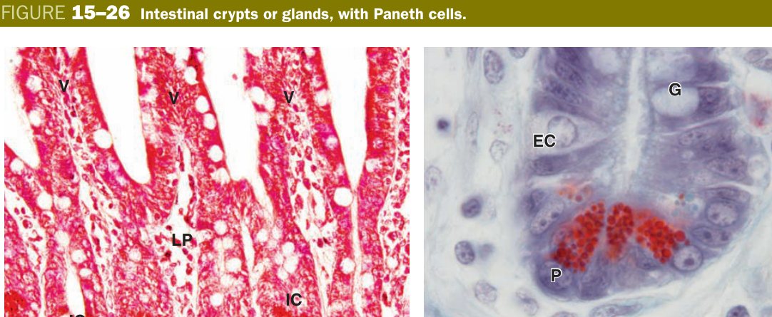
Between the bases of the villi the epithelium dips down into the lamina propria as straight tubular crypts (intestinal glands) of Lieberkühn. The crypts are the proliferative engine — this is where the stem cells live, slowly conveyor-belting new cells up onto the villus surface, where they last only 3–5 days before being shed at the tip. At the very base of each crypt sit the Paneth cells, easily recognised by their bright eosinophilic apical granules — packets of lysozyme, defensins and TNF-α that they secrete into the crypt lumen to kill bacteria and protect the stem-cell niche.
Specialised for absorption: three levels of surface amplification multiply the area ~600×:
| Amplifier | What |
|---|---|
| Plicae circulares | Circular submucosal folds (Kerckring valves) |
| Villi | Finger-like mucosal projections (epithelium + lamina-propria core) |
| Microvilli | On enterocyte apex = the brush/striated border |
If you tried to spread the absorptive surface flat, you would need a tennis court. So the gut does what a pastry chef does to make croissants: fold, then fold, then fold again. Big folds inside the wall (plicae); little fingers off the folds (villi); microscopic bristles off the fingers (microvilli). Each level of folding multiplies the surface by roughly an order of magnitude.
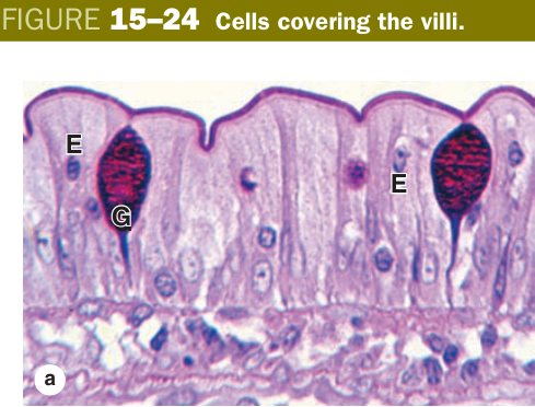
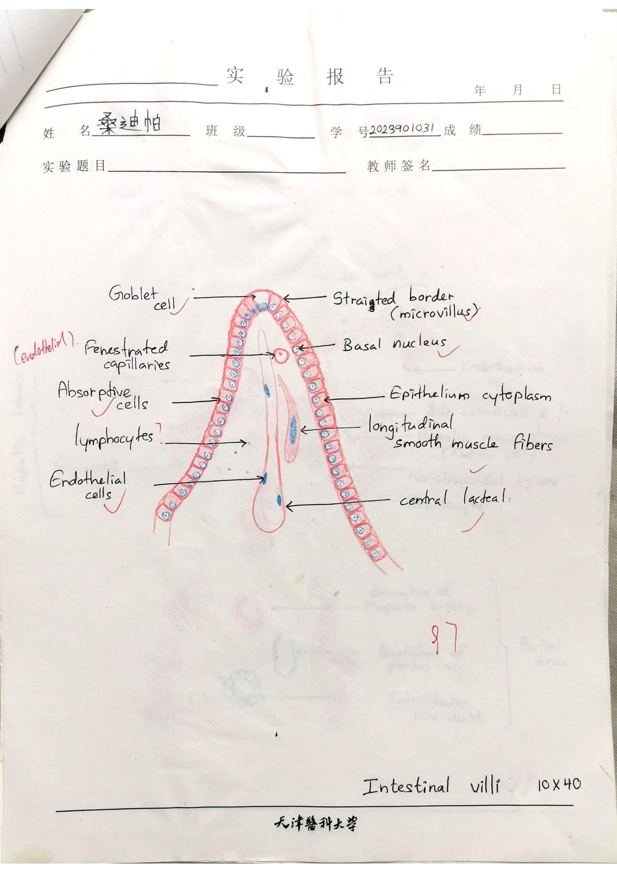
A villus is a one-cell-thick epithelial finger draped over a connective-tissue core, and you should be able to draw both. The epithelium over the villus surface is simple columnar dominated by absorptive cells (enterocytes) — tall cells with an apical brush border of microvilli and tight junctions sealing them to their neighbours, so nutrients have to enter through the cell rather than between them. Scattered between them are goblet cells (mucus, increasing in number from duodenum to ileum and becoming most abundant in the colon) and a sprinkling of enteroendocrine cells (S, I, K and EC cells releasing secretin, cholecystokinin, GIP and serotonin respectively). Over Peyer's patches in the ileum, the epithelium also includes M cells — flat-topped, microfold-bearing antigen sampling cells.
The villus core is loose lamina propria carrying a fenestrated capillary plexus (sugars and amino acids absorbed by the enterocytes drain straight into portal blood), one or two strands of smooth muscle from the muscularis mucosae that contract to milk the villus, and the all-important central lacteal — a blind-ended lymphatic capillary that picks up chylomicrons (re-packaged dietary fat) and sends them via the lymph and thoracic duct into the systemic circulation, bypassing the liver. Without a lacteal there is no fat absorption.
Down in the crypts of Lieberkühn you find five cell types: maturing absorptive and goblet cells migrating up, enteroendocrine cells, the stem cells just above the base, and at the very bottom the Paneth cells with their large acidophilic granules. The Paneth cell is the histological signature of the small-intestinal crypt — it does not appear in the stomach and only minimally in the colon, so if a question asks where Paneth cells live, the answer is always "at the base of the small-intestinal crypts."
Hold up your index finger and call its skin the enterocyte epithelium; imagine a capillary loop and a single lymph tube (the lacteal) running up its core. That is one villus. Now at the knuckle, dig a well straight down between this finger and the next — that is the crypt. New cells are minted at the bottom of the well by stem cells, slide up the side of the well, walk along the surface of the villus, and fall off the tip three days later. The Paneth cells are the security guards at the bottom of the well, keeping the stem-cell room clean.
- Villus epithelium: enterocytes with brush border + goblet cells (mucus) + enteroendocrine cells + M cells over Peyer's patches.
- Villus core (lamina propria): fenestrated capillary plexus, a central lacteal (lymphatic for fat) and a single smooth-muscle strand.
- Intestinal glands / crypts (of Lieberkühn): stem, absorptive, goblet, Paneth (eosinophilic apical granules — lysozyme/defensins; crypt base) and enteroendocrine cells.
- Regional clues: duodenum = Brunner's glands (submucosa, alkaline mucus); ileum = Peyer's patches (aggregated lymphoid nodules).
Coeliac disease: in genetically susceptible patients (HLA-DQ2/DQ8), gliadin peptides from wheat trigger a T-cell-mediated attack on the small-intestinal mucosa. The villi blunt and eventually disappear — the lining flattens out. At the same time the crypts elongate (compensatory hyperplasia) and intraepithelial lymphocytes increase. The result is loss of absorptive surface area, malabsorption, iron-deficiency anaemia, and a chronic diarrhoea/steatorrhoea. Histology of a duodenal biopsy is the gold standard, and re-villus-isation on a gluten-free diet confirms the diagnosis.
The small intestine is one continuous tube, but you are expected to tell its three segments apart on a slide. The trick is to look in three places: the submucosa, the lamina propria, and the villus shape. Two features — Brunner's glands and Peyer's patches — are pathognomonic, and they sit at opposite ends.
The duodenum is identified by Brunner's (duodenal) glands — coiled tubular mucous glands sitting in the submucosa. They are the second of the two regions where you find submucosal glands (the first being the oesophagus). Their alkaline, bicarbonate-rich mucus pours into the crypts and neutralises the acidic chyme arriving from the stomach, protecting the duodenal mucosa and bringing the lumen pH up to where pancreatic enzymes work best. The jejunum, the middle segment, has nothing distinctive in the submucosa, but it has the tallest, most finger-like villi and the most prominent plicae circulares — it is the main absorptive workhorse. The ileum is identified by Peyer's patches: large, often confluent aggregates of lymphoid follicles in the lamina propria and submucosa, with overlying M cells (no microvilli, basal pocket housing lymphocytes) that transcytose luminal antigens to the lymphoid tissue beneath. Goblet cells are also most abundant in the ileum.
| Feature | Duodenum | Jejunum | Ileum |
|---|---|---|---|
| Villi shape | Leaf-shaped, broad | Tallest (finger-like) | Shorter, fewer |
| Plicae circulares | Begin distally | Most prominent | Sparse / absent distally |
| Brunner's glands | Yes (submucosa) — alkaline mucus | No | No |
| Peyer's patches | No | Rare | Yes (LP + submucosa) |
| M cells | No | Rare | Yes — over Peyer's patches |
| Goblet cells | Fewest | Moderate | Most abundant |
Brunner's (duodenal) glands: compound tubular mucous glands located in the submucosa of the duodenum (unique — the only accessory glands in the submucosa of the small intestine). They secrete alkaline mucus rich in bicarbonate, protecting the mucosa from acidic chyme and providing the optimal pH for pancreatic enzymes.
M cells: specialised epithelial cells overlying Peyer's patches in the ileum. They have no microvilli (hence no brush border), and their basal surface is invaginated to form a pocket housing lymphocytes and macrophages. M cells transcytose antigens from the gut lumen to the underlying lymphoid tissue — initiating mucosal immune responses (secretory IgA).
Crohn's disease: a chronic transmural inflammation that classically targets the terminal ileum but can hit anywhere from mouth to anus, with characteristic skip lesions and non-caseating granulomas. Loss of terminal ileum disrupts bile-salt and vitamin B₁₂ absorption. Compare to ulcerative colitis, which is mucosal-only, continuous from the rectum upward, and stays in the colon — crypt abscesses are the histological signature. Carcinoid tumour arises from enteroendocrine cells, most commonly EC cells of the appendix or ileum, secreting serotonin; if it metastasises to the liver, serotonin escapes hepatic first-pass and produces the classic flushing-diarrhoea-wheeze of carcinoid syndrome.
Large Intestine
The colon's job is the opposite of the small intestine's. The small intestine pulled the calories out of the meal; the colon has to pull the water back out of the leftover slurry, lubricate what is left, and store it until you find a toilet. That single shift of priorities — from absorption of nutrients to absorption of water + lubrication of faeces — rewrites the histology.
The first change you notice is that the surface is flat. There are no villi at all in the large intestine — you do not need a tennis court of surface area to reabsorb water and electrolytes. The lining is simple columnar epithelium dotted with enormous numbers of goblet cells; the mucus they produce coats the increasingly solid faecal mass so it slides smoothly along the wall. Down between the goblet cells, deep straight tubular crypts of Lieberkühn dip into the lamina propria — longer and even more goblet-packed than their small-intestinal counterparts. Paneth cells are essentially absent.
The second giveaway is in the muscularis externa. Instead of a complete outer longitudinal sheet, the longitudinal layer of the colon is gathered into three discrete bands running from caecum to rectum — the teniae coli. Between them the wall puckers into the characteristic outpouchings called haustra. Both features disappear at the rectum, where the longitudinal layer regains a complete envelope.
- No villi; surface = simple columnar epithelium with long straight crypts rich in goblet cells (lubrication for faeces) & absorptive cells (water/electrolyte absorption).
- Outer longitudinal muscle gathered into three bands = teniae coli.
- Diffuse lymphoid nodules scatter the lamina propria.
If the small intestine is a sponge (folded, frilled, microvilli everywhere, made to soak up everything), the colon is a water slide — smooth-bottomed, lubricated, gathered into ribbon-like rails (teniae) on the outside so the whole thing can shorten in pulses to push the load along. No frills, just mucus and water reclamation.
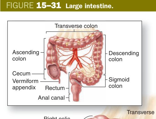
Three regions deserve their own histological identity. The appendix is the same wall plan as the colon but in miniature, with one striking change: the lamina propria and submucosa are stuffed with lymphoid follicles, often confluent enough to give the lumen an irregular, star-shaped outline on a low-power slide. Crypts are short and irregular, and in adults the lumen is often partly obliterated by fibrous tissue. Think of it as a small-but-busy lymphoid outpost — a "gut tonsil."
The anal canal is the meeting-place between the gut tube and the skin. Above the pectinate (dentate) line the lining is still simple columnar epithelium of intestinal type. At the pectinate line, that columnar epithelium transitions abruptly to non-keratinised stratified squamous (the anal transition zone). Further down, towards the anal verge, it becomes keratinised stratified squamous continuous with the skin of the perineum. Two sphincters control closure: the internal anal sphincter is a thickening of the inner circular smooth muscle of the wall (involuntary), and the external anal sphincter is skeletal muscle from the pelvic floor (voluntary).
| Region | Distinguishing histology |
|---|---|
| Appendix | Crypts shorter & fewer than colon; lymphoid follicles pack the lamina propria & submucosa (largest lymphoid aggregate in the GI tract); lumen often obliterated in adults; irregular star-shaped lumen |
| Rectum | Like colon but crypts are longer and even more goblet-rich; no teniae coli (outer longitudinal muscle is complete) |
| Upper anal canal | Anal columns of Morgagni; simple columnar epithelium transitions to stratified squamous (non-keratinised) at the pectinate line |
| Lower anal canal | Below the pectinate line: keratinised stratified squamous epithelium (continuous with perianal skin) |
| Sphincters | Internal = smooth (involuntary); external = skeletal (voluntary) |
Pectinate (dentate) line = the key landmark. Above = simple columnar (visceral innervation; no pain from internal piles). Below = keratinised stratified squamous (somatic innervation; external piles are painful). Colorectal carcinomas are adenocarcinomas (arise from glandular epithelium above the line); anal carcinomas are squamous cell carcinomas (below).
Appendicitis — acute neutrophilic inflammation of the appendix wall, often following luminal obstruction by a faecolith. Colorectal carcinoma — arises from colonic crypt epithelium through the adenoma-carcinoma sequence; histologically an adenocarcinoma. Haemorrhoids are dilated veins of the submucosal venous plexus; whether they hurt depends on the side of the pectinate line they sit on, because that decides whether their innervation is visceral or somatic.
TMU Exam Drill
📝 Open the full TMU Question Bank — 20 MCQ + 6 terms + 5 essays →
Authentic Tianjin Medical University past-paper questions (2021 Final & the multi-section Final with answer key) mapped to this unit, in the real exam format. Click Show answer to self-test.
□ Single best answer
- A. parietal cell
- B. chief cell
- C. goblet cell
- D. endocrine cell
- E. stem cell
- A. absorptive cells
- B. chief cell
- C. goblet cells
- D. Paneth cells
- E. stem cells
□ Fill in the blank
- (fill the three)
□ Structure essay
- Epithelium — simple columnar (surface mucous cells).
- Lamina propria — contains gastric glands: in fundus/body — chief cells, parietal cells, mucous neck cells, stem cells, enteroendocrine cells; plus cardiac & pyloric glands in their regions.
- Muscularis mucosae — thin smooth muscle.
Digestive tract complete
4-layer plan, stomach glands & small-intestine villi mastered. Next: Digestive Glands (liver & pancreas).