Lymphoid (Immune) Organs
Overview & Functions
Picture every barrier in your body — skin, gut lining, airway mucosa — as a leaky wall. Foreign organisms, mutated cells, and your own worn-out cells are constantly slipping through. The immune system is the patrol that recognises them and deals with them, and it does so through a network of lymphoid organs, lymphoid tissue, immune cells, and the immunoactive molecules (antibodies, cytokines, complement) they release.
Junqueira reduces all of that activity to three jobs you should be able to list before you ever open a slide. Immune defence — recognise and eliminate foreign pathogens. Immune surveillance — recognise and eliminate tumour cells and virus-infected cells. Immune homeostasis — clear aged or dead cells and the antigen-antibody complexes that accumulate from normal immune activity. Every histological structure in this unit serves one or more of those three jobs, and that is the lens you bring to every section.
Lymphoid organs are airport checkpoints arranged along the highways of lymph and blood. Lymph nodes screen the lymph draining out of every tissue bed. The spleen screens the blood. The thymus is the training academy — no passengers go through it, only new recruits (T cells) being taught who counts as “self”. Once you see this layout the rest of the unit is just architecture.
| Function | Meaning |
|---|---|
| Immune defence | Recognise & eliminate foreign pathogens |
| Immune surveillance | Recognise & eliminate tumour / virus-infected cells |
| Immune homeostasis | Remove aged/dead cells & immune complexes |
The working cells of immunity come in two families. Lymphocytes (T, B, NK) are the specific arm — each one carries a single antigen receptor and only fires when it meets its match. Antigen-presenting cells are the bridge — dendritic cells in tissues capture antigen and carry it back to the lymphocytes, and the wider mononuclear phagocyte system (MPS) mops up debris everywhere blood goes.
The MPS is one of TMU's favourite traps. Every member is descended from a blood monocyte that has settled into a tissue and changed its name. In the liver the monocyte becomes a Kupffer cell; in the central nervous system, microglia; in bone, an osteoclast; in the lung alveolus, a dust cell; in the epidermis and lymph node, a Langerhans/dendritic cell. Same lineage, different postcodes. Granulocytes (neutrophils, eosinophils, basophils) are not part of the MPS — they are a different developmental branch.
MPS: the body-wide family of phagocytes derived from blood monocytes — tissue macrophages, Kupffer cells (liver), microglia (CNS), osteoclasts (bone), dust cells (lung), and dendritic/Langerhans cells.
Lymphoid Tissue & Lymphoid Nodule
Wherever lymphocytes live in bulk, they need scaffolding. That scaffolding is reticular tissue — reticular cells producing fine type-III collagen (reticular fibres) that form a three-dimensional net. Lymphocytes settle into the holes of this net like fish in coral. So when a histologist says “lymphoid tissue” they mean reticular framework + lymphocytes, plus a few macrophages and dendritic cells.
The same tissue comes in two architectures depending on how organised the lymphocytes look. Diffuse lymphoid tissue has no clear edge — lymphocytes are scattered through the reticular mesh, often around small postcapillary venules; you see it in the lamina propria of every mucosa. Nodular (follicular) lymphoid tissue packs lymphocytes into a sharp little sphere with a definite boundary — the lymphoid nodule.
Lymphoid tissue = a specialised connective tissue with a framework of reticular tissue (reticular cells + reticular fibres) housing large numbers of lymphocytes and other immune cells. Two forms: diffuse (no clear boundary, often with postcapillary venules) and nodular.
A lymphoid nodule (follicle) is a 0.2–1 mm sphere of densely packed lymphocytes with a sharp edge, and it is essentially a B-cell structure. A primary nodule is uniformly dark — just small resting B cells crowded together, no antigen yet seen. A secondary nodule has been hit by antigen and now contains a pale central germinal centre surrounded by a dark rim (the mantle or corona) of small B cells.
The germinal centre is where the B-cell response actually happens. In its dark zone activated B cells called centroblasts proliferate furiously and mutate their immunoglobulin genes (somatic hypermutation). They drift into the light zone as smaller centrocytes, where follicular dendritic cells (FDCs) present antigen on their long processes; only B cells whose mutated receptor binds well survive, the others die by apoptosis and are eaten by tingible-body macrophages. Survivors exit as either plasma cells or memory B cells, having class-switched to a more useful antibody isotype. That entire process — proliferation, mutation, selection, class switch — is what makes a germinal centre pale on H&E.
Think of the germinal centre as an audition. Centroblasts in the dark zone churn out thousands of slightly different versions of the same B-cell (mutating their receptor with every division). Centrocytes then walk out into the light zone where the follicular dendritic cell — the judge — holds the antigen. Only B cells whose new receptor binds the antigen tightly get a callback (survive and differentiate); the rest are removed. The pale colour you see on H&E is the audition floor; the dark mantle is the queue of small resting B cells waiting outside.
Lymphoid nodule: a spherical, 0.2–1 mm aggregate of densely-packed lymphocytes with a clear boundary; mainly B-lymphocytes. A primary nodule lacks, and a secondary nodule has, a pale germinal centre (site of B-cell proliferation after antigen stimulation).
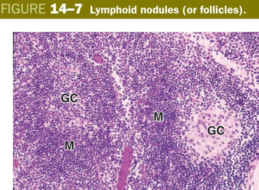
Follicular lymphoma is a B-cell malignancy of germinal-centre origin. The t(14;18) translocation moves the anti-apoptotic BCL-2 gene next to the immunoglobulin heavy-chain enhancer, so centrocytes that should have died in the “audition” instead survive indefinitely — the node fills with monomorphic, back-to-back follicles that have lost their tingible-body macrophages. Histologically the give-away is follicles everywhere, including the medulla, all looking the same.
Thymus — the T-cell training academy
Before you study any single organ, fix the central/peripheral distinction. Primary (central) lymphoid organs are where lymphocytes are made and educated: the bone marrow (B cells finish development here) and the thymus (T cells finish development here). Secondary (peripheral) lymphoid organs are where mature lymphocytes meet antigen and mount responses: lymph nodes, spleen, and the mucosa-associated lymphoid tissue (tonsils, Peyer's patches, appendix).
| Class | Organs | Role |
|---|---|---|
| Central (primary) | Thymus, bone marrow | Lymphocytes develop & mature (T in thymus, B in marrow) |
| Peripheral (secondary) | Lymph nodes, spleen, tonsils (MALT) | Sites of the immune response |
The thymus has a capsule sending septa inward to divide it into lobules, but inside every lobule the stroma is something you will see nowhere else in this unit. It is not reticular connective tissue. Instead, the framework is a meshwork of epithelial reticular cells — star-shaped epithelial cells joined to each other by desmosomes, leaving little spaces between them where developing T cells (thymocytes) sit. That single fact — epithelial scaffold, not connective scaffold — is the cleanest distinguishing point between thymus and every other lymphoid organ, and TMU loves asking about it.
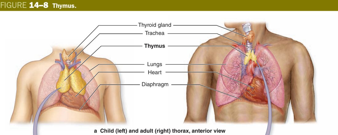
Each lobule has a dark cortex and a paler medulla. The cortex is dark because it is jammed with immature thymocytes that are dividing and being tested for self-reactivity; the medulla is paler because there are fewer, mostly mature, thymocytes plus more visible epithelial reticular cells. In the medulla you also see the diagnostic Hassall's (thymic) corpuscles — concentric whorls of degenerating, keratinised epithelial reticular cells. If you see them on a slide, you are looking at thymic medulla. There is one more rule with no exception: the thymus has no B-cell nodules and no afferent lymphatics, because it is a school, not a filter.
The thymus is a boarding school for T cells. The dark cortex is the crowded freshman dormitory — thousands of new recruits packed in, most of whom will flunk the entrance exam (positive + negative selection) and be removed. The pale medulla is the senior common room — the few who passed are spacious, mature, and ready to graduate into the blood. Hassall's corpuscles are the old school trophies in the corner: layered, keratinised, unmistakeable.
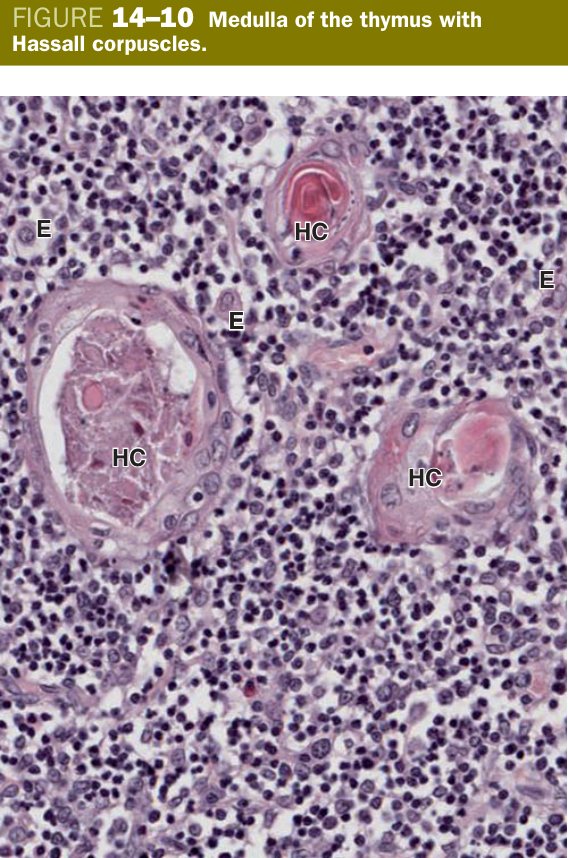
Star-shaped epithelial cell joined to neighbours by desmosomes that forms the stroma of the thymus (replacing the reticular CT of every other lymphoid organ). Subtypes line the cortex, form the blood-thymus barrier, and ultimately build Hassall's corpuscles in the medulla.
A developing thymocyte must learn to ignore self while it is still naive, which means it cannot be exposed to circulating antigens. The blood-thymus barrier in the cortex provides that isolation. Three layers separate blood from thymocyte: (1) the continuous capillary endothelium with its basal lamina, (2) a layer of macrophages patrolling the perivascular space, and (3) a sheath of epithelial reticular cells with their own basal lamina. There is no equivalent barrier in the medulla, which is why mature T cells leave through medullary venules. The barrier exists in the cortex only.
The thymus is also unique in being a regressing organ. It is largest, relative to body weight, around birth and peaks in absolute size at puberty. From then on it undergoes involution — lymphocytes are progressively replaced by adipose tissue and Hassall's corpuscles persist as the last clue you are still looking at thymus. By old age very little functional cortex remains, and the gland is mostly fat.
In DiGeorge syndrome (22q11.2 deletion), the third and fourth pharyngeal pouches fail, so the thymus (and parathyroids) do not develop. With no thymic epithelium there is no T-cell education — the patient has profoundly reduced circulating T cells, recurrent viral and fungal infections, and on biopsy of a lymph node the paracortex (T-zone) is empty while B-cell follicles are preserved.
Lymph Node (essay-grade)
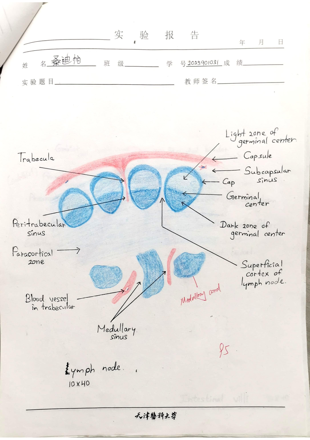
A lymph node is bean-shaped, with a convex surface and a concave indent called the hilum. The whole organ is wrapped in a dense connective-tissue capsule that sends trabeculae inward to carry blood vessels and partition the parenchyma. Afferent lymphatics pierce the convex surface from many points (each one with its own valve) so lymph from the drainage field always flows in at the convex side. A single (sometimes two) efferent lymphatic leaves at the hilum carrying filtered lymph onward; the hilum is also where blood vessels enter and leave.
The direction of lymph flow inside the node follows that geometry: afferent vessel → subcapsular sinus → cortical (peritrabecular) sinus → medullary sinus → efferent vessel at the hilum. As lymph creeps through these sinuses, macrophages sitting in the reticular mesh of the sinus walls phagocytose antigen, debris, and the occasional tumour cell. That is why a node draining an infected wound becomes swollen and tender, and why metastatic cancer cells from a primary tumour first lodge in the regional nodes.
A lymph node is a customs post at a town with many roads leading in but only one road leading out. Many afferent vessels (the back roads) pour lymph onto the convex perimeter. One efferent vessel (the highway) leaves at the hilum carrying lymph after it has been screened. If something is going to be caught (infection, tumour cell, immune complex), it is caught between those two points.
Lymph flow: afferent vessel → subcapsular sinus → peritrabecular sinus → (cortical tissue) → medullary sinus → efferent vessel at hilum.
Inside the capsule, the parenchyma divides into cortex (outer) and medulla (inner, near the hilum), and the cortex itself splits into a superficial cortex and a paracortex (deep cortex). This three-zone plan — superficial cortex · paracortex · medulla — is the structural backbone of the essay answer, and each zone is functionally a different specialist: superficial cortex = B-cell zone; paracortex = T-cell zone; medulla = antibody-secreting output zone.
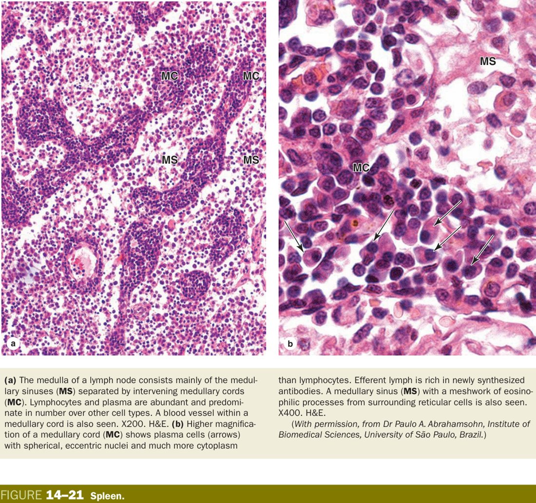
The superficial cortex contains the lymphoid nodules (primary and secondary, with germinal centres). The paracortex is diffuse lymphoid tissue dominated by T cells; its defining histological feature is the high endothelial venule (HEV) — a postcapillary venule lined by tall cuboidal endothelium. Circulating lymphocytes use L-selectin and the chemokine CCL21 to bind these HEVs and squeeze across into the node; this is the route by which naive lymphocytes enter to look for antigen. The medulla is built of medullary cords (anastomosing strings of B cells, plasma cells, and macrophages embedded in reticular tissue) separated by wide medullary sinuses draining toward the efferent vessel. Plasma cells generated in the germinal centres above migrate down into these cords and dump antibody straight into the efferent lymph.
| Region | Structure |
|---|---|
| Superficial cortex | Lymphoid nodules (B-cells) + internodular diffuse tissue |
| Paracortex (deep cortex) | Diffuse lymphoid tissue = T-cell zone; contains postcapillary (high-endothelial) venules where blood lymphocytes enter |
| Cortical sinuses | Subcapsular + peritrabecular sinuses |
| Medulla | Medullary cords (B-cells, plasma cells, macrophages) + medullary sinuses (like cortical sinuses, more macrophages) |
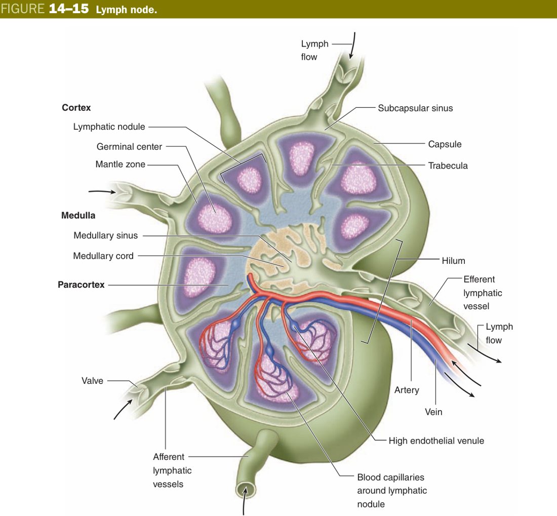
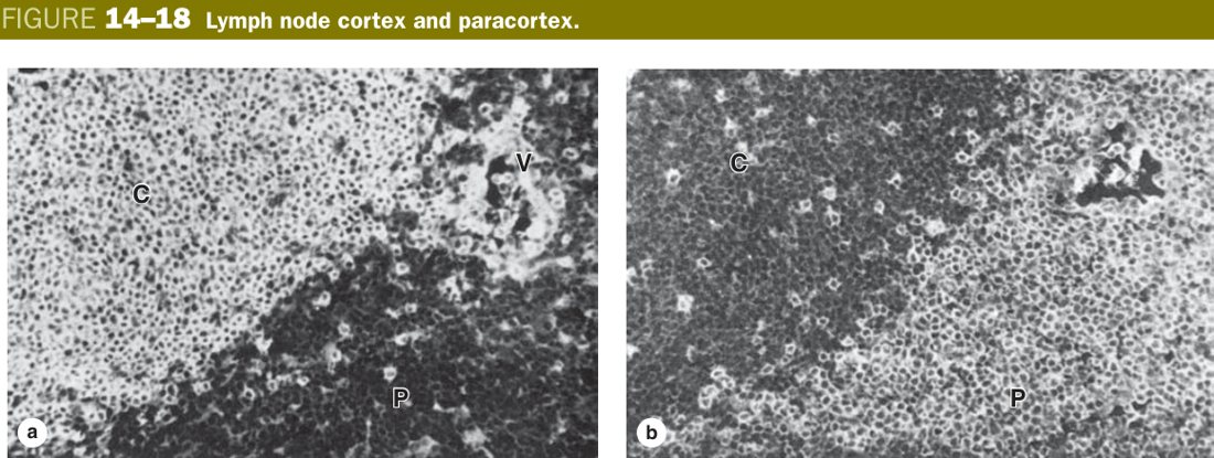
Lymphocyte recirculation: a lymphocyte's life is not stationary. Naive lymphocytes ride the blood into a node through the HEV, sweep through the cortex looking for their antigen, exit in the efferent lymph, return to the venous circulation via the thoracic duct, and then enter another node hours later. They do this cycle continuously until one of them happens to meet its matching antigen — at which point it stays put in that node and starts dividing.
HIV infects CD4+ T cells, the dominant cell of the paracortex. Late in disease the paracortex collapses on biopsy — T zones are empty, germinal centres become disrupted, and the patient becomes susceptible to intracellular pathogens (Pneumocystis, mycobacteria, CMV). Hodgkin lymphoma is recognised by giant binucleate Reed-Sternberg cells in a background of reactive lymphocytes; node architecture is effaced and the disease spreads stepwise from one node group to the next along lymphatic drainage routes.
Spleen (essay-grade)
The single sentence that organises every fact about the spleen is this: the spleen filters blood, not lymph. There are no afferent lymphatics on a spleen. Blood enters through the splenic artery at the hilum, branches along the trabeculae, and eventually arrives as a central artery which becomes wrapped by the lymphoid tissue of the white pulp. Blood, antigen, and senescent red cells therefore pass through the lymphoid tissue itself, not alongside it as in a node.
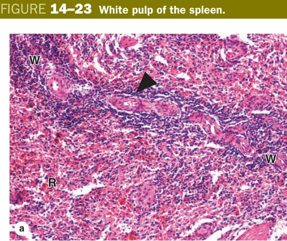
On a low-power slide the parenchyma is unmistakable: scattered violet dots (the white pulp — lymphocytes around central arteries) sit inside a sea of red (the red pulp — cords and blood-filled sinusoids). Between them is a thin transitional layer called the marginal zone, where blood first decants out of the central artery's side-branches and is screened by marginal-zone B cells and macrophages.
Capsule (mesothelium + dense CT + smooth muscle) → trabeculae. Parenchyma = white pulp + red pulp, with a marginal zone between:
Picture each white-pulp area as a small village built around a well (the central artery). The villagers nearest the well are T cells — this is the PALS. Off to one side of the well sits a second cluster of houses, the splenic corpuscle, populated by B cells. Beyond the village edge is the wilderness of the red pulp — loose chains of houses (the cords of Billroth) separated by river channels (the sinusoids) that are constantly full of blood.
| Region | Structure |
|---|---|
| White pulp — PALS | Periarterial lymphatic sheath: diffuse T-lymphocytes around the central artery |
| White pulp — splenic corpuscle | A lymphoid nodule, mainly B-lymphocytes |
| Marginal zone | Between white & red pulp; B, T & macrophages; marginal sinus = entry point for blood antigen/lymphocytes |
| Red pulp — splenic cords | (Billroth cords) lymphoid tissue rich in blood: B-cells, plasma cells, macrophages |
| Red pulp — splenic sinuses | Endothelial cells + incomplete basement membrane + reticular fibres (filter blood) |
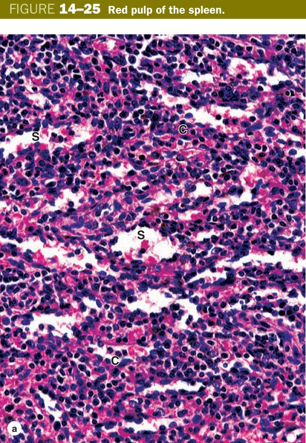
A splenic sinusoid is a strange vessel. Its endothelial cells are long, rod-shaped, and lie parallel to the long axis of the sinusoid — the so-called stave cells — with persistent slits between them. The basal lamina is incomplete and rings the sinus like the hoops of a barrel. The result is a vessel whose wall is full of gaps just wide enough for a flexible red cell to squeeze through. Blood from the central artery is partly emptied directly into the cords (open circulation) and must then crawl back into the sinusoids through these slits.
That mechanical filtration is how the spleen catches worn-out red cells. A young, flexible RBC deforms and slips between the stave cells back into the sinus; an old, stiff, or oxidatively damaged RBC cannot, gets stuck in the cord, and is eaten by a cordal macrophage — which then recycles its iron back into transferrin. This is also how Howell-Jolly bodies are normally removed, and why their reappearance in a peripheral smear signals a non-functioning or absent spleen.
Functions: filter blood, immune defence, haemopoiesis (fetal & stem-cell reserve), blood storage.
Lymph node filters LYMPH; spleen filters BLOOD. In both, T-cells sit in the diffuse zones (paracortex / PALS) and B-cells in the nodules (cortical nodule / splenic corpuscle). The spleen's central artery is wrapped by the T-cell PALS.
After splenectomy (or functional asplenia from sickle-cell autosplenectomy — repeated micro-infarcts of the red pulp), the patient loses two critical defences: the mechanical filter and the marginal-zone B cells that respond to polysaccharide capsules. The clinical consequence is OPSI (overwhelming post-splenectomy infection) by encapsulated organisms — Streptococcus pneumoniae, Haemophilus influenzae type b, Neisseria meningitidis — which is why these patients must be vaccinated. Smear shows Howell-Jolly bodies because nothing is pitting them out anymore.
MALT — tonsils, Peyer's patches & appendix
Most antigens never reach the spleen or a deep lymph node. They are met first at mucosal surfaces — the gut, airways, urogenital tract — by the diffuse and nodular lymphoid tissue collectively called MALT (mucosa-associated lymphoid tissue). MALT is everywhere the body opens to the outside world, and three of its concentrations have proper anatomical names you must recognise on a slide: the tonsils, the Peyer's patches of the ileum, and the lymphoid tissue of the appendix.
Each tonsil is identified by its overlying epithelium. The palatine tonsil sits between the palatoglossal and palatopharyngeal arches and is covered by non-keratinised stratified squamous epithelium dipping in as deep crypts; lymphoid nodules with germinal centres line the crypt walls. The lingual tonsils on the posterior tongue are also covered by stratified squamous epithelium but the crypts are shallower. The pharyngeal tonsil (adenoid) on the roof of the nasopharynx is covered instead by pseudostratified ciliated columnar (respiratory) epithelium with folds rather than true crypts. A useful exam shortcut: the only tonsil with respiratory epithelium is the pharyngeal one.
Peyer's patches are aggregates of 10–200 lymphoid nodules in the lamina propria of the distal ileum that bulge into the lumen. The mucosa overlying them lacks villi and is studded with M (microfold) cells — flat enterocytes specialised to endocytose luminal antigen and pass it straight to the lymphocytes underneath. The appendix is essentially a small intestine with confluent lymphoid tissue all the way around its lamina propria and submucosa. Both Peyer's patches and the appendix are core surveillance for gut bacteria.
Want to tell which tonsil? Look up at the epithelium. Stratified squamous with deep crypts → palatine. Stratified squamous with shallow crypts on tongue base → lingual. Ciliated columnar with folds in the nasopharynx → pharyngeal/adenoid. M cells sitting on flat domes over follicles in the ileum → Peyer's patch.
TMU Exam Drill
📝 Open the full TMU Question Bank — 20 MCQ + 6 terms + 5 essays →
Authentic Tianjin Medical University past-paper questions (2021 Final & the multi-section Final with answer key) mapped to this unit, in the real exam format. Click Show answer to self-test.
□ Fill in the blank
- (fill cortex ×3 and medulla ×2)
□ True or false
□ Structure essay
- Capsule — mesothelium + dense CT with smooth muscle; trabeculae extend in.
- White pulp — periarterial lymphatic sheath (PALS, T cells) around the central artery + splenic (lymphoid) nodules (B cells).
- Marginal zone — between white & red pulp; screens blood antigens.
- Red pulp — splenic cords (of Billroth) + splenic (venous) sinuses filled with blood.
- Cortex: superficial cortex with lymphoid nodules (B cells, germinal centres) + paracortex (T cells) + cortical (subcapsular & trabecular) sinuses.
- Medulla: medullary cords (B cells, plasma cells, macrophages) + medullary sinuses.
- Lymph flows: afferent → subcapsular → cortical → medullary sinuses → efferent at hilum.
Lymphoid organs complete
Lymph node & spleen essays, PALS vs nodule, thymus mastered. Next: Digestive Tract.