Blood
Composition & the Blood Smear
Open Junqueira chapter 12 and the very first sentence reframes everything you learned in the last unit: blood is a connective tissue. That sounds wrong — you cannot stretch it, you cannot palpate it, it runs out of a needle. But the definition holds. Connective tissue is cells suspended in extracellular matrix, and blood is exactly that: it is just that the ECM (the plasma) happens to be a liquid rather than a gel or a mineralised plate.
If you spin a tube of anticoagulated blood in a centrifuge, the components separate into three honest layers. The bottom 45% is a deep red sludge of erythrocytes — this number, the haematocrit, is what the lab measures every time you order an FBC. The top 55% is a straw-coloured fluid: that is the plasma, the liquid ECM. And sitting between the two is a thin white-grey ribbon barely a millimetre thick — the buffy coat, holding all of your leukocytes and platelets. The fact that an entire defensive army takes up only 1% of the column volume tells you something about how rare these cells really are.
You study blood not in a block of paraffin but on a peripheral smear: a drop spread thin across a slide, air-dried, and stained with a Romanowsky cocktail — Wright's, Giemsa, or Leishman. The mix is methylene blue (basic, picks up acidic nuclei and RNA) plus eosin (acidic, picks up basic structures like haemoglobin and eosinophil granules). On a good slide every cell type has a signature colour, and that is the whole point of the stain — you do not need a microscope eyepiece reticle, just educated eyes.
Think of a spun tube as a tiny lava lamp standing still. Heavy red cells fall to the bottom, light plasma floats on top, and the rare white cells and platelets — lighter than RBCs but heavier than plasma — trap themselves in between as a thin pale band. The proportions never lie: a haematocrit below 35% screams anaemia; a buffy coat thicker than usual hints at leucocytosis.
| Element | Normal count | Fraction |
|---|---|---|
| Plasma (liquid ECM) | water + albumin/globulins/fibrinogen + electrolytes | ~55% |
| Erythrocytes (RBC) | ♂ ~4.0–5.5 × 10¹²/L; ♀ ~3.5–5.0 × 10¹²/L | ~45% (haematocrit) |
| Leukocytes (WBC) | 4–10 × 10⁹/L | part of the buffy coat <1% |
| Platelets | 150–400 × 10⁹/L | part of the buffy coat |
Plasma minus the clotting proteins (chiefly fibrinogen consumed during coagulation) is called serum. That distinction is not pedantic: every biochemistry test you will order in clinics is done on serum, while a coagulation screen is done on plasma. Same fluid, different proteins still on board.
Erythrocyte (Red Blood Cell)
The mature erythrocyte is the strangest cell in the body, and that strangeness is the whole point. During its final differentiation in the bone marrow it ejects its nucleus and digests its mitochondria, its Golgi, its rough ER — every organelle it ever had. What is left is a plasma membrane wrapped around a concentrated solution of haemoglobin. That is not an accident or an oversight; it is the design specification. A nucleus would take up space that could carry oxygen. Mitochondria would consume the oxygen the cell is meant to deliver. So the RBC throws them out and rides through your capillaries as a pure gas-transport sack.
Shape-wise it is a biconcave disc about 7.5 µm across — thin in the middle, slightly thicker at the rim. On an H&E smear this shows up as a uniform pink ring with a pale centre (the famous central pallor); the thin middle stains less because there is simply less haemoglobin between the two membranes there. The biconcavity is brilliant geometry: it maximises surface-to-volume ratio (good for gas exchange) and lets the cell fold like an umbrella to squeeze through capillaries narrower than itself.
That deformability is held together by a membrane cytoskeleton you must know by name: a hexagonal mesh of spectrin dimers tied to the inner membrane by ankyrin and band 4.1, anchored to integral proteins like band 3. Knock out any of those links and the cell loses its shape — it rounds up into a sphere, becomes brittle in narrow capillaries, and gets ripped apart by the spleen. That single sentence is the entire pathophysiology of hereditary spherocytosis.
Picture a soft jelly doughnut pressed thin in the middle from both sides but stopping just before the two surfaces touch. That is your erythrocyte. The two depressed faces make gas exchange fast because no haemoglobin molecule is far from the membrane, and the squishy edge lets the cell fold itself in half to enter a 3 µm capillary. A rigid sphere of the same volume could not even enter.
- LM: biconcave anucleate disc, D ≈ 7.5 µm (a built-in ruler — if a nucleus on the slide is the same size as an RBC, it is small).
- EM: no organelles; cytoplasm filled with Hb; membrane backed by a spectrin/ankyrin/band-4.1 cytoskeleton.
- Function: reversible binding & transport of O₂ (to tissues) and CO₂ (back to the lung). Lifespan ~120 days.
- Death: aged RBCs are pulled out of circulation by splenic macrophages; Hb is recycled (iron → transferrin; haem → bilirubin).
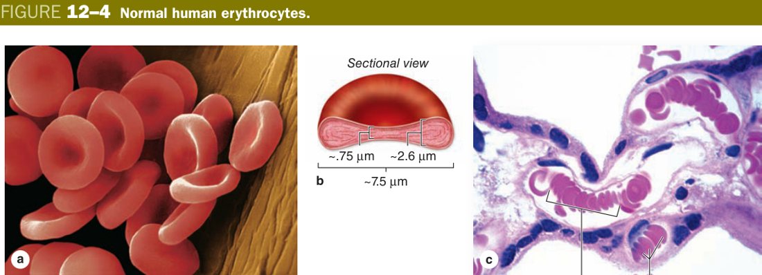
Sickle-cell disease — a single Glu→Val substitution makes HbS polymerise when deoxygenated; the cell deforms into a rigid crescent, blocks capillaries, and lyses early. Hereditary spherocytosis — spectrin or ankyrin defect → spherical RBC → splenic destruction → haemolytic anaemia. Thalassaemia — underproduction of an α or β globin chain. G6PD deficiency — oxidative stress (fava beans, primaquine) precipitates Hb as Heinz bodies, the spleen bites them out, and the cell lyses.
Leukocytes — Overview & Comparison
Where the erythrocyte has stripped itself down to a single purpose, the leukocytes have specialised in five different directions. They all share one feature: they keep their nucleus, because every one of them still has work to do that needs gene expression on demand. The classical split is by cytoplasmic granules: granulocytes (neutrophil, eosinophil, basophil) carry conspicuous specific granules; agranulocytes (lymphocyte, monocyte) do not, although they carry a few small azurophilic lysosomes that look like fine dust.
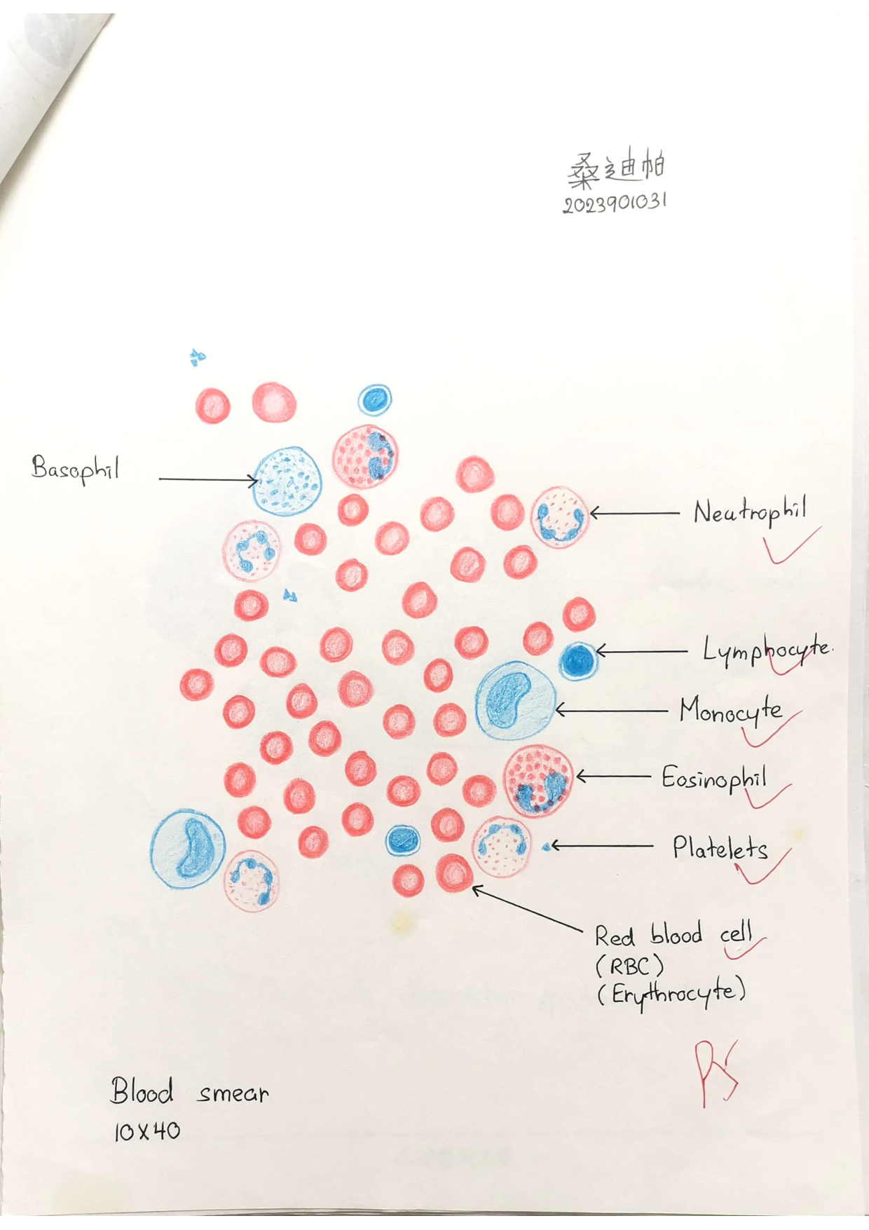
The single most testable fact about leukocytes is their relative abundance on a normal smear — the differential. You will hear it as the NLMEB sequence, top to bottom: Neutrophil > Lymphocyte > Monocyte > Eosinophil > Basophil. Memorise that order because every CBC report you will ever read uses it, and every shift away from the order has a clinical meaning.
| Leukocyte | Diameter | Nucleus | Cytoplasm / granules | % | Function |
|---|---|---|---|---|---|
| Neutrophil | 10–12 µm | 2–5 lobes joined by thin threads | Fine lilac specific granules (lysozyme, lactoferrin) + azurophilic lysosomes | 60–70% | First-responder phagocyte against bacteria |
| Eosinophil | 12–15 µm | Bilobed (spectacles) | Coarse crimson refractile granules with crystalline core (major basic protein) | 1–4% | Anti-parasitic; allergy modulator |
| Basophil | 10–14 µm | Irregular S-shape, often obscured | Large dark blue-purple metachromatic granules (histamine + heparin) | <1% (rarest) | Immediate hypersensitivity |
| Lymphocyte | 6–8 µm (small) | Large, round, densely stained — nearly fills the cell | Thin sky-blue rim; no specific granules | 20–30% | B / T / NK adaptive immunity |
| Monocyte | 14–20 µm (largest) | Kidney / horseshoe | Bluish-grey, fine azurophilic granules (lysosomes) | 3–8% | Leaves blood → tissue macrophage |
Treat the five leukocytes as a fire-station roster. Neutrophils are the engine crew — most numerous, first on scene, sprays bactericidal foam, dies on the job (pus = dead neutrophils). Eosinophils are the specialist anti-parasite squad, bringing crystalline-core grenades. Basophils are the alarm callers — rare, but they pull the histamine bell and bring the whole allergic cascade overhead. Lymphocytes are the intelligence officers — small, quiet, dense, remember every face. Monocytes are the heavy-lifting cleanup truck — biggest, leave the station, become macrophages that camp in the tissue.
Abundance high→low: “Never Let Monkeys Eat Bananas” — Neutrophil > Lymphocyte > Monocyte > Eosinophil > Basophil. Nucleus shapes: neutrophil = multilobed; monocyte = kidney/horseshoe; lymphocyte = round & dense; eosinophil = bilobed (spectacles); basophil = obscured by dark granules.
Granulocytes
The neutrophil is the workhorse of innate immunity and the most abundant nucleated cell in your blood. Its nucleus is its name-card: 2 to 5 dense lobes connected by thread-thin chromatin bridges. The lobulation is functional — a compact spherical nucleus would jam in a capillary, but a string of lobes folds like a chain and slips through. In a young (band) neutrophil the nucleus is still a single horseshoe; the lobes appear as the cell matures. On a smear, a sudden flood of band forms (“left shift”) tells you the marrow is dumping immature cells because demand has overwhelmed supply — a sepsis signature.
Its cytoplasm hides two granule populations. The specific (secondary) granules are small, neutral-staining (hence the name neutrophil) and packed with lysozyme, lactoferrin, collagenase — weapons that work outside the cell on bacterial walls and tissue scaffolds. The azurophilic (primary) granules are larger lysosomes carrying myeloperoxidase and acid hydrolases — weapons that work inside the phagosome. Together they execute the entire kill cycle: chemotaxis → diapedesis → phagocytosis → oxidative burst → degranulation → apoptosis. A dying neutrophil with the bacterium it caught is what you see as pus.
Picture a fire-engine crew that arrives within minutes, sprays foam (specific granules outside the cell), drags the burning material inside the truck and incinerates it with bleach (the oxidative burst), then dies on shift. The lobed nucleus is the segmented hose of the truck — long, flexible, threads through any gap. That is a neutrophil from start to finish.
The eosinophil sits next in line: a bilobed nucleus that looks like a pair of spectacles, and a cytoplasm crammed with coarse crimson refractile granules. Under EM each granule shows a striking crystalline core of major basic protein embedded in a less dense matrix — pure pathognomonic morphology. MBP is a parasiticide: it punches holes in helminth cuticles. Eosinophils also dampen allergy by neutralising histamine and inactivating leukotrienes — hence the saying that eosinophils “both fight parasites and clean up after basophils.”
The basophil is the rarest cell in blood (often <1%). Its nucleus is irregular and S-shaped, but you rarely see it clearly because the cytoplasm is jammed with large, dark blue-purple granules that are metachromatic (they shift the colour of certain dyes). Inside those granules: histamine and heparin, the same payload as the tissue mast cell. When IgE crosslinks on the surface receptor, the basophil degranulates and triggers immediate (type I) hypersensitivity — the urticaria, wheeze and anaphylaxis you must always rule out at A&E.
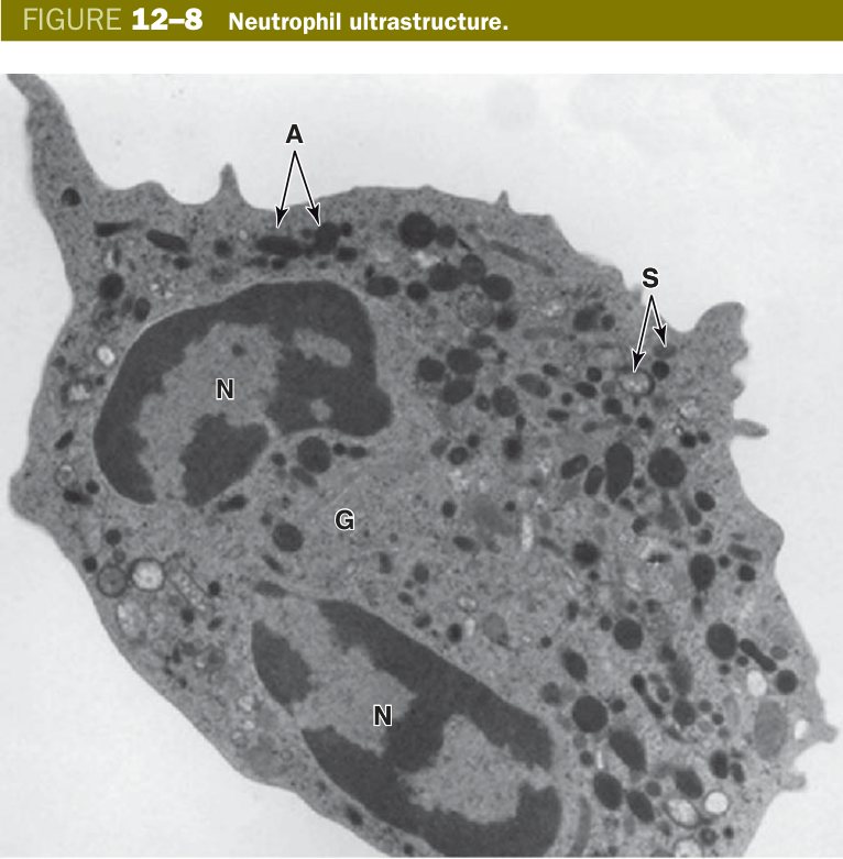
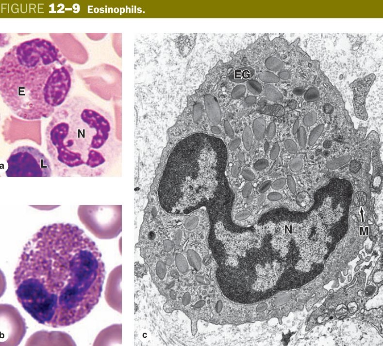
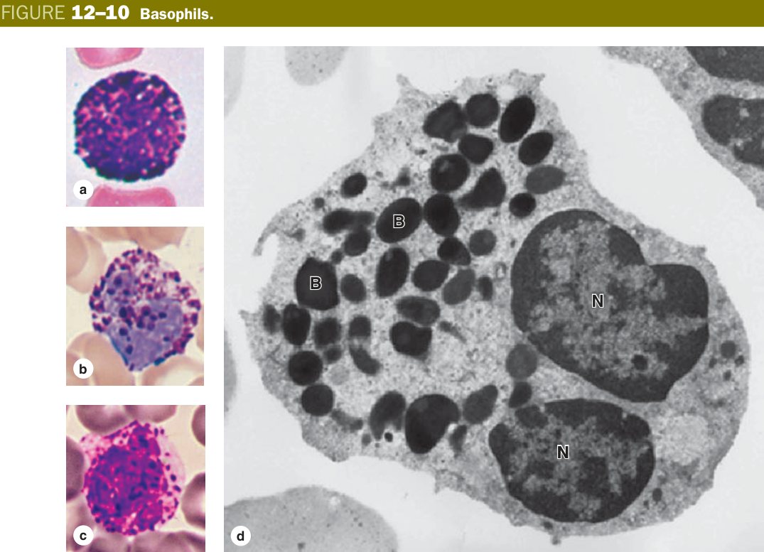
Neutrophilia — acute bacterial infection, MI, steroids. Neutropenia — chemo, marrow failure; counts <0.5 × 10⁹/L are a febrile-neutropenia emergency. Eosinophilia — allergy, asthma, drug reactions, helminth infection (the “NAACP” mnemonic: Neoplasia, Allergy/Asthma, Connective-tissue disease, Parasites). Basophilia is one of the textbook clues to chronic myeloid leukaemia (BCR-ABL / Philadelphia chromosome).
Agranulocytes
The lymphocyte is small, quiet, and disproportionately important. On a smear it looks almost like a nucleus with a coat painted around it — a dense, round, deeply basophilic nucleus that nearly fills the cell, leaving only a thin sky-blue rim of cytoplasm and no specific granules. That sparse appearance is misleading: every adaptive immune memory you possess, every antibody you make, every viral peptide you ever recognise lives inside this morphologically dull cell. On the smear you cannot tell a B from a T cell — you need flow cytometry for that — but you can compare it to a neighbouring RBC: a small lymphocyte is barely larger than an erythrocyte (~6–8 µm); a large lymphocyte is bigger because it is activated.
Functionally lymphocytes split into three lineages: B cells mature in the bone marrow and become antibody-producing plasma cells; T cells mature in the thymus and split into helpers (CD4+) and cytotoxics (CD8+); NK cells are innate killers that lyse virus-infected and tumour cells without needing prior sensitisation. None of this is visible on light microscopy — the smear lymphocyte is the single dimmest cell on a CBC and the brightest cell in immunology.
Lymphocytes look boring on the smear because their work is mental, not muscular. A neutrophil is a bouncer; a lymphocyte is a librarian who has memorised every pathogen face in your life. The cytoplasm is sparse because the cell is mostly nucleus — the archive is what matters.
The monocyte is the largest cell on a normal smear (14–20 µm). Its nucleus is unmistakable: a deep kidney or horseshoe shape, often with one side notched as if a bite were taken. The cytoplasm is pale grey-blue with fine azurophilic dust — those granules are lysosomes, ready for action. A monocyte in blood is a cell in transit: it leaves the vessel by diapedesis, enters tissue, swells, and differentiates into a tissue macrophage. Once it does, it gets a tissue-specific local name: Kupffer cell in liver, microglia in brain, alveolar macrophage in lung, osteoclast (with fusion) in bone, dust cell in lung, Langerhans cell in skin. All of these are members of the mononuclear phagocyte system and they all started life as a monocyte.
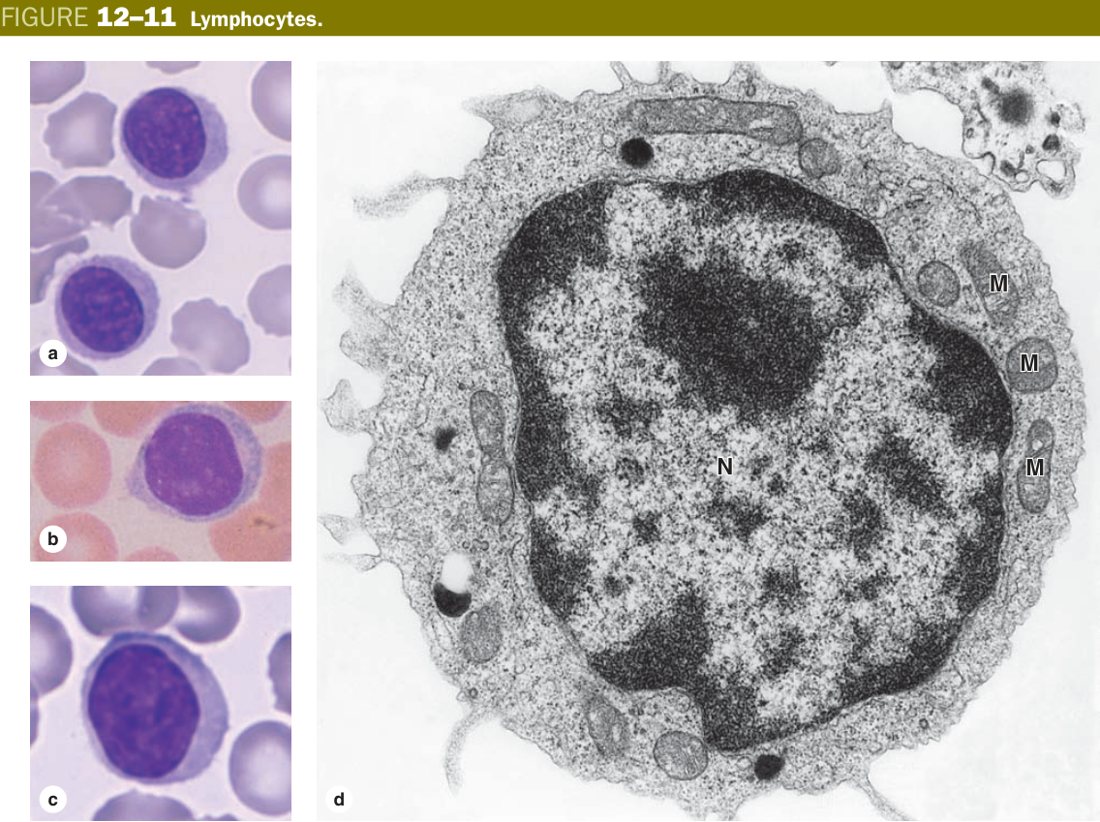
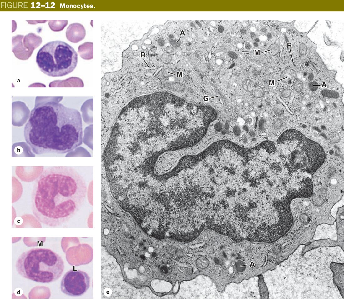
Lymphocytosis — viral infections (EBV, CMV), chronic lymphocytic leukaemia (smudge cells on smear), pertussis. Lymphopenia — HIV (CD4 destruction), steroids, sepsis. Acute lymphoblastic leukaemia (ALL) — the commonest paediatric cancer; bone-marrow blast crisis with cytopenias. Monocytosis — chronic infection (TB, endocarditis), connective-tissue disease. Acute myeloid leukaemia (AML) with Auer rods is the malignant counterpart of the granulocyte/monocyte line.
Platelets (Thrombocytes)
Platelets are not whole cells — they are cytoplasmic fragments shed from megakaryocytes in the bone marrow. A single megakaryocyte extends long pseudopodial processes through a marrow sinusoidal endothelium, and the shear of blood flow snaps off thousands of platelet-sized packets. The result is a 2–4 µm anucleate disc that circulates for 8–10 days. On a smear they look like blue-purple specks, often clumped — each speck carries a peripheral pale zone (the hyalomere, mostly microtubules and actin) and a darker granular centre (the granulomere, where the granules live).
Two granule populations are testable. The α-granules contain the heavy adhesive and growth-factor cargo: von Willebrand factor, fibrinogen, factor V, platelet-derived growth factor (PDGF). The dense (δ) granules carry the small signalling molecules that recruit more platelets: ADP, ATP, serotonin, calcium. When a vessel is breached, exposed collagen and von Willebrand factor capture platelets via the GPIb / GPVI receptors; the platelets activate, change shape into spiked spheres, dump their granules, and recruit a flood of new platelets — the primary haemostatic plug. The coagulation cascade then weaves fibrin around the plug to make it stable.
Think of platelets as sandbags pre-positioned in the bloodstream. The moment a vessel wall tears, the first sandbags stick to the exposed collagen (vWF is the glue), then call radio for thousands more (ADP). Within seconds you have a wall of sandbags — the primary plug — while the engineering team (clotting cascade) shows up to pour fibrin cement and turn the sandbag wall into a permanent dam.
- LM: 2–4 µm anucleate fragments; pale hyalomere at the edge, granular granulomere in the centre.
- EM: α-granules (vWF, fibrinogen, PDGF) and dense δ-granules (ADP, ATP, serotonin, Ca²⁺); marginal microtubule band gives the disc shape.
- Origin: shed from megakaryocytes of bone marrow (thrombopoiesis driven by thrombopoietin from liver/kidney).
- Function: primary haemostasis — adhere, activate, aggregate, secrete; provide the surface for the coagulation cascade.
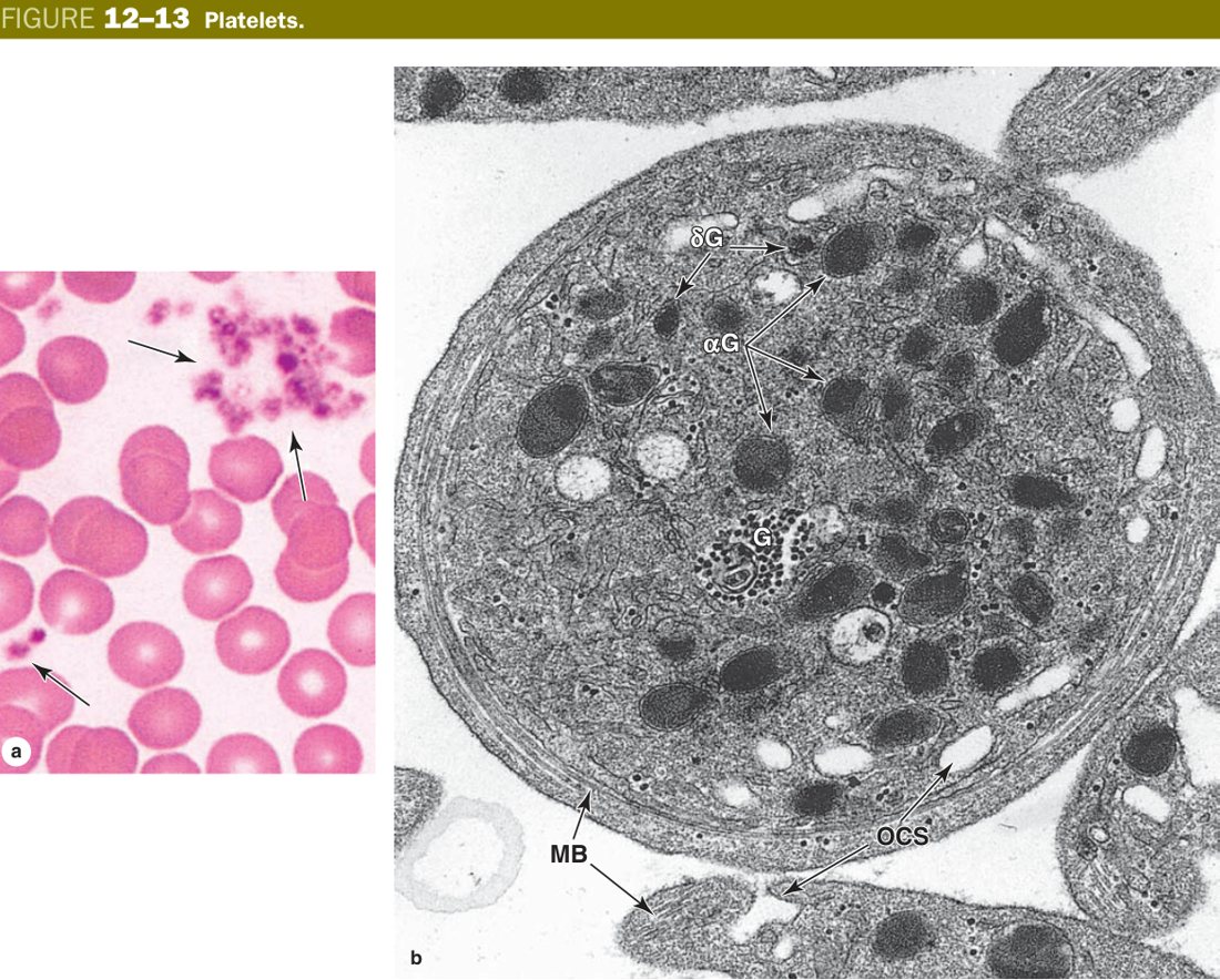
Thrombocytopenia (<150 × 10⁹/L) → petechiae, mucosal bleeding. Causes: ITP (autoantibody-mediated destruction), DIC (consumption), marrow failure, hypersplenism. Thrombocytosis — reactive (iron deficiency, inflammation) or clonal (essential thrombocythaemia, JAK2). Drugs: aspirin irreversibly blocks COX-1, clopidogrel blocks the ADP receptor — both used as antiplatelet therapy.
Haemopoiesis
No blood cell circulating in your veins today is more than a few months old. The whole blood compartment is renewed constantly, and the factory keeps moving locations as you grow. In the first trimester haemopoiesis happens in the yolk sac — primitive nucleated RBCs that look more like a frog's than a human's. In the second trimester the work moves to the liver and spleen; you can still find marrow-style islands in fetal liver biopsies. From the seventh month onwards the bone marrow takes over, and at birth essentially every bone in the body is filled with red (active) marrow.
As you grow the red marrow retreats. In the adult, only the axial skeleton — vertebrae, sternum, ribs, pelvis, skull, proximal femur and humerus — still produces blood. The long-bone marrow turns yellow with fat. That is why a bone-marrow biopsy is taken from the posterior iliac crest: it is the most accessible bit of red marrow left in an adult. Under severe stress (haemolysis, marrow failure) the liver and spleen can reactivate as extramedullary haemopoiesis, which is why they may enlarge in thalassaemia and myelofibrosis.
Imagine a state-owned factory that is born inside a tent (yolk sac), expands into a warehouse (liver/spleen), then settles permanently into a vast complex (bone marrow). As the worker (you) grows, the factory closes its peripheral branches (long bones turn to yellow fat marrow) and consolidates in the head office (axial skeleton). In emergencies the warehouse is reopened — extramedullary haemopoiesis.
The red marrow is a soft, vascular tissue squeezed between bone trabeculae. Architecturally it is two interlocking compartments: haemopoietic cords (islands of developing cells supported by a reticular-fibre meshwork and fat cells) and vascular sinusoids (wide, thin-walled, fenestrated capillaries that drain into a central vein). Mature blood cells leave the cords by squeezing through the endothelial fenestrations into the sinusoid — the same trans-endothelial migration trick a leukocyte will later use in the opposite direction at sites of infection.
All blood lineages descend from a single rare ancestor: the pluripotent haemopoietic stem cell (HSC, CD34+). The HSC commits first to either a common myeloid progenitor (erythroid, granulocyte/monocyte, megakaryocyte) or a common lymphoid progenitor (T, B, NK). Each downstream commitment is steered by specific cytokines — erythropoietin from kidney drives the erythroid line, thrombopoietin drives megakaryocytes, G-CSF / GM-CSF push the granulocyte line, IL-3 / IL-7 / SCF support multiple lineages. The marrow stroma physically provides the niche; the cytokines provide the instructions.
Picture the marrow as marshland: dry “cord” islands where the cells grow up, separated by “sinusoid” rivers that carry them away. When a young cell is ready, it walks to the riverbank and steps through the porous wall straight into the current. The current empties into the central vein and from there into your circulation.
Watch a developing erythroid cell across a week and you see a single relentless story: the cell shrinks, the nucleus condenses, the cytoplasm shifts from blue (RNA-rich, making haemoglobin) to pink (haemoglobin-filled), and finally the nucleus is extruded. Five named stages mark this journey — you must know them in order.
| Stage | Diameter | Cytoplasm | Nucleus | Defining feature |
|---|---|---|---|---|
| Proerythroblast | 14–19 µm | deeply basophilic | large, round, fine chromatin, nucleoli | first committed cell |
| Basophilic erythroblast | 12–17 µm | deep blue (RNA) | condensing — “clock-face” | Hb synthesis just beginning |
| Polychromatophilic erythroblast | 10–15 µm | blue-grey (RNA + Hb mix) | more condensed | last stage that divides |
| Orthochromatic erythroblast (normoblast) | 8–12 µm | pink (mostly Hb) | small, dense, pyknotic — about to be extruded | last nucleated stage |
| Reticulocyte | ~8 µm | pink with residual ribosomal “reticulum” | none (nucleus already extruded) | enters blood; matures in ~1 day |
| Erythrocyte | 7.5 µm | uniformly pink | none | mature, ready to work |
The whole sequence is paced by erythropoietin (EPO), a glycoprotein made in the kidney peritubular fibroblasts in response to tissue hypoxia. Anaemia or altitude → renal HIF-1α stabilises → EPO transcription rises → more proerythroblasts commit and survive. That single feedback loop is why patients on chronic dialysis need recombinant EPO injections (their kidneys cannot sense or produce it).
The granulocyte line follows the same story arc — condensation of the nucleus, accumulation of granules — in six named stages: myeloblast → promyelocyte → myelocyte → metamyelocyte → band → mature granulocyte. The transition you must remember: azurophilic (primary) granules appear at the promyelocyte stage; specific (secondary) granules appear at the myelocyte stage. From the metamyelocyte onwards the cell can no longer divide; the nucleus indents, becomes a band (horseshoe), then segments into the mature lobed shape. Under bacterial stress the marrow ships out band forms early — the left shift you will quote in clinical write-ups.
The platelet line is the most peculiar. A single megakaryocyte progenitor undergoes endomitosis — it replicates its DNA without dividing the cell — producing a giant cell with a single huge polyploid nucleus (up to 64N) and a cytoplasm crammed with future-platelet granules. The cell then extends pro-platelet processes through the sinusoidal endothelium and shears off thousands of platelets directly into the blood. Thrombopoietin from liver and kidney is the regulator.
The granulocyte and platelet stories show two opposite ways to mass-produce: the granulocyte line is a long Ford-style assembly belt, dividing again and again until enough copies exist. The platelet line is a single giant 3-D printer (the megakaryocyte) that copies its blueprints internally (endomitosis) and shears off all the products at once from a single body.
Anaemia — iron deficiency (microcytic), B12/folate (macrophages), CKD (lack of EPO), haemolytic (sickle, spherocytosis, G6PD, thalassaemia). Acute myeloid leukaemia (AML) — myeloblast proliferation; Auer rods. Chronic myeloid leukaemia (CML) — Philadelphia chromosome (BCR-ABL); basophilia; treated with imatinib. Acute lymphoblastic leukaemia (ALL) — commonest paediatric leukaemia. Chronic lymphocytic leukaemia (CLL) — smudge cells. Polycythaemia vera — JAK2 mutation, raised RBC mass. Essential thrombocythaemia — JAK2/CALR, platelet excess. ITP — autoimmune platelet destruction. DIC — uncontrolled clotting consumes platelets and factors, then bleeding.
TMU Exam Drill
📝 Open the full TMU Question Bank — 20 MCQ + 6 terms + 5 essays →
Authentic Tianjin Medical University past-paper questions (2021 Final & the multi-section Final with answer key) mapped to this unit, in the real exam format. Click Show answer to self-test.
□ Single best answer
- A. erythrocyte
- B. basophil
- C. eosinophil
- D. lymphocyte
- E. monocyte
- A. Most abundant leukocyte.
- B. The nucleus is kidney-shaped.
- C. Neutrophils are spherical.
- D. Special granules in cytoplasm.
- E. Phagocytic & bactericidal.
- A. Small lymphocyte is spherical.
- B. Very little cytoplasm.
- C. Basophilic cytoplasm.
- D. Special granules in the cytoplasm.
- E. Main immune cell of the body.
□ True or false
Blood complete
All formed elements + the Wright-stain drawing mastered. Next: Lymphoid Organs.