Circulatory System
Overview & General Vessel Wall
Open Junqueira at chapter 11 and the first thing he reminds you is that “circulatory” is really two parallel networks. The cardiovascular system — heart, arteries, capillaries, veins — pumps blood in a closed loop. The lymph vascular system — lymph capillaries, lymph vessels, ducts — is a one-way drainage tree that picks up the small fraction of plasma that escapes the capillary bed and returns it to the venous side. You will meet both on the same slide and you need to read the wall to know which one you are looking at.
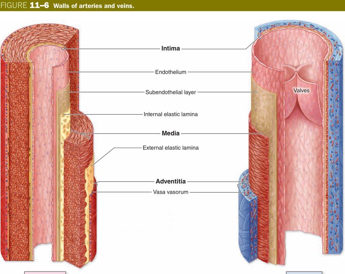
Almost every vessel, from the aorta down to the largest lymphatic, is built on the same three-tunic plan. The closer you get to the capillary, the more the tunics simplify; the closer you get to the heart, the more they specialise. Get the three-tunic scaffold solid now and the rest of the unit becomes a story of which layer is thick and why.
| Tunic | Contents |
|---|---|
| Tunica intima (inner) | Endothelium (simple squamous) + subendothelial CT (± internal elastic membrane) |
| Tunica media (middle) | Circular smooth muscle ± elastic/collagen fibres |
| Tunica adventitia (outer) | CT with vessels (vasa vasorum) & nerves (nervi vasorum) ± external elastic membrane |
The endothelium deserves its own line because it is more than a passive lining. It is the simple squamous epithelium that runs from the inside of the left atrium, out through the aorta, through every capillary, back through every vein, and into the right atrium — one continuous sheet, one phenotype. It is anti-thrombotic, semi-permeable, and the source of vasoactive signals such as nitric oxide and endothelin. When you write “endothelium” on an exam paper, you are naming the most clinically important epithelium in the body.
Think of every vessel as a winter jacket. The smooth nylon liner that touches your skin is the intima — thin, slippery, low-friction. The padded middle that does the actual work of keeping the shape and holding pressure is the media. The tough outer canvas that anchors the jacket to your trousers and survives the weather is the adventitia. An aorta is just a jacket with extra padding; a vein is the same jacket with thin padding and a baggy fit.
Endothelium: the simple squamous epithelium lining the heart, all blood vessels and all lymphatic vessels. It is anti-thrombotic, controls selective exchange, and secretes vasoactive mediators (NO, endothelin, von Willebrand factor).
• Epithelium of endothelium? → Simple squamous
• Where is the internal elastic membrane? → Between intima and media
• What runs in the adventitia? → Vasa vasorum + nervi vasorum
• Two parallel circulations? → Cardiovascular + lymph vascular
Heart Wall (essay-grade)
The heart is not a special organ that broke the rules of vessel design — it is a giant vessel that learned to beat. The three layers of the heart wall mirror the three tunics of a blood vessel, layer for layer, and that is the cleanest way to memorise them. Reading from inside the chamber outward you meet the endocardium (= intima), the myocardium (= media), and the epicardium (= adventitia + a serous covering). Examiners ask you to describe the heart wall almost every year; they want the three layers in order, with the cells and the contents named.
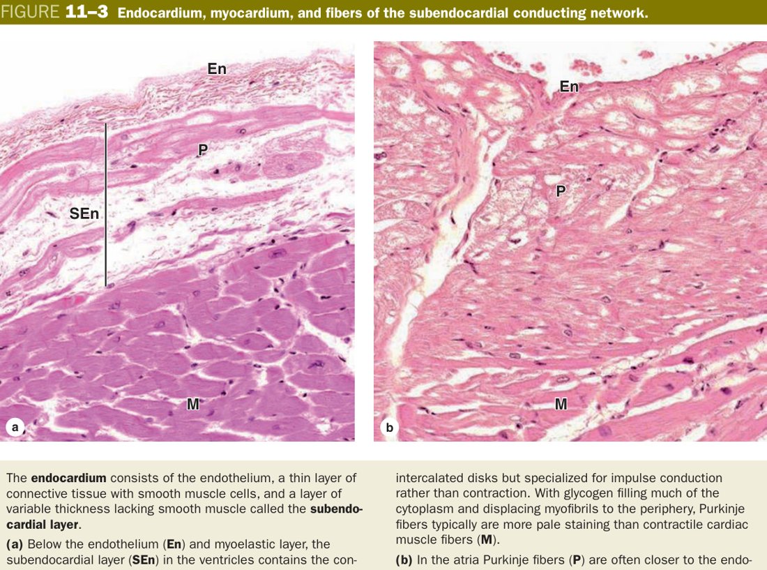
The endocardium is itself a three-part layer. Touching the blood is an endothelium — simple squamous, continuous with the vessel intima at the heart valves. Beneath it sits a thin subendothelial layer of fine collagen and a few smooth muscle cells. Deepest comes the subendocardial layer: loose connective tissue carrying small vessels, nerves, and the heart's own wiring — the Purkinje fibres. You can locate Purkinje fibres on a slide because they are large, pale, and sit exactly here, immediately under the endocardium.
The myocardium is the working bulk. Bundles of branching, striated cardiac muscle spiral around the chambers, joined end-to-end by intercalated discs that act as both mechanical anchors and electrical low-resistance bridges. The wall is thickest in the left ventricle, thinner in the right ventricle, and thinnest in the atria — pressure work matched to muscle mass. Between the muscle fibres runs a dense capillary network that keeps the cardiomyocytes aerobic at every contraction.
The epicardium is the visceral layer of the serous pericardium. From inside out it is connective tissue containing fat, the coronary arteries and their accompanying veins, and lymphatics, all wrapped in a single layer of mesothelium — simple squamous epithelium of mesodermal origin. The mesothelium produces a thin film of serous fluid into the pericardial cavity, which lets the heart slide against the parietal pericardium with almost no friction tens of millions of times a year.
| Layer | Structure |
|---|---|
| Endocardium | Endothelium (simple squamous) + subendothelial layer (thin CT + a few smooth muscle) + subendocardial layer (loose CT with vessels, nerves and Purkinje fibres) |
| Myocardium | The thickest layer; branching cardiac muscle fibres with intercalated discs; rich capillary bed; thickest in LV |
| Epicardium | Visceral serous pericardium: CT with fat, coronary vessels, nerves; outer covering of mesothelium |
Strip a piece of aorta and a piece of left ventricle, lay them side by side, and the layers line up: endothelium opposite endothelium, smooth muscle opposite cardiac muscle, outer connective tissue opposite outer connective tissue + mesothelium. The heart did not invent a new wall plan — it just thickened the middle layer, switched the muscle from smooth to striated, and tucked itself inside a serous sac for friction-free movement.
Sitting between the muscular chambers is a quiet but exam-relevant skeleton you can not see grossly: the cardiac skeleton — dense fibrous CT forming the annuli fibrosi around the four valves, the trigones between them, and the membranous interventricular septum. It electrically insulates atria from ventricles (forcing the impulse through the AV node) and gives the valve leaflets and chordae tendineae something to anchor onto.
A myocardial infarction kills cardiomyocytes, and because adult cardiac muscle has almost no regenerative capacity, the dead area is replaced by fibrous scar. The scar does not contract and does not conduct — that is why a large infarct produces heart failure and ventricular arrhythmias. Endocarditis (often rheumatic or infective) damages the valves; pericarditis inflames the epicardial/parietal mesothelium and can produce a friction rub on auscultation.
• Where do Purkinje fibres sit? → Subendocardial layer of endocardium
• What lines the epicardial surface? → Mesothelium (visceral serous pericardium)
• Thickest layer of heart wall? → Myocardium (LV thickest)
• Cardiac skeleton parts? → Annuli fibrosi, trigones, membranous IVS
Conducting System & Purkinje Fibres
The heart beats because a small population of cardiomyocytes can depolarise spontaneously and a larger population can pass that depolarisation on at high speed. Follow the impulse: the sinoatrial (SA) node in the wall of the right atrium near the entry of the superior vena cava fires first. The wave spreads cell-to-cell through atrial myocardium until it reaches the atrioventricular (AV) node in the interatrial septum, where conduction slows deliberately so that atrial systole finishes before ventricular systole begins. From the AV node the impulse races down the bundle of His, splits into right and left bundle branches on either side of the interventricular septum, and finally fans out through the Purkinje fibre network in the subendocardium of both ventricles, triggering a near-simultaneous contraction from apex upwards.
Histologically the conducting system is built from three specialised cardiac-muscle cell types. Pacemaker (P) cells are small, pale, spindly cells clustered in the SA and AV nodes; they have few myofibrils because their job is to depolarise rhythmically, not to contract powerfully. Transitional cells are intermediate in size and link the P cells to ordinary myocardium. Purkinje fibres are the unmistakable ones: broader and shorter than working cardiomyocytes, pale because they are stuffed with glycogen and have their myofibrils pushed to the periphery, with one or two central nuclei and well-developed intercalated discs. On H&E they look like fat, washed-out cardiomyocytes — that pale wash is the giveaway.
| Cell | Features / location / function |
|---|---|
| Pacemaker (P) cell | Pale, small, spindle; centre of SA & AV nodes; generates the impulse |
| Transitional cell | Elongated; periphery of SA/AV nodes; conducts impulses to working myocardium |
| Purkinje fibre | Shorter & broader; 1–2 central nuclei; pale glycogen-rich sarcoplasm; few peripheral myofibrils; prominent intercalated discs; in subendocardium; conducts impulse to working myocardium |
Ordinary cardiomyocytes are the city streets — they connect everywhere but conduction crawls cell-to-cell. Purkinje fibres are the motorway that loops through the ventricle subendocardium: fewer myofibrils (less “cargo”), more glycogen (fuel), bigger diameter, and many gap junctions. That is why the ventricle contracts as one synchronous squeeze from apex up rather than as a slow wave.
Purkinje fibre: a specialised conducting cardiac-muscle cell of the impulse-conducting system, lying in the subendocardium; broader and shorter than ordinary cardiac muscle, pale, rich in sarcoplasm (glycogen + mitochondria) with myofibrils pushed to the periphery and 1–2 central nuclei. Function: rapidly conducts impulses to the working myocardial cells.
Block anywhere along this path produces an arrhythmia named for the level. SA-node disease → sick sinus syndrome. AV-node block (1st–3rd degree) → bradycardia and dropped beats; complete heart block requires a pacemaker. Bundle-branch block widens the QRS on ECG. Ischaemia of the subendocardium (where Purkinje fibres live) is a classic source of ventricular arrhythmias post-MI.
• Three conducting cell types? → P cells, transitional cells, Purkinje fibres
• Why are Purkinje fibres pale on H&E? → Glycogen-rich, few peripheral myofibrils
• Layer where Purkinje fibres lie? → Subendocardial layer
• Why does AV node delay? → Let atria finish emptying before ventricular systole
Arteries
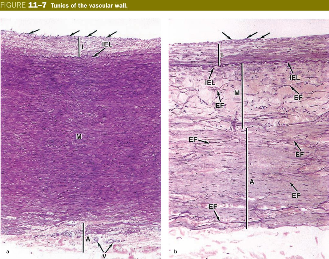
All arteries share the three-tunic plan, but the contents of the media change as you move away from the heart. Right at the aorta the wall must store the energy of left-ventricular ejection and release it during diastole — this is the Windkessel effect — so the media is packed with elastic laminae. By the time you reach a named artery in the limb the job has changed to distributing blood to a particular region, so the media is now mostly smooth muscle that the autonomic nervous system can constrict or relax. Down at the arteriole the job is to set regional resistance — one or two muscle layers can pinch the lumen and double or halve flow into the capillary bed.
That is the whole story of arteries in one sentence: as calibre falls, elastic laminae give way to smooth muscle, and smooth muscle in turn thins out at the arteriole. Knowing where you are on this continuum lets you identify any artery in a microscope field, and it tells you the physiology — conducting, distributing, or resistance.
| Type | Calibre | Key feature |
|---|---|---|
| Large (elastic) | e.g. aorta, pulmonary trunk, brachiocephalic | Media thickest = 40–70 fenestrated elastic laminae (+ some smooth muscle); thick subendothelial intima; Windkessel function |
| Medium (muscular) | >1 mm, the named arteries (radial, femoral, coronary) | Media = 10–40 layers of circular smooth muscle; prominent internal & external elastic membranes |
| Small artery | 0.3–1 mm | Media 3–9 muscle layers; internal elastic membrane present; external elastic membrane usually absent |
| Arteriole | <0.3 mm | Media 1–2 muscle layers; no elastic membranes; principal resistance vessel (sets TPR) |
An elastic artery is a balloon — it inflates with each beat and recoils between beats, smoothing the pulse. A muscular artery is a garden hose with a clamp around it — the clamp opens or closes to send blood to the region that needs it. An arteriole is the tap on the end of the hose — a tiny turn changes the flow enormously. Three calibres, three jobs, three media compositions.
Atherosclerosis begins in the intima of elastic and large muscular arteries: LDL enters, oxidises, macrophages eat it and become foam cells, a fibrous cap forms over a lipid core — the atheroma. Aortic aneurysm is a disease of the media: degraded elastic laminae let the wall stretch and tear. Marfan syndrome (fibrillin-1 mutation) weakens the elastic media and predisposes to aortic dissection. Giant cell (temporal) arteritis inflames the media of the temporal artery, causing headache and visual loss in the elderly. Arteriolar tone, set by the media, is the chief determinant of peripheral resistance — that is what antihypertensives work on.
• Media of muscular artery? → 10–40 smooth-muscle layers
• Arteriole defining feature? → 1–2 muscle layers, no elastic membranes
• Resistance vessel? → Arteriole
• Where does atherosclerosis start? → Intima
• Where does aneurysm form? → Media
If you are going to memorise one vessel by heart for the histology exam, make it the muscular artery in cross-section. Examiners love it because it shows every feature of the three-tunic plan cleanly and it pairs perfectly with its companion vein, letting them ask compare-and-contrast questions for easy marks.
Reading the wall from the lumen outward: an endothelium sits on a delicate subendothelial CT; immediately deep to that lies a clear, refractile, wavy pink line on H&E — the internal elastic membrane. This wavy line is your signpost: see it and you are almost certainly looking at a muscular artery, not a vein. Across the membrane begins the tunica media, 10 to 40 layers of circular smooth muscle wrapped tight around the lumen, with thin elastic and collagen fibres laid down by the muscle cells themselves (these smooth-muscle cells double as fibroblasts in the vessel wall). Outside the media a less obvious external elastic membrane marks the boundary with the tunica adventitia — loose CT carrying the vasa vasorum (small vessels that feed the outer wall of a vessel too thick to be nourished by luminal diffusion alone) and nervi vasorum (autonomic axons that drive vasoconstriction).
| Tunic | Structure |
|---|---|
| Intima | Endothelium + thin subendothelial CT + a clear, wavy pink internal elastic membrane (elastin) |
| Media | 10–40 layers of circular smooth muscle + collagen & elastic fibres (smooth muscle cells synthesise these fibres & ground substance themselves) |
| Adventitia | External elastic membrane + CT with vasa vasorum & nervi vasorum |
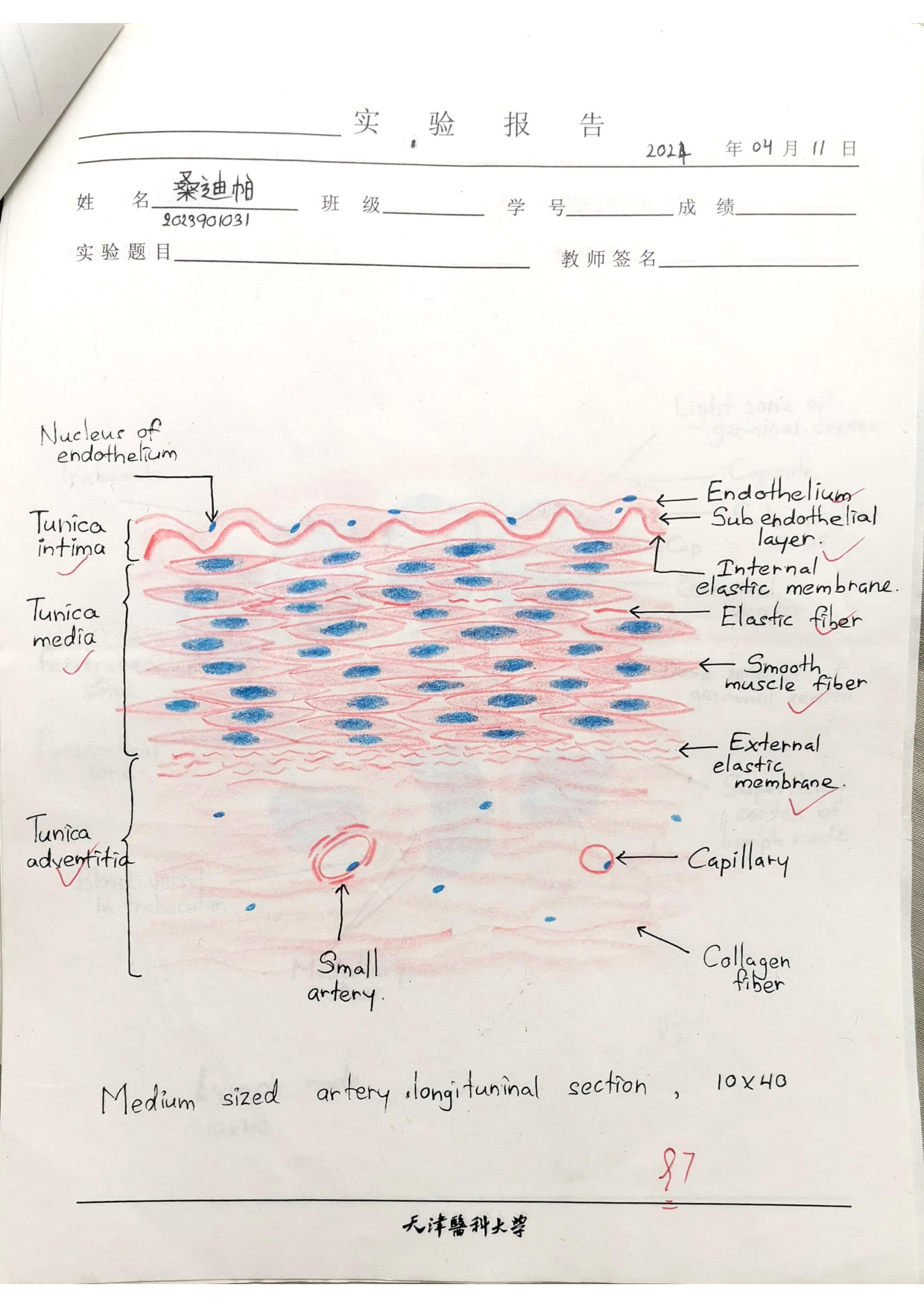
When a muscular artery is fixed and cut, its smooth muscle contracts slightly while the elastic internal membrane recoils elastically — that mismatch is what gives the membrane its wavy appearance. A vein does not have a thick internal elastic membrane, so its intima looks flat. Train your eye for that pink scribble at the edge of the lumen and you have your answer to “artery or vein?” in under a second.
• How many smooth-muscle layers in media? → 10–40
• Who makes the elastic & collagen in the media? → The smooth muscle cells themselves
• What runs in the adventitia? → Vasa vasorum + nervi vasorum
• Quickest artery-vs-vein call? → Round lumen + thick muscular wall + wavy IEL = artery
Veins
Veins live at low pressure, so their wall does not need to be thick to keep its shape. The same three tunics are there, but the boundaries are blurred, the media is thin, and the adventitia is the dominant layer — the opposite of the muscular artery. The lumen is wide and often collapsed into an irregular shape on a slide, and you will usually see a vein paired with its companion artery: small thick-walled round vessel = artery, large thin-walled irregular vessel = vein.
The defining feature of medium and large limb veins is the valve: a paired fold of intima projecting into the lumen, covered on both sides by endothelium and reinforced by a thin core of CT. Valves face the heart and prevent backflow; they are what lets the skeletal-muscle pump (calf muscles squeezing the deep veins during walking) push venous blood uphill against gravity. They are absent in the great veins of the trunk and in cerebral veins.
Just upstream of the smallest veins are the postcapillary venules, the site where leukocytes leave the bloodstream during inflammation — they roll along the endothelium, stick, and squeeze between endothelial cells (diapedesis). Histamine and other inflammatory mediators act here to open the endothelial junctions, which is why a wheal forms after an insect bite.
- More numerous than arteries; large, irregular lumen.
- Thinner wall with three layers but indistinct boundaries; less smooth muscle & elastic fibre, more connective tissue in the adventitia.
- Valves (folds of intima covered by endothelium) prevent backflow, especially in limb veins.
- Postcapillary venules = site of leukocyte diapedesis in inflammation.
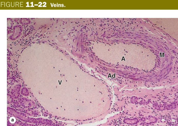
Pressure inside a vein is so low that the wall would collapse if it were stiff. So nature builds the vein as a soft rubber tube and adds one-way doors (valves) inside. Walking pumps the doors open in sequence and the blood climbs against gravity. Stand still all day in heat and the doors stretch open — that is varicose veins.
Incompetent venous valves + prolonged standing → varicose veins. Stasis + endothelial injury + hypercoagulability (Virchow's triad) → deep vein thrombosis (DVT), which can embolise to the lungs. Failure of lymphatic drainage (the “other” venous return) produces lymphoedema, classically after filarial infection or axillary node dissection in breast-cancer surgery.
• Valves of veins are folds of which tunic? → Intima (endothelium-covered)
• Site of leukocyte diapedesis? → Postcapillary venule
• Three components of Virchow's triad? → Stasis, endothelial injury, hypercoagulability
• Why are great veins valveless? → Pressure gradient + size already do the job
Capillaries — the Three Types
A capillary is the simplest vessel in the body and the most important one in the chapter: it is the only place where exchange happens. Strip away everything and you are left with three components — an endothelial tube (a single rolled-up squamous cell or a few cells joined edge-to-edge), a basal lamina around it, and the occasional pericyte tucked into a split in that basal lamina. Pericytes are contractile, mildly stem-cell-like, and offer mechanical support; they hug capillaries and postcapillary venules and are absent from large vessels.
Where capillaries differ is in how leaky they are, and Junqueira classifies them into three types by what their endothelium and basal lamina do. The differences are tiny on an LM slide but very important on EM, and even more important physiologically — they explain why some organs filter, some retain large proteins, and some let whole cells across.
| Type | Wall feature | Distribution |
|---|---|---|
| Continuous | Continuous endothelium sealed by tight junctions; continuous basal lamina; plasmalemmal vesicles ferry molecules across | Muscle, lung, skin, CT, and the CNS (where the tight junctions form the blood-brain barrier) |
| Fenestrated | Pores (fenestrae) in the endothelium ± thin diaphragm; continuous basal lamina | Gut mucosa, renal glomerulus (fenestrae without diaphragms), endocrine glands (with diaphragms) |
| Sinusoidal (discontinuous) | Wide irregular lumen; large gaps between endothelial cells; incomplete or absent basal lamina | Liver, spleen, bone marrow, and some endocrine glands |
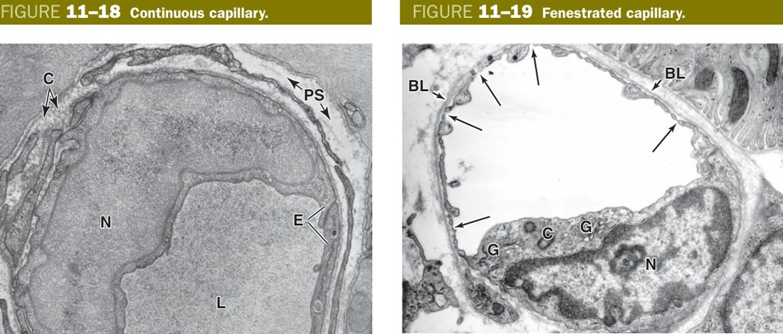
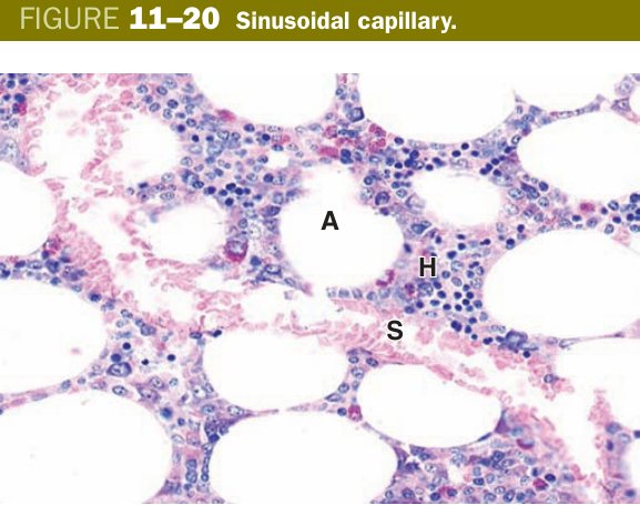
Read the three types as a gradient of permeability. The continuous capillary is the most selective: nothing crosses without going through an endothelial cell or being shuttled in a vesicle — the BBB is the extreme version. The fenestrated capillary punches small windows in the endothelium so that water and small solutes pass freely while plasma proteins are usually retained — ideal for the kidney glomerulus, where you want bulk filtration but no protein loss. The sinusoid throws permeability wide open: gaps in the endothelium and a broken basal lamina mean whole cells can cross — perfect for liver (hepatocytes meet whole blood), spleen (red cell quality control), and bone marrow (new red cells leave for the circulation).
Three types, three doors. Continuous capillary = a sealed door with a postman who carries packages one at a time (vesicular transport). Fenestrated capillary = a screen window: air and small things pass, but the cat stays in (small molecules out, plasma proteins retained). Sinusoidal capillary = an open garage door: people, furniture, anything walks through (whole cells transit). Match the door to the organ's job and you remember the distribution.
Continuous = sealed, continuous BL → brain/muscle/skin/lung. Fenestrated = pores + intact BL → gut, glomerulus, endocrine. Sinusoidal = big gaps + discontinuous/absent BL → liver, spleen, bone marrow (lets whole cells cross).
The blood-brain barrier (continuous capillaries + tight junctions + astrocyte end-feet) is why most antibiotics fail to enter the CNS and why intrathecal injection is sometimes needed. Disruption of the glomerular fenestrated endothelium plus podocyte slit diaphragms produces proteinuria (nephrotic syndrome). In the liver, sinusoidal endothelial damage with fibrosis (cirrhosis) reduces exchange and raises portal pressure.
• BBB capillary type? → Continuous + tight junctions
• Glomerular capillary type? → Fenestrated (no diaphragm)
• Liver/spleen/marrow capillary type? → Sinusoidal
• Three components of a capillary wall? → Endothelium + basal lamina + pericyte
• Where do lymphatics begin? → Blind-ended thin-walled capillaries with anchoring filaments
TMU Exam Drill
📝 Open the full TMU Question Bank — 20 MCQ + 6 terms + 5 essays →
Authentic Tianjin Medical University past-paper questions (2021 Final & the multi-section Final with answer key) mapped to this unit, in the real exam format. Click Show answer to self-test.
□ Fill in the blank
- (fill the three types)
□ True or false
□ Explain the following terms
□ Structure essay
- Endocardium — endothelium + subendothelial CT + subendocardial layer (containing Purkinje fibers).
- Myocardium — thickest; spiral/layered cardiac muscle, intercalated discs; thicker in ventricles.
- Epicardium — visceral pericardium: mesothelium + CT with fat, vessels & nerves.
Circulatory system complete
Heart wall, Purkinje fibres, vessel tunics & the 3 capillary types mastered. Next: Blood.