Nervous Tissue
General Features & Components
Every thought you have, every breath you take, every reflex that pulls your hand off a hot stove — all of it is nervous tissue at work. What looks like a single grey-pink mass under the dissection lamp is in fact a vast electrical network built from only two cell families, and once you learn to tell those two families apart on a slide, the entire nervous system starts to make histological sense.
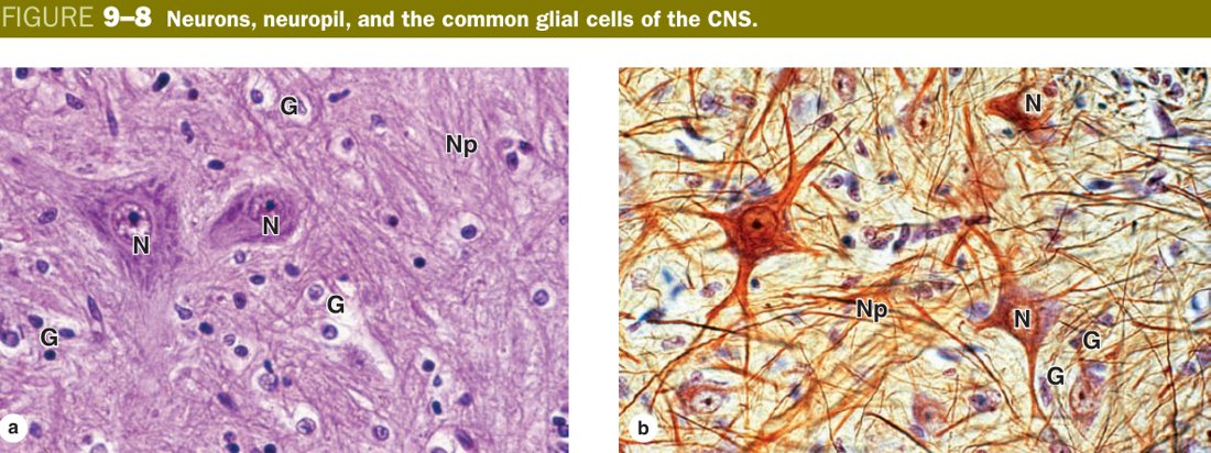
The first family is the neuron — the excitable, signal-carrying cell. It is the “wire” of the system, the one that actually receives a stimulus, integrates it at the cell body, and conducts an impulse to the next cell. The second family is the neuroglia — ten-times more numerous than neurons, non-conducting, but absolutely essential. Glia hold neurons in place, feed them, insulate their axons with myelin, line the ventricles, and act as the brain’s immune system. Junqueira puts it well: neurons do the talking, glia do everything else.
Where do you find each? Cell bodies cluster in the grey matter of the CNS (the cortices, the brain-stem nuclei, the spinal-cord grey horns) and in the ganglia of the PNS. The processes — the long axons — gather into the white matter tracts of the CNS and into the named peripheral nerves outside. Grey looks grey because cell bodies and Nissl substance absorb the stain; white looks white because myelin lipid does not.
| Component | Role |
|---|---|
| Neuron (nerve cell) | The structural & functional unit; has numerous processes; reception, integration & conduction of stimuli (some have endocrine function) |
| Neuroglia (glial cells) | Support, protect, nourish & insulate neurons; form myelin; defence |
Picture a city’s telecom grid. The neurons are the copper wires that actually carry the signal. The glia are the linemen, the insulators, the cooling fans, the security guards and the cleaning crew. Cut the wire and nothing flows; remove the support team and the wire melts inside a week. You need both for the system to work, and on a slide you must learn to recognise both.
• Which one conducts impulses? → Neuron
• Cell bodies live where in the CNS? → Grey matter (and PNS ganglia)
• Why is white matter white? → Myelin lipid
• Three parts of every neuron? → Soma, dendrite, axon
The Neuron
A neuron is built like a little tree with a workshop in the middle. The soma (cell body or perikaryon) is the workshop — this is where the nucleus sits and where almost every protein the neuron will ever need is manufactured. Branching off the soma you have the dendrites, short tapering processes that act as antennas collecting signals from upstream cells. And running away from the soma is the axon — usually a single, long, thin cable that carries the impulse downstream to the next neuron or to a muscle or gland.
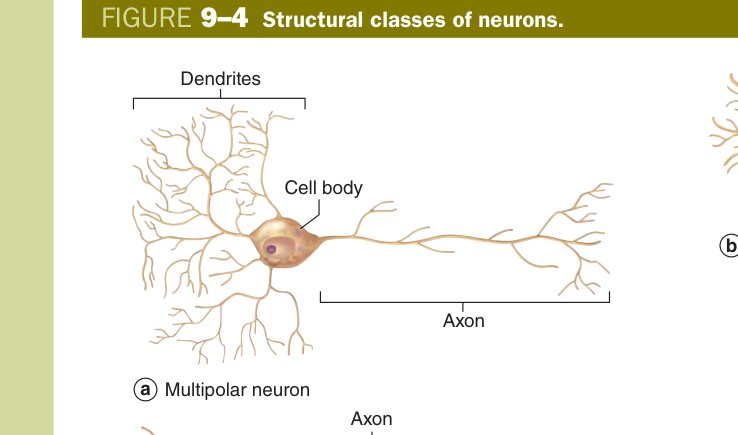
Junqueira classifies neurons by how many processes leave the cell body, and this is one of the most exam-favoured pieces of histology in the whole semester. A multipolar neuron has many dendrites and one axon — this is the typical look (spinal motor neuron, cortical pyramidal cell, cerebellar Purkinje cell). A bipolar neuron has one dendrite at one pole and one axon at the other — the look reserved for the special senses (retinal bipolars, olfactory receptors, the cochlear and vestibular ganglion cells). A pseudounipolar neuron looks like it has only one process leaving the soma, but that process quickly forks into a peripheral branch (going to the receptor) and a central branch (going into the cord) — the textbook example is the dorsal root ganglion neuron. True unipolar neurons are vanishingly rare in vertebrates.
You will also see neurons classified by function (sensory, interneuron, motor) and by neurotransmitter (cholinergic, noradrenergic, peptidergic, GABA-ergic, etc.). For your histology paper, however, the morphological classification is what gets tested.
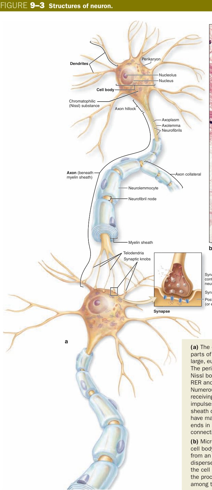
| Classification (by number of processes) | Example |
|---|---|
| Multipolar (many dendrites + 1 axon) | Motor neuron (spinal cord), pyramidal cell (cortex), Purkinje cell (cerebellum) |
| Bipolar (1 dendrite + 1 axon) | Cochlear/vestibular ganglion, retina, olfactory |
| Pseudounipolar (single process that divides) | Spinal (dorsal root) ganglion |
Walk up to the cell body as if it were a small house and count the cables coming out of it. Many cables out and one of them long? Multipolar — the workhorse design. Exactly two cables, one each side? Bipolar — the slim special-sensory design. One cable that splits in two further along? Pseudounipolar — the sensory ganglion design where peripheral and central branches must stay continuous so a finger-tip impulse can run uninterrupted into the spinal cord.
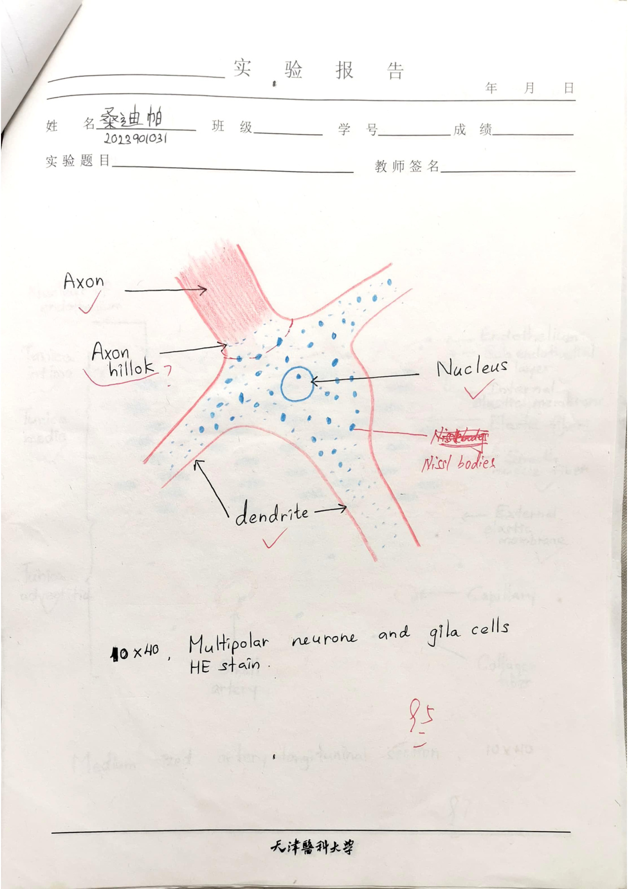
Open up the soma under high power and the first thing that strikes you is the nucleus. It is large, round, pale-staining, and carries one prominent nucleolus — the classic “vesicular nucleus” or “open-faced” nucleus. Why does it look so pale? Because the chromatin is largely euchromatic — the neuron is transcribing flat-out, day and night, to keep its huge cytoplasmic volume supplied with proteins. A cell with a long axon is essentially running a small factory shipping goods to a warehouse a metre away.
Around that nucleus sits the perikaryon, the cytoplasm of the cell body, and this is the home of two structures the examiner loves: Nissl bodies and neurofibrils. Nissl bodies are basophilic, tigroid-looking clumps that on EM resolve into stacks of rough endoplasmic reticulum studded with ribosomes, plus pools of free polyribosomes between the stacks. They are the protein-synthesis machinery of the neuron, and they are present in the perikaryon and in the dendrites — but they are conspicuously absent from the axon hillock and the axon. That single fact explains why the axon hillock stains pale and why the axon itself appears as a clear cable on light microscopy.
Neurofibrils are the second structure. With a silver stain they appear as fine brown filaments running through the soma, into the dendrites and right down the axon. On EM these “neurofibrils” are actually bundles of microtubules, microfilaments and neurofilaments — the cytoskeleton of the neuron. They give the cell its shape and they form the rails on which axonal transport runs, carrying vesicles forwards (anterograde) and recycled material backwards (retrograde) along the axon.
Nissl body: Location — perikaryon and dendrites (absent from the axon & axon hillock). LM — basophilic granular (tigroid) masses. EM — stacks of rough ER + free ribosomes. Function — protein synthesis (enzymes for neurotransmitters/neuromodulators).
Think of the soma as a printing press, the dendrites as the loading dock, and the axon as the delivery truck. The presses (Nissl/RER) stay in the workshop and on the dock — you would never bolt a printing press onto a moving truck. So the dock and the workshop are dark with ink (basophilic), and the truck and the ramp it drives out on (the axon hillock and the axon) are conspicuously clean. That contrast is your diagnostic clue on a slide.
| Nissl body | Neurofibril | |
|---|---|---|
| Location | Perikaryon + dendrites (not axon) | Perikaryon, dendrites and axon |
| LM | Basophilic granules | Brown filaments (silver stain) |
| EM | RER + free ribosomes | Microtubules, microfilaments, neurofilaments |
| Function | Protein synthesis | Cytoskeleton + axonal transport |
After a peripheral axon is cut, the soma reacts by swelling, the nucleus moves to the periphery and the Nissl substance disperses — this is chromatolysis. The pattern is so reliable that pathologists use it to time axonal injury. Rabies takes the opposite route: the virus highjacks retrograde axonal transport to climb from a bite wound back to the CNS, and once there it leaves cytoplasmic Negri bodies in the soma of hippocampal and Purkinje cells.
• Where are Nissl bodies absent? → Axon hillock + axon
• Stain that reveals neurofibrils? → Silver
• Cytoplasmic body left by rabies in Purkinje/hippocampal neurons? → Negri body
• Soma reaction to axonal injury? → Chromatolysis
• Nucleus appearance? → Large, pale, vesicular with prominent nucleolus
Dendrites and the axon may both be processes of the same neuron, but on histology and physiology they are nearly opposites. Dendrites are usually multiple, short, taper rapidly and branch like a tree. Their cytoplasm is essentially perikaryon stretched out — they still carry Nissl substance, mitochondria and Golgi — and many of them are studded with little knobs called dendritic spines where excitatory synapses land. Their job is to receive signals from many upstream neurons and pass that summed input into the soma.
The axon is the opposite. There is usually exactly one per neuron. It is long, thin, of uniform diameter, branches sparsely until it reaches its target, and arises from a specialised cone of cytoplasm called the axon hillock. The axon is wrapped in its own plasma membrane (axolemma) and its cytoplasm (axoplasm) contains neurofilaments, microtubules and mitochondria but, as we said, no Nissl bodies. Its job is to conduct the impulse away from the soma to the next cell, often over remarkable distances.
Because the axon has no ribosomes, every protein it needs — from vesicle membranes to ion-channel subunits to the housekeeping enzymes at the terminal — has to be manufactured in the soma and trucked down the axon. That is what axonal transport does. Anterograde transport (soma → terminal) is driven by kinesin motors walking along microtubules, carrying vesicles full of neurotransmitter and membrane. Retrograde transport (terminal → soma) is driven by dynein, recycling membrane and returning growth signals — but, as the rabies and herpes viruses have learned to exploit, also unwelcome cargo.
Picture a radio tower. Many feeder antennas point in different directions to catch signals from all around (those are the dendrites). One thick transmission cable runs from the base of the tower across the city to the next station (that is the axon). The tower’s antennas and base have all the electronics needed to do work; the long cable is just an insulated conductor — it relies on the base station for every component. Take away the cable’s support trucks (axonal transport) and the transmission silently fails within days.
- Dendrites: usually multiple, branched; cytoplasm similar to perikaryon (contain Nissl); bear dendritic spines; receive stimuli.
- Axon: usually one, long, thin, few branches; arises at the axon hillock; covered by axolemma; axoplasm has neurofibrils & mitochondria but no Nissl bodies; conducts impulses away from the soma.
- Axonal transport: anterograde (soma→terminal, kinesin) & retrograde (terminal→soma, dynein) — exploited by rabies, tetanus and herpes.
• Process that conducts away from the soma? → Axon
• Where does the axon begin? → Axon hillock
• Anterograde motor protein? → Kinesin
• Retrograde motor protein? → Dynein
Synapse
A neuron in isolation is useless. What turns a collection of neurons into a nervous system is the synapse — the specialised contact where one neuron hands its signal to the next cell. That next cell does not have to be another neuron; it can equally be a muscle fibre (the neuromuscular junction) or a gland cell. What is fixed is that the synapse only runs in one direction, presynaptic → postsynaptic, and that there are only two architectural designs in the body: the chemical synapse (overwhelmingly the common one) and the electrical synapse.
In the chemical synapse the impulse arrives at the presynaptic terminal, the cell releases a packet of neurotransmitter into a narrow gap, and the next cell’s receptors read it. There is always a delay, but there is also extraordinary flexibility — you can amplify, you can inhibit, you can modulate, you can fatigue. In the electrical synapse the two cells are physically coupled by gap junctions; ions flow directly from one cytoplasm to the next with essentially no delay, but you give up almost all that flexibility. Electrical synapses dominate where speed and synchrony matter more than control — cardiac muscle, smooth muscle, parts of the brain stem.
Synapse: a specialised junction where a neuron contacts another neuron or an effector cell (muscle/gland) to transmit a signal in one direction. Types: electrical (gap junctions) and chemical (the common type).
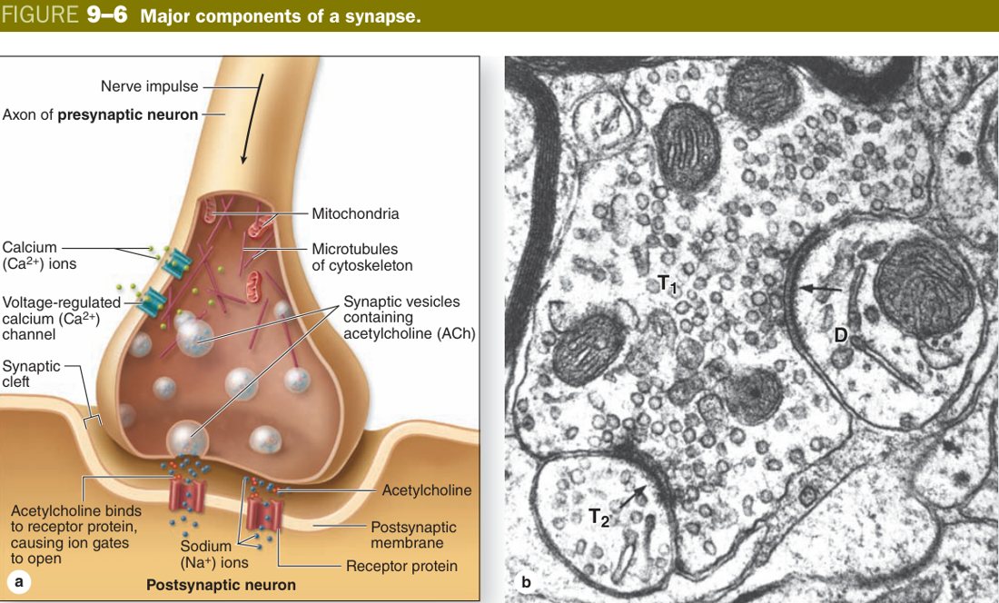
Look closely at a chemical synapse on EM and you will always see three components. The presynaptic element is usually a swelling at the end of the axon — the “terminal button” or bouton. Its membrane is thickened on the cytoplasmic side (the active zone), it is packed with synaptic vesicles full of neurotransmitter, and it carries plenty of mitochondria to fund the cycle. The synaptic cleft is a precise 15–30 nm gap that holds adhesion molecules to keep the two cells aligned. And the postsynaptic element is the receiving cell’s membrane, marked by a dark postsynaptic density that anchors the receptors and the scaffolding proteins.
| Part | Structure |
|---|---|
| Presynaptic element | Axon terminal (terminal button): presynaptic membrane (dense), synaptic vesicles with neurotransmitter, mitochondria |
| Synaptic cleft | 15–30 nm gap |
| Postsynaptic element | Postsynaptic membrane with receptors + postsynaptic density |
The transmission sequence is worth memorising as a chain of events: the impulse reaches the terminal → voltage-gated Ca²⁺ channels open → Ca²⁺ enters → synaptic vesicles fuse with the active zone and dump neurotransmitter into the cleft by exocytosis → transmitter binds postsynaptic receptors → a new potential (excitatory or inhibitory) is generated. Stop calcium entry and you stop transmission — this is exactly how botulinum and tetanus toxins paralyse their victims, by cleaving the SNARE proteins that allow vesicle fusion.
A chemical synapse is a postman. The presynaptic terminal is the post office, full of envelopes (vesicles) loaded with letters (neurotransmitter). The synaptic cleft is the gap between your house and the post office’s wall. The postman only delivers when the bell rings (calcium enters), and only the houses with the right mailbox (receptors) actually receive the letter. The postman never crosses back — one-way only. Now imagine an electrical synapse instead: the post office and your house share a wall with a hole in it, and information just leaks through — instant, but you have lost all delivery control.
Synapse disorders are a high-yield clinical theme. Myasthenia gravis attacks the post-synaptic ACh receptor at the neuromuscular junction. Lambert-Eaton attacks the pre-synaptic P/Q-type Ca²⁺ channel — same end result (weakness) but a different door. Botulinum toxin cleaves SNARE proteins so vesicles cannot fuse (paralysis). Tetanus toxin hits inhibitory synapses in the cord, producing the opposite picture of unrelenting muscle spasm. The synapse is also where most psychiatric drugs work — SSRIs, antipsychotics, benzodiazepines all act at this 30-nm gap.
• Width of the cleft? → 15–30 nm
• Trigger for vesicle release? → Ca²⁺ entry
• Mechanism of release? → Exocytosis at the active zone
• Electrical synapse structure? → Gap junction
• Toxin that cleaves SNARE proteins? → Botulinum
Neuroglia
If neurons are the wires, the neuroglia are the support staff — and they outnumber neurons roughly ten to one. They never conduct impulses, but without them the neurons could not survive a single day. There are six glial types you must know cold, four in the central nervous system and two in the peripheral nervous system, and on a TMU exam paper this list reappears in some form every year.
The four CNS glia are the astrocyte, the oligodendrocyte, the microglia and the ependymal cell. The astrocyte is the most numerous and most versatile — it is star-shaped (hence the name), it expresses the intermediate filament GFAP (your immuno-marker for it), and it does many jobs. Its foot processes ensheath capillaries and contribute to the blood-brain barrier; it buffers extracellular K⁺ and recycles neurotransmitter; and after CNS injury it lays down the glial scar. Two flavours: protoplasmic astrocytes in grey matter (short, leafy processes) and fibrous astrocytes in white matter (long, slender processes).
The oligodendrocyte is the myelin-maker of the CNS. One oligodendrocyte sends out several flat extensions, and each extension wraps an internode on a different nearby axon. So one oligodendrocyte myelinates several axons — the opposite of the Schwann cell pattern. The microglia are the brain’s immune cells. Unlike every other glial type, they are mesodermal in origin (a yolk-sac macrophage lineage); the rest are ectodermal. They surveil the parenchyma, phagocytose debris and present antigen. The ependymal cells are cuboidal-to-columnar cells with cilia and microvilli that line the cerebral ventricles and the spinal central canal; they help generate cerebrospinal fluid (with the choroid plexus) and their cilia keep CSF moving along.
The two PNS glia are the Schwann cell and the satellite cell. The Schwann cell myelinates PNS axons, but with a critical difference from the oligodendrocyte: one Schwann cell myelinates one internode of one axon (1:1 ratio), and unlike CNS glia it lays down its own basal lamina. The satellite cells are the small cuboidal cells you see ringing the cell bodies of neurons inside a sensory or autonomic ganglion — the PNS equivalent of an astrocyte for the soma, providing support and electrical insulation.
| Glial cell | Location | Function |
|---|---|---|
| Astrocyte (fibrous = white matter; protoplasmic = grey) | CNS | Support, nutrition, glia limitans & blood-brain barrier, glial scar; contain GFAP |
| Oligodendrocyte | CNS | Forms myelin in the CNS (one cell myelinates several axons) |
| Microglia | CNS | Phagocytosis / immune defence (CNS macrophage; mesodermal origin) |
| Ependymal cell | CNS cavity lining | Cuboidal/columnar, cilia/microvilli; line ventricles & central canal; help produce & circulate CSF |
| Schwann cell | PNS | Forms myelin in the PNS (1 cell : 1 internode); has a basal lamina |
| Satellite cell | PNS ganglia | Surround ganglion cell bodies; support & insulation |
CNS glia = “A.O.M.E.”: Astrocyte, Oligodendrocyte, Microglia, Ependymal. PNS = Schwann + Satellite. Myelin makers: Oligodendrocyte (CNS), Schwann (PNS). Microglia = the brain’s macrophage (mesodermal — the only glial outsider).
An oligodendrocyte is a school teacher: it sits in one classroom and sends arms out to wrap a sheet of paper around several nearby students’ books. A Schwann cell is a private tutor: one tutor, one student, one book. So in the CNS the ratio is one teacher to many internodes; in the PNS the ratio is one tutor to one internode. The same logic explains why PNS axons can regenerate (the tutor is still sitting there waiting to re-myelinate) and CNS axons largely cannot (lose the teacher and you have lost everyone’s wrapping).
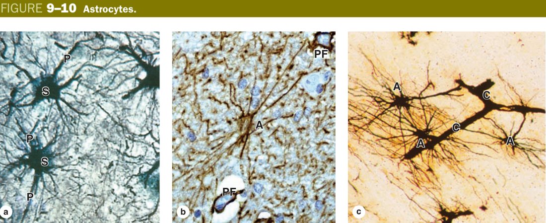
Multiple sclerosis is the CNS demyelinating disease — oligodendrocytes are attacked, producing white-matter plaques (optic neuritis, internuclear ophthalmoplegia, sensory and motor deficits in young adults). Guillain-Barré syndrome is its PNS counterpart: an autoimmune attack on Schwann-cell myelin, producing ascending paralysis. Charcot-Marie-Tooth is a hereditary PNS demyelination (PMP22). Astrocytes form the glial scar and account for the majority of primary CNS tumours (astrocytoma → glioblastoma multiforme). Microglial activation is now a central player in Alzheimer’s and other neurodegenerations.
• Two PNS glia? → Schwann + satellite
• CNS myelin maker? → Oligodendrocyte (1 cell : many axons)
• PNS myelin maker? → Schwann cell (1 cell : 1 internode)
• Astrocyte immuno-marker? → GFAP
• Only mesoderm-derived glia? → Microglia
• CSF lining cells? → Ependymal
Nerve Fibres & Nerves
A nerve fibre is not just an axon — it is an axon plus the glial cells wrapping it. In the PNS that means an axon plus its Schwann cells; in the CNS it means an axon plus its oligodendrocyte processes. Fibres come in two flavours: myelinated (with a spiral lipid sheath) or unmyelinated (with only a thin glial covering and no insulation). Myelinated fibres conduct fast and are typical of motor neurons and large sensory fibres; unmyelinated fibres conduct slowly and carry pain, temperature and autonomic signals.
A nerve fibre = an axon + the glial cells surrounding it; myelinated or unmyelinated.
This is the one structure in the whole unit you can almost guarantee will appear as a long-essay question. So learn it as a story rather than as a list. Start at the centre and work outwards. In the middle of every PNS myelinated fibre is an axon — the actual conductor. Wrapped tightly around that axon is the myelin sheath, made from the plasma membrane of a single Schwann cell that has rolled itself around the axon dozens of times to produce a compact spiral of lipid. The Schwann-cell nucleus and the thin rim of cytoplasm that contains it sit on the outside of the sheath and form the neurolemma.
Now run your eye along the fibre. The Schwann cells line up like beads on a string, and where two adjacent Schwann cells meet there is a tiny unmyelinated gap a micron or so wide called the node of Ranvier. At the node the axolemma is exposed and densely packed with voltage-gated Na⁺ channels — this is where the action potential is regenerated. The length of myelinated axon between two nodes (one Schwann cell’s worth) is the internode. Inside the myelin you will sometimes see oblique pale clefts; these are Schmidt-Lanterman incisures, small pockets of Schwann-cell cytoplasm trapped inside the spiral that keep the sheath supplied and flexible. The narrow connection from the outer cytoplasm into the spiral is called the mesaxon.
All of this anatomy exists for one reason: saltatory conduction. Because myelin is a great electrical insulator, the impulse cannot leak out along the internode; it can only regenerate at the next node. So it appears to leap from node to node, vastly faster than a continuous wave would travel. Strip the myelin and conduction collapses — which is exactly what happens in multiple sclerosis and in Guillain-Barré.
A quick note on the CNS counterpart and on unmyelinated fibres, because the contrast is high-yield. In the CNS the myelin is made by an oligodendrocyte — one oligodendrocyte myelinates several different axons, there is no neurolemma, no basal lamina, and the cell body sits in the parenchyma rather than against the axon. In the PNS, unmyelinated fibres are not bare; instead, one Schwann cell wraps itself loosely around several thin axons, parking each one in a groove on its surface. This bundle is sometimes called a Remak bundle. No spiral, no compact myelin, no nodes, no saltatory conduction — just slow continuous conduction.
Picture a long electrical extension cord that has been wrapped tightly in rubber tape every metre, with one centimetre of bare copper left exposed between each wrap. Current cannot leak through the rubber, but at every bare patch the wire can pick up a fresh push from a small booster station. That is exactly the myelinated nerve fibre: rubber = myelin internodes, bare copper = nodes of Ranvier, booster stations = the Na⁺ channels that regenerate the impulse. Now picture an unmyelinated cord with no tape at all — current trickles down it the whole way, slowly, leaking the whole time. That is the unmyelinated fibre.
- Axon: in the centre.
- Myelin sheath: at the periphery, formed by the wrapped plasma membrane of a Schwann cell (lipid + protein); neurolemma = outer Schwann-cell cytoplasm + nucleus.
- Node of Ranvier: the narrow gap between adjacent Schwann cells where the axolemma is exposed; densely packed with Na⁺ channels.
- Internode: the myelinated segment between two nodes (= one Schwann cell).
- Schmidt-Lanterman incisures: small clefts of Schwann-cell cytoplasm within the myelin, giving flexibility & aiding turnover.
- Mesaxon: the membrane connection between the outer Schwann-cell surface and the start of the myelin spiral.
- Function: insulation + saltatory conduction (impulse jumps node-to-node → faster).
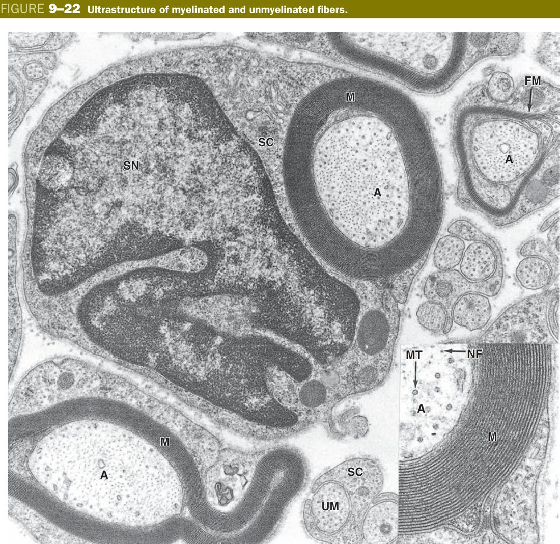
If the Schwann cell or its myelin is attacked, the patient develops a peripheral neuropathy. Guillain-Barré (autoimmune Schwann attack), Charcot-Marie-Tooth (hereditary PMP22 problem), and leprosy (Schwann cells are M. leprae’s favourite home) all produce loss of saltatory conduction with weakness and sensory loss. After a clean PNS cut the picture is different and remarkable: the distal segment dies (Wallerian degeneration) but the Schwann cells survive, form a hollow endoneurial tube and guide the proximal axon’s sprouts back to the target at about 1 mm a day. That regenerative capacity is precisely what CNS axons lack, because oligodendrocyte myelin is laced with growth inhibitors (Nogo, MAG, OMgp) and the astrocyte scar is impassable.
• Cell forming CNS myelin? → Oligodendrocyte (1 : many)
• Gap with bare axolemma? → Node of Ranvier
• Myelinated segment? → Internode
• Pale clefts within the sheath? → Schmidt-Lanterman incisures
• Why is conduction fast? → Saltatory (jumps node to node)
• PNS unmyelinated bundle name? → Remak bundle
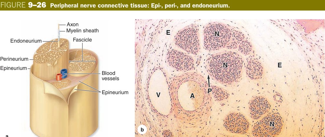
A peripheral nerve is not a single fibre — it is hundreds or thousands of fibres bound together in connective tissue. Histology gives you three concentric wrappings, and on a transverse section you can name all three by where you find them. Innermost is the endoneurium, a delicate layer of reticular fibres and a basal lamina that hugs each individual nerve fibre (one axon plus its Schwann cells). A group of these fibres is bundled into a fascicle by a tougher, layered sleeve of flattened epithelioid cells called the perineurium — and the perineurium does something special: its tight junctions form the blood-nerve barrier, the peripheral analogue of the BBB. Several fascicles are then bundled together by a thick dense-irregular outer coat called the epineurium, which carries the larger blood vessels of the nerve (vasa nervorum) and gives the nerve its mechanical strength.
A second high-yield piece of nerve histology is the ganglion. A ganglion is a cluster of neuronal cell bodies in the PNS, and you must be able to tell two kinds apart. The sensory ganglion (dorsal root ganglion of a spinal nerve, or a sensory cranial ganglion) holds round pseudounipolar cell bodies, neatly arranged with satellite cells around each one, and no synapses inside. The autonomic ganglion holds multipolar cell bodies, scattered rather than arranged, with eccentric nuclei, fewer satellite cells — and synapses do occur inside (this is where preganglionic fibres terminate). Grey matter of the brain is not called a ganglion (basal ganglia are an old-school exception); proper ganglia are PNS structures.
| Layer | Wraps |
|---|---|
| Endoneurium | Each individual nerve fibre |
| Perineurium (epithelioid cells; tight junctions = blood-nerve barrier) | A bundle (fascicle) of fibres |
| Epineurium (dense irregular CT; carries vasa nervorum) | The whole peripheral nerve |
Imagine a thick telephone cable cut across. Each tiny copper wire (axon + Schwann) is wrapped in its own thin plastic sleeve — that is the endoneurium. A handful of these wires is bound into a coloured ribbon by a tougher tape — that is the perineurium, and that tape is also waterproof (blood-nerve barrier). Several ribbons are wrapped together inside one fat outer black jacket containing the cable’s power line — that is the epineurium with its vasa nervorum. Three layers, three loyalties.
• Layer wrapping a fascicle? → Perineurium
• Layer wrapping the whole nerve? → Epineurium
• Blood-nerve barrier formed by? → Perineurial tight junctions
• DRG neuron type? → Pseudounipolar (no internal synapses)
• Autonomic ganglion neuron type? → Multipolar (synapses inside)
TMU Exam Drill
📝 Open the full TMU Question Bank — 20 MCQ + 6 terms + 5 essays →
Authentic Tianjin Medical University past-paper questions (2021 Final & the multi-section Final with answer key) mapped to this unit, in the real exam format. Click Show answer to self-test.
□ Single best answer
- A. astrocytes
- B. oligodendrocytes
- C. microglia
- D. Schwann cells
- E. satellite cells
□ Explain the following terms
Nervous tissue complete
Neuron, Nissl, synapse, 6 glia & the myelinated-fibre essay mastered. Next: Circulatory System.