Muscle Tissue
General Features & Classification
Muscle is the body's tissue specialised for conversion of chemical energy (ATP) into mechanical work. Wherever something in the body has to shorten on command — a limb, a heart chamber, the wall of the gut, the iris of the eye — the histology you are looking at is a sheet, bundle or syncytium of elongated contractile cells crammed with two filament proteins, actin and myosin.
Because these cells are so specialised, anatomists gave their normal organelles new names that all begin with the prefix sarco- (Greek sarx = flesh). When you read the textbook, mentally translate as you go: sarcolemma is just the plasma membrane, sarcoplasm is the cytoplasm, and the sarcoplasmic reticulum (SR) is a modified smooth endoplasmic reticulum that has been reorganised into a calcium store. The cell itself, especially the long skeletal one, is called a muscle fibre — an unfortunate term, because every histology student first hears “fibre” and pictures collagen. Here a muscle fibre is a whole cell, not a thread of extracellular matrix.
There are exactly three histological flavours of this tissue, and the slide-room game is to tell them apart from a single field of view. The three are skeletal, cardiac and smooth, and they differ in two visible features and one functional one:
- Sarcolemma = cell membrane · Sarcoplasm = cytoplasm · Sarcoplasmic reticulum (SR) = smooth ER (stores Ca²⁺).
| Type | Striation | Control |
|---|---|---|
| Skeletal muscle | Striated | Voluntary |
| Cardiac muscle | Striated | Involuntary |
| Smooth muscle | Non-striated | Involuntary |
Think of muscle as three ways of solving the same engineering problem — how to make a cell that shortens reliably. Skeletal = a long parallel cable optimised for fast, voluntary, short-burst force. Cardiac = an electrically wired meshwork that has to beat for eighty years without rest. Smooth = a slow, energy-cheap tonic squeezer that lines hollow organs. The striations you see (or don't) under the microscope are just a visible read-out of how tightly the actin–myosin filaments are packed into repeating sarcomeres.
• Which two are striated? → Skeletal & cardiac.
• Which two are involuntary? → Cardiac & smooth.
• What does “sarco-” mean? → Flesh; sarcolemma = plasma membrane, sarcoplasm = cytoplasm, SR = smooth ER (Ca²⁺ store).
• Is a muscle fibre a cell or an organelle? → A whole cell.
Skeletal Muscle — Light Microscopy
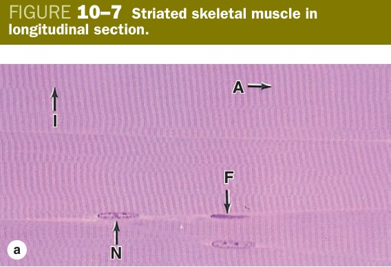
When you put a longitudinal section of a biceps under the microscope, the first thing that strikes you is the length and parallelism of the cells. A single skeletal muscle fibre can be 10–100 µm thick yet several millimetres — in the sartorius, up to about 40 mm — long. It is one of the longest cells in the body, and it has to be: a sarcomere is barely 2 µm, so producing a useful contraction means stacking thousands of them end-to-end inside one cell.
To pack that much contractile machinery into one cell, the embryo fuses dozens of mononuclear myoblasts. The result is a syncytium with many flattened nuclei pushed out to the periphery, lying just beneath the sarcolemma. This is one of the diagnostic features in the slide room: if the nuclei are peripheral and there are several of them along the fibre, you are looking at skeletal muscle. The sarcoplasm itself is strongly acidophilic because it is stuffed with eosinophilic myofibrils 1–2 µm thick, all running parallel, and their precise register from one myofibril to the next is what gives the fibre its cross-striations — the alternating dark A-bands and light I-bands you can resolve even at medium power.
Crucially, skeletal fibres are not joined to one another end-to-end: there are no intercalated discs. Each fibre is a separate cell that takes orders from its own motor end-plate. Tucked between the sarcolemma and the surrounding basal lamina sit small, dormant satellite cells — quiescent stem cells which wake up after injury, divide, and fuse into the damaged fibre. This is why skeletal muscle, unlike cardiac muscle, can regenerate after a tear or a crush.
Around the fibre the connective-tissue investments come in three nested layers, and Junqueira makes a point of the names. The endomysium is a delicate reticular sleeve around each individual fibre and carries the capillaries and fine nerves; the perimysium is denser, bundles fibres into fascicles, and carries the larger vessels and nerves; the epimysium is the dense irregular collagen coat around the whole muscle that is continuous with its tendon. The whole arrangement transmits force from the sarcomere to the bone.
- Shape: long, cylindrical fibres; diameter 10–100 µm, up to 1–40 mm long.
- Nucleus: multinucleate, many flattened nuclei at the periphery (just under the sarcolemma).
- Sarcoplasm: acidophilic; packed with myofibrils (1–2 µm, parallel) showing distinct cross-striations.
- No intercalated discs.
- Satellite cells lie between sarcolemma and basement membrane — regenerative stem cells (differentiate into myoblasts after injury).
- CT coverings: epimysium (whole muscle) → perimysium (fascicles) → endomysium (each fibre).
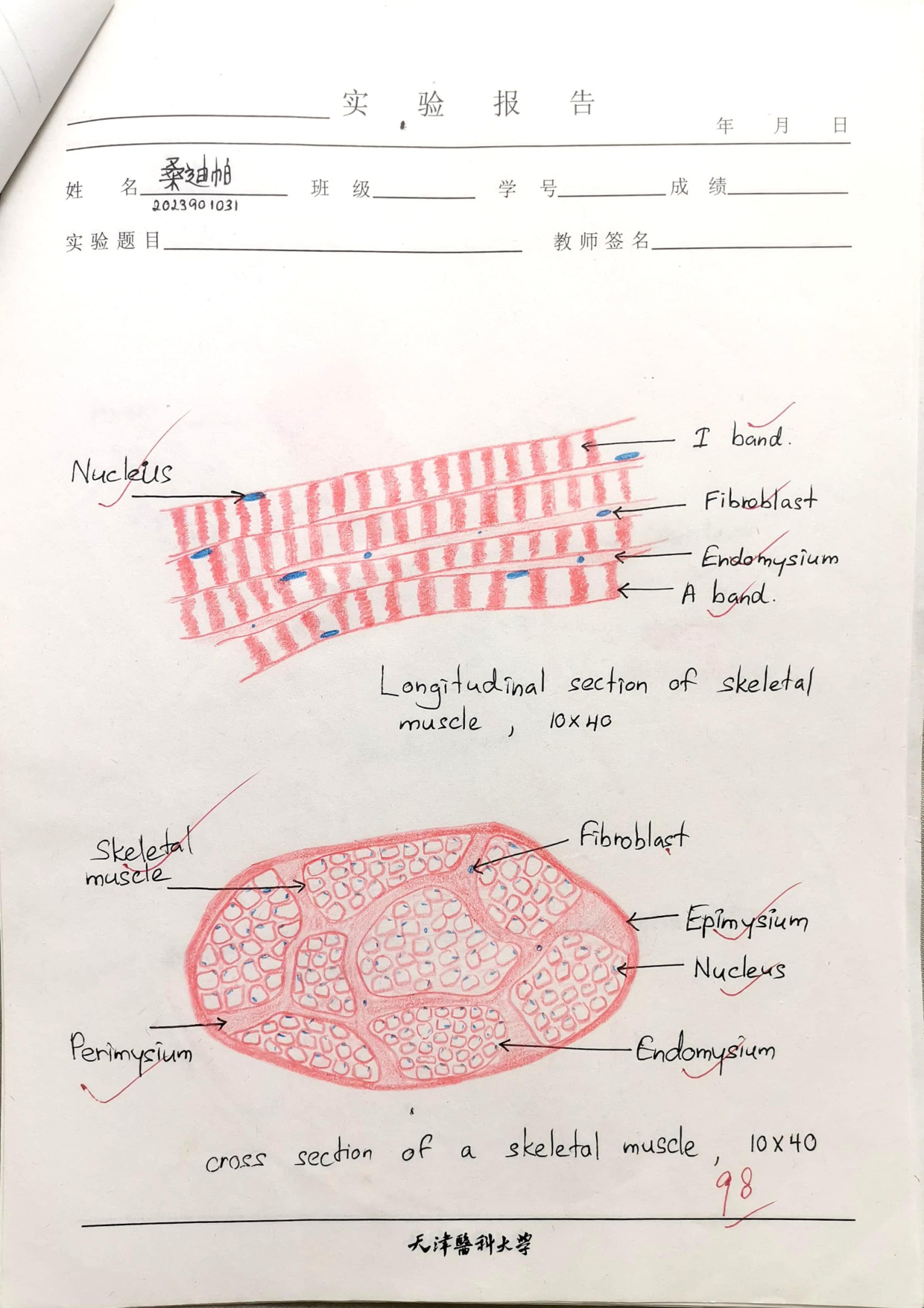
Picture a skeletal fibre as a fibre-optic cable: a single long sheath (sarcolemma) wrapping thousands of identical glassy strands (myofibrils) running perfectly in parallel. The connective-tissue layers — endo-, peri-, epimysium — are the kevlar braids that hold the bundle together so the force generated at one end is delivered cleanly to the tendon at the other.
• Why so many nuclei per cell? → The fibre forms by fusion of myoblasts (syncytium).
• Three CT sheaths, outside in? → Epimysium → perimysium → endomysium.
• Which cell regenerates skeletal muscle? → Satellite cell, between sarcolemma and basal lamina.
• Why do you see cross-striations? → Sarcomeres of adjacent myofibrils are in register.
Myofibril, Bands & the Sarcomere
A myofibril is not a uniform rod. Look at it lengthwise in the EM and you see a repeating pattern of alternating dark and light bands — the same pattern responsible for the cross-striations of the whole fibre. Each repeating unit is a sarcomere, the smallest functional contractile machine in the body, and it is bounded at each end by a dense transverse line called the Z-line.
The dark band straddling the centre of every sarcomere is the A-band ("A" for anisotropic — it is birefringent under polarised light). It is dark because it is where the thick myosin filaments live, and at its outer edges the thick filaments overlap with thin actin filaments coming in from each Z-line. The lighter I-band ("I" for isotropic) lies on either side of the Z-line and contains thin filaments only — that is why it stains lighter; there is no thick myosin in it.
Inside the A-band, right at the centre of the sarcomere, you can sometimes resolve a still paler stripe — the H-band. This is the zone where thick filaments are not overlapped by thin ones. Bisecting the H-band is the dark M-line, a transverse scaffold of proteins that links the thick filaments to one another and holds them in register. Each sarcomere therefore reads, from Z to Z: ½ I-band → A-band (with H-band and M-line) → ½ I-band.
This geometry is what makes the sliding-filament theory visible. When the sarcomere contracts, the thin filaments slide inwards over the thick filaments; the A-band stays the same length (the thick filaments don't change), but the I-band and H-band narrow because the thin filaments encroach on them. Examiners love this point — it lets them ask a one-line question that tests whether you actually understand the architecture or just memorised the band letters.
| Band / line | What it is |
|---|---|
| I-band (light) | Thin (actin) filaments only; bisected by the Z-line |
| A-band (dark) | Thick (myosin) filaments (+ overlap of thin); contains H-band & M-line |
| Z-line | Anchors thin filaments; boundary of the sarcomere |
| H-band | Central part of A-band with thick filaments only |
| M-line | Middle of the H-band; links thick filaments |
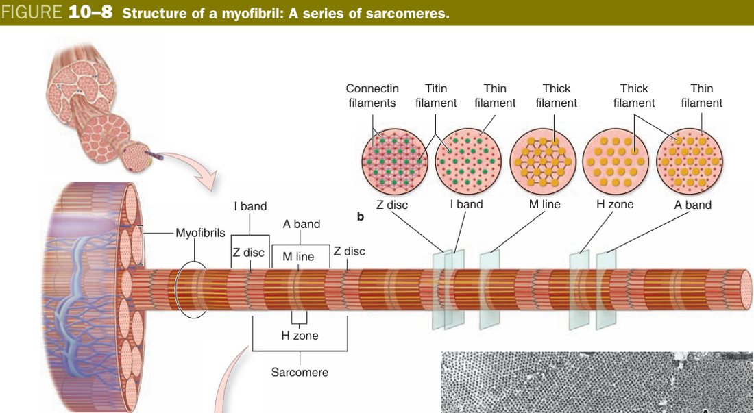
Picture two combs facing one another and locked together teeth-first. The handles (Z-lines) sit at the outside; the teeth from each comb (thin actin filaments) reach inwards and slide past a third row of central rods (thick myosin filaments). The A-band is the territory of the central rods. When the combs pull their handles toward the centre, the rods don't move — only the teeth slide deeper, and the gap between the handles (the sarcomere) shortens.
Sarcomere: the segment of a myofibril between two adjacent Z-lines; it consists of ½ I-band + A-band + ½ I-band. It is the structural and functional (contractile) unit of the myofibril.
• A-band contains? → Thick filaments (and thin-filament overlap at the edges).
• I-band contains? → Thin filaments only, bisected by the Z-line.
• H-band? → Central part of A-band, thick filaments only (no overlap).
• What does the M-line do? → Links thick filaments transversely at the centre of the H-band.
• On contraction, which bands narrow? → I and H; A stays constant.
Skeletal Muscle — EM, Myofilaments & Triad
Under the electron microscope the “rods” and “teeth” of the comb analogy resolve into two specific protein assemblies. The thick filament is a bundle of about 300 myosin II molecules. Each myosin has a long rod-like tail that lies in the bundle and a pair of globular heads that project outwards. Those heads are where the action is: each carries an ATPase site and an actin-binding site, and during contraction the head will reach across, grab the thin filament, swivel, and let go — the cross-bridge cycle.
The thin filament is a twisted double-helical polymer of F-actin, with two regulatory proteins draped along it. Tropomyosin is a long fibrous protein that lies in the groove of the actin helix and physically covers the myosin-binding sites. Troponin is a small complex bound to tropomyosin at regular intervals along the filament. It has three subunits, each named for what it does: TnT binds tropomyosin, TnI is inhibitory (it locks tropomyosin over the actin site at rest), and TnC binds the Ca²⁺ ion that throws the switch. When TnC binds calcium, the whole troponin–tropomyosin complex tilts off the binding sites, and myosin heads can now attach.
For this to work as a coordinated millisecond event across the entire cell, the depolarisation that opens the calcium gates must reach every sarcomere at once. The skeletal fibre solves this with two membrane systems that you must be able to draw in the exam. The transverse (T) tubules are narrow invaginations of the sarcolemma that dive into the depth of the fibre and run perpendicular to its long axis. The sarcoplasmic reticulum wraps the myofibrils as a fine longitudinal mesh, and at intervals it widens into pancake-shaped terminal cisternae that abut the T-tubule.
In skeletal muscle, this geometry is the triad: a central T-tubule flanked by two terminal cisternae, one on each side, positioned precisely at the A–I band junction. There are therefore two triads per sarcomere. Each cisterna's membrane is studded with ryanodine receptors (RyR1) coupled to voltage-sensing dihydropyridine receptors in the T-tubule. When the depolarisation arrives, the gates open, calcium floods the sarcoplasm, troponin C binds it, and every sarcomere in the cell contracts together — that is the meaning of excitation–contraction coupling.
| Myofilament | Protein | Detail |
|---|---|---|
| Thick | Myosin | Globular head = ATPase + actin-binding site (forms the cross-bridge); rod-like tail |
| Thin | Actin + tropomyosin + troponin | Troponin has 3 subunits: TnT (binds tropomyosin), TnI (inhibitory), TnC (binds Ca²⁺) |
- Transverse (T) tubules: sarcolemma invaginates into the fibre at the A–I junction — conducts the impulse rapidly to all myofibrils.
- Sarcoplasmic reticulum (SR): longitudinal tubules forming a net around myofibrils; expanded terminal cisternae flank each T-tubule (store/release Ca²⁺ via calcium pump & channel).
Imagine the SR as a soaker hose wrapped tightly around the myofibrils, filled with calcium under pressure. The T-tubule is an electrical wire that dives in from the surface and runs alongside the hose at every A–I junction. When the wire fires, the valves on the hose pop open at exactly that point — calcium floods out, the sarcomeres on either side contract, and the wire–hose contact is so close (the “triad”) that the response is essentially instantaneous.
Triad: in skeletal muscle, two terminal cisternae of the SR + one central T-tubule, located at the A–I band junction. (Contrast: cardiac muscle has a diad — one T-tubule + one cisterna — located at the Z-line.)
• Which troponin subunit binds Ca²⁺? → TnC.
• What does TnI do? → Inhibits actin–myosin interaction at rest.
• Triad = what + where? → T-tubule + 2 terminal cisternae, at the A–I junction.
• How many triads per sarcomere? → Two.
• What couples the T-tubule signal to SR Ca²⁺ release? → Dihydropyridine receptor (T-tubule) talks to ryanodine receptor RyR1 (SR).
Once you can see the architecture, the contraction story is just a sequence of switches. The motor neuron fires, releasing acetylcholine onto the motor end-plate; the sarcolemma depolarises and the wave runs down every T-tubule. Where the T-tubule meets the SR at the triad, the SR terminal cisternae dump their stored calcium into the sarcoplasm. Calcium binds TnC, troponin tilts, tropomyosin slides off the myosin-binding sites, and the myosin heads — already cocked — latch onto actin.
Each head then performs a single power stroke, swivelling and dragging the thin filament toward the M-line. ATP binds the head, the head releases, hydrolyses the ATP to re-cock itself, and the cycle repeats as long as calcium and ATP are available. Because thin filaments slide inwards but the thick filaments don't change length, the A-band stays constant while the I-band and H-band narrow, and the Z-lines are pulled toward one another. The whole sarcomere shortens, and along with it the whole fibre, the whole muscle, and the joint it crosses.
When the nerve impulse stops, the SR calcium pump (SERCA) sucks calcium back into the cisternae against a 10 000-fold gradient. Sarcoplasmic calcium falls, troponin C lets go, tropomyosin re-covers the actin site, and the fibre relaxes. If ATP is unavailable — as after death — myosin cannot detach, the cross-bridges lock, and the body goes into rigor mortis.
Summary trigger chain: Impulse → T-tubule → SR releases Ca²⁺ → Ca²⁺ binds TnC → tropomyosin shifts, exposing actin's myosin-binding site → myosin heads (cross-bridges) pull the thin filaments past the thick filaments → sarcomere shortens. The A-band stays constant while the I-band and H-band narrow.
Duchenne muscular dystrophy (dystrophin defect) → progressive skeletal-muscle degeneration. Myasthenia gravis — autoantibodies against the ACh receptor on the motor end-plate (sarcolemma) → fatigable weakness. Cardiac troponin (TnI/TnT) released into blood is the key marker of myocardial infarction. Malignant hyperthermia — mutation in the skeletal RyR1 calcium-release channel; volatile anaesthetics trigger uncontrolled SR Ca²⁺ release, sustained contraction, and a runaway rise in body temperature.
• Which bands change length on contraction? → I and H narrow; A is constant.
• What returns Ca²⁺ to the SR? → The SERCA pump.
• Why rigor mortis? → No ATP → myosin heads cannot detach from actin.
• Which receptor mutation causes malignant hyperthermia? → RyR1.
Cardiac Muscle
Cardiac muscle is built to a different brief from skeletal. It has to contract roughly once a second for a lifetime, propagate its own rhythm without a motor neuron, and resist fatigue. Histology shows the consequences. The cells are short, roughly cylindrical columns (about 15 µm thick and 100 µm long), and they branch — you will see Y-shaped profiles in a good section. Each cell has one or two central nuclei, never the peripheral palisade of skeletal muscle. The sarcoplasm is striated, because cardiomyocytes also use sarcomeres, but the striations look fuzzier than in skeletal because the myofibrils are not sharply demarcated — they tend to branch and merge across the cell.
The other dead-giveaway feature, and the one that examiners love, is the intercalated disc. End-to-end, cardiac cells are joined by these dark transverse step-like lines that you can see at light microscopy. The disc is not a single junction but a composite of three classical junctional types, each with a job:
- Fascia adherens — the transverse face anchors actin filaments of the terminal sarcomere of each cell, transmitting pull.
- Macula adherens (desmosomes) — spot-welds that bind the desmin intermediate filaments and stop the cells from pulling apart under load.
- Gap junctions — sit on the longitudinal face and electrically couple neighbours, so depolarisation jumps cell to cell and the myocardium contracts as a single functional syncytium.
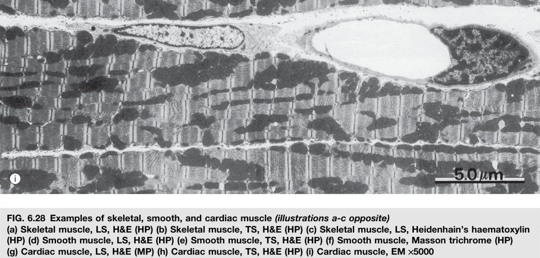
Because the heart never rests, cardiac muscle is loaded with mitochondria (they take up roughly a third of the cell volume) and with myoglobin, both of which support continuous aerobic metabolism. The membrane system is also adapted: T-tubules are wider than in skeletal muscle and are positioned at the Z-line rather than the A–I junction, and they meet one SR cisterna instead of two. That arrangement is called a diad, and there is one per sarcomere instead of the skeletal two. The simpler coupling is fine because cardiac contraction also depends on extracellular calcium entering through the T-tubule itself (calcium-induced calcium release), so the SR doesn't have to do all the work alone.
Finally, the cardiac cell does not regenerate. There are no satellite cells in the myocardium; after a myocardial infarction the dead cardiomyocytes are replaced by collagenous scar laid down by fibroblasts, not by new muscle. That irreversibility is the reason every clinical decision around acute coronary syndromes is dominated by the clock.
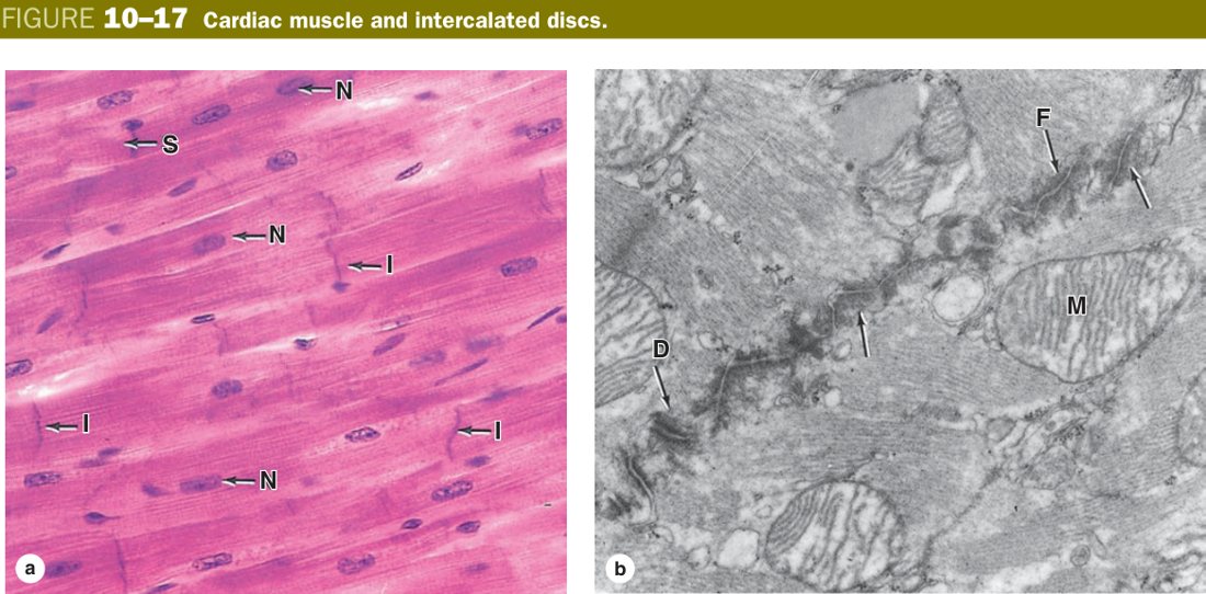
- LM: short, branching columns (~15 µm × ~100 µm); 1–2 central nuclei; cross-striated; joined end-to-end by intercalated discs (transverse dark/step-like lines).
- EM: myofibrils incomplete (not sharply demarcated); diads (T-tubule + 1 cisterna) at the Z-line; abundant mitochondria.
The intercalated disc does two jobs in one structure — it is a mechanical weld on the transverse face (fascia adherens + desmosomes hold cells together against the systolic pull) and an electrical short-circuit on the longitudinal face (gap junctions let the action potential leap from cell to cell). That is why a piece of myocardium beats as one sheet even though it is made of millions of independent cells.
Intercalated disc: the specialised end-to-end junction between cardiac muscle cells. LM: a transverse dark-staining, step-like line. EM: its transverse portion has desmosomes + adherens (intermediate) junctions (mechanical anchorage); its longitudinal portion has gap junctions (electrical coupling, so the heart contracts as a syncytium).
After a myocardial infarction, the ischaemic cardiomyocytes die and release their contents into blood — cardiac troponin I and T are the specific serum markers used to confirm the diagnosis. Because cardiac muscle cannot regenerate, the infarcted zone heals by fibrous scar, which is non-contractile and prone to aneurysm. Mutations in desmosomal proteins of the intercalated disc underlie arrhythmogenic right ventricular cardiomyopathy, where mechanical failure of the disc lets cells pull apart and triggers lethal arrhythmias.
• Do cardiac cells branch? → Yes.
• Three components of intercalated disc? → Fascia adherens, desmosomes, gap junctions.
• Which component electrically couples cells? → Gap junctions (longitudinal face).
• Triad or diad? Where? → Diad at the Z-line (one T-tubule + one SR cisterna).
• Can cardiac muscle regenerate? → No — injury heals by fibrous scar.
• Blood marker of MI? → Cardiac troponin I / T.
Smooth Muscle
Smooth muscle is the involuntary muscle of hollow viscera — the wall of the gut, the bladder, the bronchi, the uterus, and the tunica media of every artery. Its job is slow, sustained, energy-cheap contraction that can hold for minutes or hours, the opposite design problem from skeletal muscle. The histology reflects that brief in three ways: the cells are spindle-shaped, the nucleus is single and central, and there are no cross-striations.
A typical smooth muscle cell is 5–20 µm wide and 60–100 µm long, but it can stretch enormously — the smooth muscle cell in a term-pregnant uterus may exceed 500 µm. Its sarcoplasm is acidophilic and homogeneous: no myofibrils are visible at light microscopy because the actin and myosin are not arranged in sarcomeres. Instead, the thin filaments are anchored at dense bodies — small electron-dense plaques scattered through the cytoplasm and attached to the inner face of the sarcolemma. The dense body is the smooth-muscle equivalent of the Z-line.
Because there are no sarcomeres, smooth muscle has no troponin. Calcium still triggers contraction, but it works through a different switch: it binds the cytoplasmic protein calmodulin, the Ca–calmodulin complex activates myosin light-chain kinase (MLCK), MLCK phosphorylates the myosin regulatory light chain, and only then can myosin engage actin. The whole pathway is slower than the troponin switch, which is exactly why smooth muscle contracts and relaxes over seconds rather than milliseconds.
Two other differences are worth flagging. Smooth muscle has no T-tubules — instead the sarcolemma forms small invaginations called caveolae that bring extracellular Ca²⁺ close to a modest SR. And smooth muscle cells are linked by gap junctions, so a wave of contraction can spread cell to cell across a sheet of gut wall as a functional syncytium. Finally, smooth muscle retains the capacity to divide: cells can undergo mitosis (uterus in pregnancy, vessel wall in atheroma), and they can also secrete the surrounding matrix — a property no striated muscle has.
- LM: spindle-shaped cells (5–20 µm × 60–100 µm, longer in pregnant uterus); a single, central, elongated (cigar-shaped) nucleus; acidophilic sarcoplasm; no visible myofibrils, no cross-striation, no intercalated disc.
- EM: actin & myosin present but not in sarcomeres; anchored to dense bodies; caveolae instead of T-tubules; gap junctions couple cells.
- Location: walls of GI tract, blood vessels, uterus, bronchi, bladder. Involuntary.
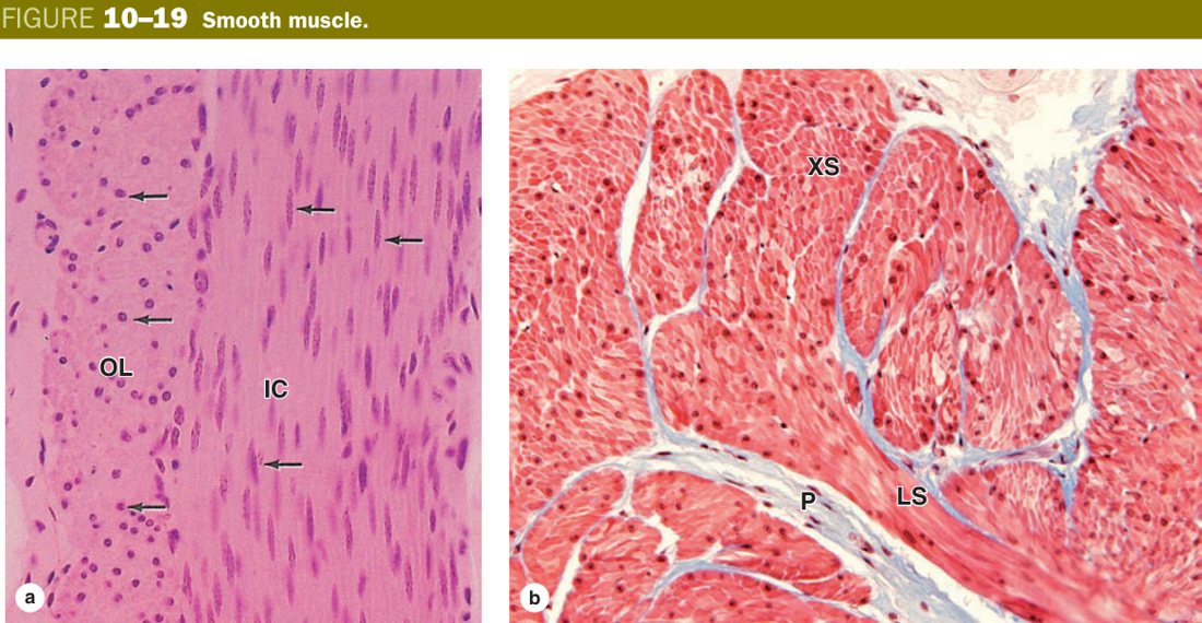
Think of smooth muscle as a net of overlapping spindles rather than a row of cables. When the cells contract, each spindle shortens along its long axis, and because the dense bodies inside are linked to the sarcolemma and to one another by an intermediate-filament cytoskeleton, the whole sheet purses inward like a draw-string bag. The slow Ca–calmodulin–MLCK switch is the price you pay for being able to hold that purse-string closed for hours without exhausting ATP.
Benign tumours of smooth muscle are called leiomyomas — uterine fibroids are the commonest example. Their malignant counterpart is the leiomyosarcoma. (The skeletal-muscle equivalents are the rhabdomyoma and the paediatric rhabdomyosarcoma.) In atherosclerosis, smooth muscle cells of the arterial media migrate into the intima and secrete matrix — they are central to plaque growth, illustrating both their proliferative capacity and their secretory role.
• Why no striations? → Actin & myosin are not arranged in sarcomeres.
• Z-line equivalent? → Dense body.
• Calcium sensor? → Calmodulin → MLCK (no troponin).
• T-tubule equivalent? → Caveolae.
• Can it regenerate / divide? → Yes — smooth muscle retains mitotic capacity.
• Benign tumour name? → Leiomyoma (e.g. uterine fibroid).
Three-Way Comparison (the 15-mark essay)
This is the question TMU has put on essentially every Histology final. The skill being tested is not memory of one feature but the ability to read three columns at once — shape, nucleus, striations, junctions, membrane system, control — and to recognise the engineering trade-off each muscle type embodies. The table below is your spine; if you can write it from memory and add one sentence of justification per row, you have the 15 marks.
A practical slide-room shortcut: look at the nuclei first. Many flat peripheral nuclei = skeletal. One or two central nuclei in a branching cell = cardiac. One central cigar-shaped nucleus in a spindle cell = smooth. From there, the presence/absence of striations and intercalated discs nails the diagnosis.
| Feature | Skeletal | Cardiac | Smooth |
|---|---|---|---|
| Shape | Long cylindrical | Short column, branched | Spindle (fusiform) |
| Nucleus | Multinucleate, peripheral | 1–2, central | One, central |
| Cross-striation | Distinct | Less distinct | None |
| Intercalated disc | No | Yes | No |
| T-tubule junction | Triad (A–I junction) | Diad (Z-line) | None (caveolae) |
| Ca²⁺ sensor | Troponin C | Troponin C | Calmodulin → MLCK |
| Regeneration | Good (satellite cells) | Essentially none (fibrosis) | Retained (cells divide) |
| Control | Voluntary | Involuntary | Involuntary |
Nuclei tell the type: many + peripheral = skeletal; 1–2 + central + branching + intercalated disc = cardiac; single + central + spindle + no striation = smooth. Triad (skeletal) at the A–I junction; Diad (cardiac) at the Z-line.
• Striated & voluntary? → Skeletal.
• Striated & involuntary, branching? → Cardiac.
• Non-striated, spindle? → Smooth.
• Triad vs diad location? → Triad at A–I junction (skeletal); diad at Z-line (cardiac).
• Which type cannot regenerate? → Cardiac — heals by scar.
TMU Exam Drill
📝 Open the full TMU Question Bank — 20 MCQ + 6 terms + 5 essays →
Authentic Tianjin Medical University past-paper questions (2021 Final & the multi-section Final with answer key) mapped to this unit, in the real exam format. Click Show answer to self-test.
□ Single best answer
- A. skeletal muscle fibers
- B. nerve fibers
- C. cardiac muscle fibers
- D. elastic fibers
- E. smooth muscle fibers
□ Explain the following terms
Muscle complete
Sarcomere, triad, intercalated disc & the 3-way comparison mastered. Next: Nervous Tissue.