Cartilage & Bone
Cartilage — General Features
Cartilage is the body's first answer to the problem of supporting weight without being brittle. Picture the cartilage at the tip of your nose, the springy ring of your ear, the smooth cap on the head of your femur — firm enough to hold shape, flexible enough to bend, smooth enough to glide. The whole tissue is just three ingredients: a single resident cell type (the chondrocyte), a hydrated gel-like extracellular matrix (collagen fibres embedded in proteoglycan ground substance), and an outer connective-tissue jacket called the perichondrium.
Three features define every cartilage you will ever see on a slide. First, the only cell in the tissue is the chondrocyte, sitting inside a small cavity called a lacuna. Second, the matrix is semi-rigid — firm but flexible, like a stiff jelly. Third, cartilage is avascular and contains no nerves or lymphatics; chondrocytes survive only by diffusion of nutrients from blood vessels in the surrounding perichondrium. This last point is why cartilage heals so slowly after injury and why osteoarthritis is so unforgiving — once the matrix is damaged, there is no rapid vascular repair to call on.
Cartilage = a hydrated gel that has learnt to push back. Bone (later) = the same gel after it has been reinforced with concrete and steel rods. Cartilage trades hardness for resilience; bone trades resilience for hardness. Your skeleton uses each where its trade-off pays off.
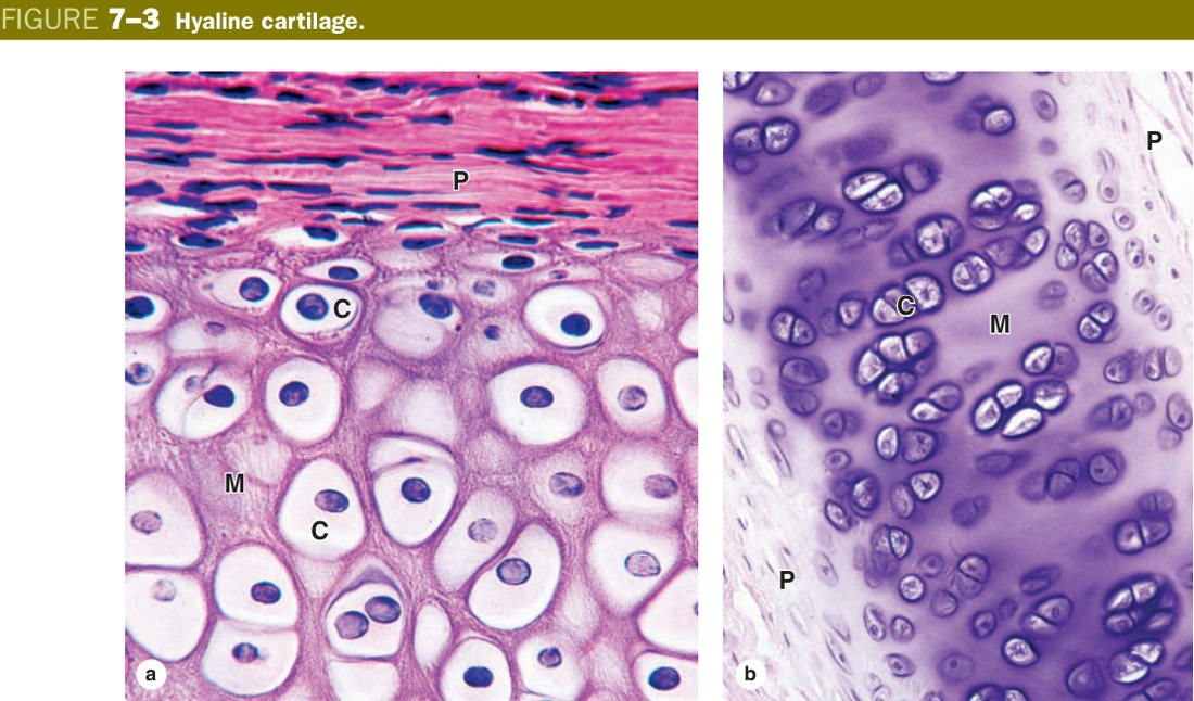
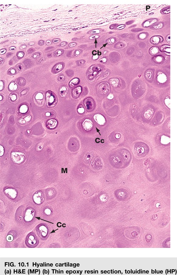
A chondrocyte is, mechanically, a protein factory marooned in its own gel. It lives inside a lacuna, secretes the surrounding matrix, then becomes locked inside what it has built. Under the microscope the location of the cell within the slice tells you its age. Near the perichondrium, where new cartilage is being added, chondrocytes are young: smaller, flattened, single, with weakly basophilic cytoplasm — these are sometimes called chondroblasts. Deeper in the matrix, the cells are older, larger, rounder, and lie in small clusters — isogenous groups.
On EM the chondrocyte is unmistakably a secretory cell — abundant rough endoplasmic reticulum, prominent Golgi, free ribosomes — all the machinery needed to manufacture and export type II collagen and the proteoglycan aggrecan. Every gram of cartilage matrix has, at some point, been extruded through one of these cells.
| Position | LM appearance |
|---|---|
| Periphery (young) | Smaller, flattened, single, less mature; basophilic cytoplasm |
| Deeper part (mature) | Larger, spherical, in groups (isogenous groups), more mature |
An isogenous group is what happens when one chondrocyte deep inside the matrix decides to divide. Because the surrounding matrix is stiff, the daughter cells cannot drift apart — they stay packed together in the same neighbourhood, often as 2–8 cells huddled inside a slightly enlarged shared cavity. This is the engine of interstitial growth: the cartilage expands from within by clonal division, not by adding new cells at the surface.
Isogenous group: a small cluster of 2–8 chondrocytes lying together within the deeper cartilage matrix, all derived by mitosis from a single parent chondrocyte. They represent interstitial growth of cartilage.
• The small cavity housing a chondrocyte is called? → a lacuna
• Why does cartilage heal so slowly? → it is avascular — nutrients reach the chondrocytes only by diffusion from perichondrial vessels
• Define an isogenous group in one line. → 2–8 chondrocytes derived by mitosis from one parent, sitting together deep in the matrix
• Which growth mechanism does an isogenous group represent? → interstitial growth
• What does the EM of a chondrocyte show? → abundant RER, Golgi, free ribosomes — a busy secretory cell
If the chondrocyte is the worker, the matrix is everything else — and the matrix is what gives cartilage its mechanical personality. It has two parts: collagen fibrils for tensile strength, and a hydrated ground substance that resists compression. The ground substance is roughly 75% water trapped inside a sponge of negatively charged proteoglycans (the chondromucoprotein complex: a core protein decorated with chondroitin sulfate, keratan sulfate and hyaluronic acid). Press on a piece of cartilage and you are squeezing water out of that sponge; release it and the negative charges pull the water back in. This water-pump mechanism is what makes articular cartilage shock-absorbent.
Because the ground substance is loaded with sulphated acidic groups, it stains basophilic with H&E. The collagen here is type II — the fine, hyaline-cartilage version — and the fibrils are so thin and so well matched in refractive index to the ground substance that you cannot see individual fibres on a routine slide. That is why hyaline cartilage matrix looks deceptively glassy and homogeneous.
Look closely around each lacuna and you will notice a darker, more intensely basophilic ring of matrix — the cartilage capsule or territorial matrix. This is freshly secreted, sulphated-GAG-rich matrix laid down by the chondrocyte itself, so it has the highest concentration of basic-dye-binding groups. The lighter, paler matrix between cell nests is older interterritorial matrix.
Wrapping the whole sheet of cartilage (except on articular surfaces and fibrocartilage) is the perichondrium, a dense connective-tissue jacket with two layers. The outer fibrous layer is protective and vascular — this is where the blood vessels live that feed the avascular cartilage by diffusion. The inner chondrogenic layer contains spindle stem cells that can wake up, become chondroblasts, and lay down new matrix on the existing surface — this is appositional growth. So cartilage uses two growth strategies in parallel: interstitial (from inside, via isogenous groups) and appositional (from outside, via the perichondrium).
- Fibres embedded in ground substance (collagenous fibrils; in hyaline cartilage these are type II collagen).
- Ground substance: water (~75%) + chondromucoprotein (protein + chondroitin sulfate + hyaluronic acid + keratan sulfate). It is basophilic.
- Cartilage capsule (territorial matrix): the ring of matrix immediately around each lacuna is intensely basophilic (rich in sulphated GAGs).
- Perichondrium: a dense CT sheath with an outer (fibrous, protective, vascular) zone and an inner (chondrogenic) zone whose cells form new chondroblasts → appositional growth. (Articular & fibrocartilage lack a perichondrium.)
Cartilage matrix = a charged sponge. Press it — water leaves. Release — the trapped negative charges of the proteoglycans suck water back in. That elastic recoil is your knee cartilage cushioning every step.
• Which collagen type dominates hyaline-cartilage matrix? → type II
• What is the cartilage capsule, and why is it the darkest part of the matrix? → the freshly-secreted territorial matrix around each lacuna, richest in sulphated GAGs
• Which layer of the perichondrium drives appositional growth? → the inner chondrogenic layer
• Two cartilages with NO perichondrium? → articular hyaline cartilage and fibrocartilage
• The proteoglycan aggregate (core protein + chondroitin sulfate + keratan sulfate + hyaluronan) of cartilage is collectively called? → chondromucoprotein
The Three Types of Cartilage
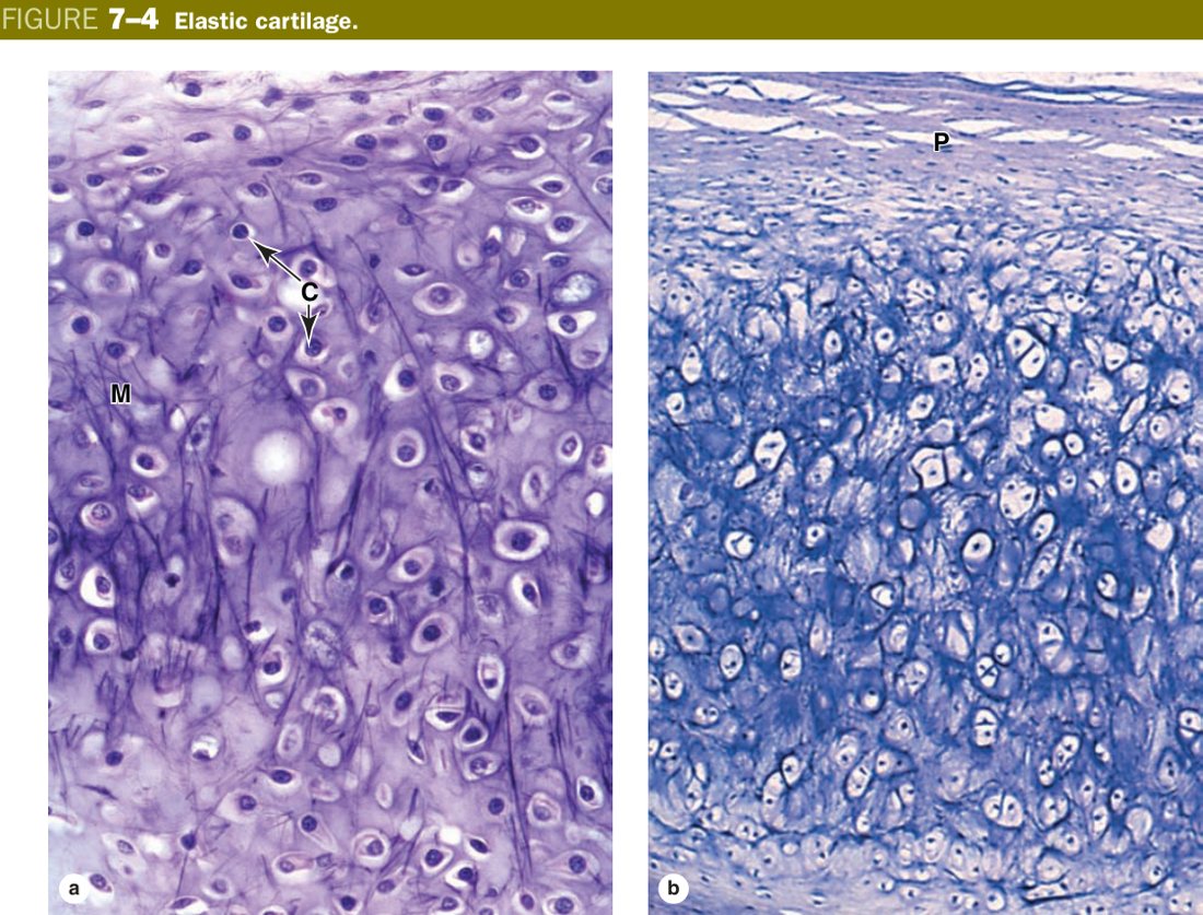
There are exactly three types of cartilage, and the easiest way to keep them straight is to ask: what fibre is in the matrix, and what mechanical job does it do? Hyaline cartilage uses fine type II collagen and behaves like a smooth, slippery cushion — ideal for joint surfaces and the respiratory tract. Elastic cartilage is hyaline with abundant elastic fibres woven in — it springs back, perfect for the ear and epiglottis. Fibrocartilage swaps the fine type II for bundles of stout type I collagen — the strongest, designed for sites of tension and shear like the intervertebral disc and pubic symphysis.
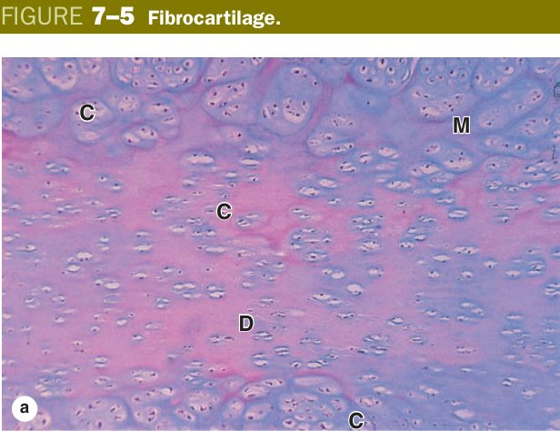
Two more clues sharpen the diagnosis. Look for a perichondrium: hyaline (except articular) and elastic both have one, but fibrocartilage does not — it sits as a transitional tissue blending into the dense regular CT of tendons and ligaments. And look for isogenous groups: spherical clusters of chondrocytes are easy to see in hyaline and elastic; in fibrocartilage the cells are squeezed into single-file rows between parallel collagen bundles.
| Type | Fibre in matrix | Key feature | Perichondrium | Location |
|---|---|---|---|---|
| Hyaline | Type II collagen (fine, masked) | Commonest; glassy basophilic matrix; isogenous groups | Yes (except articular) | Articular surfaces, costal cartilage, nose, trachea/bronchi, larynx, epiphyseal (growth) plate, fetal skeleton |
| Elastic | Type II + abundant elastic fibres | Flexible, springy; elastic fibres seen on special stain | Yes | Auricle (external ear), epiglottis, auditory (Eustachian) tube, some laryngeal cartilages |
| Fibrocartilage | Type I collagen (large bundles) | Many collagen fibres, little ground substance; chondrocytes in rows; NO perichondrium | No | Intervertebral discs, pubic symphysis, menisci, tendon/ligament insertions |
A practical clinical aside on the joint: the articular cap on the end of a long bone is hyaline cartilage with no perichondrium. Once it is worn down, there is no chondrogenic layer to regenerate it — this is the central problem of osteoarthritis. Rheumatoid arthritis attacks the same cartilage from a different angle, via inflammatory pannus eroding from the synovium inward. Either way, the underlying weakness is the avascular, slow-turnover nature of the tissue you have just learnt about.
Hyaline = type II, “glass”, the default. Elastic = Ears & Epiglottis (flexible). Fibrocartilage = type I, no perichondrium, lives where there's stress & shear — discs, symphysis, menisci. Only fibrocartilage uses type I (the “strong” collagen).
Because cartilage is avascular it repairs poorly after injury. Degeneration of articular hyaline cartilage = osteoarthritis. A gain-of-function mutation in the FGFR3 receptor freezes growth-plate hyaline cartilage and causes achondroplasia — the commonest form of dwarfism, with short limbs but a normal-sized trunk. Intervertebral disc herniation (a tear in the fibrocartilage annulus letting nucleus pulposus prolapse) compresses spinal nerve roots.
• Elastic cartilage sits in which two classic locations? → auricle (ear) and epiglottis
• The only cartilage with type I collagen and no perichondrium is? → fibrocartilage
• How are chondrocytes arranged in fibrocartilage versus hyaline? → single-file rows (fibrocartilage) vs. spherical isogenous groups (hyaline)
• Which receptor, mutated, freezes the growth-plate hyaline cartilage and causes achondroplasia? → FGFR3 (gain-of-function)
• What pathology is "wear-and-tear of articular hyaline cartilage"? → osteoarthritis
Bone — General Features & Matrix
Bone solves the same problem as cartilage — support — but commits to the opposite trade-off. Instead of a soft hydrated gel, the matrix is permeated by crystals of hydroxyapatite, a calcium-phosphate mineral that turns the tissue into something with the compressive strength of cast iron. Yet bone is not brittle: the same matrix contains a dense weave of type I collagen that gives it tensile strength. Mineral resists compression, collagen resists tension — together they make a composite material as effective as reinforced concrete.
An anatomist's "bone" is an organ — osseous tissue + periosteum + endosteum + marrow cavity + articular cartilage. A histologist's "bone" is just the osseous tissue: hard, rigid, and made of two parts — bone cells embedded in a calcified bone matrix. The matrix itself comes in two flavours. The organic phase (which, when first laid down by an osteoblast and still uncalcified, is called osteoid) is type I collagen plus a small but important cast of non-collagenous proteins: osteocalcin, osteonectin, sialoproteins, and proteoglycans. The inorganic phase (~65% by weight) is calcium-phosphate hydroxyapatite crystallised along the collagen fibrils.
| Bone matrix | Components |
|---|---|
| Organic (= osteoid when uncalcified) | Type I collagen fibres + ground substance (glycosaminoglycans, osteocalcin, osteonectin) |
| Inorganic (~65%) | Calcium phosphate salts as hydroxyapatite crystals — give hardness |
Bone = reinforced concrete. The collagen fibrils are the steel rebar; the hydroxyapatite crystals are the cement. Take away the mineral (decalcify a bone in acid) and it bends like rubber — only collagen left. Take away the collagen (incinerate the bone) and it crumbles to chalk at a touch — only mineral left. You need both.
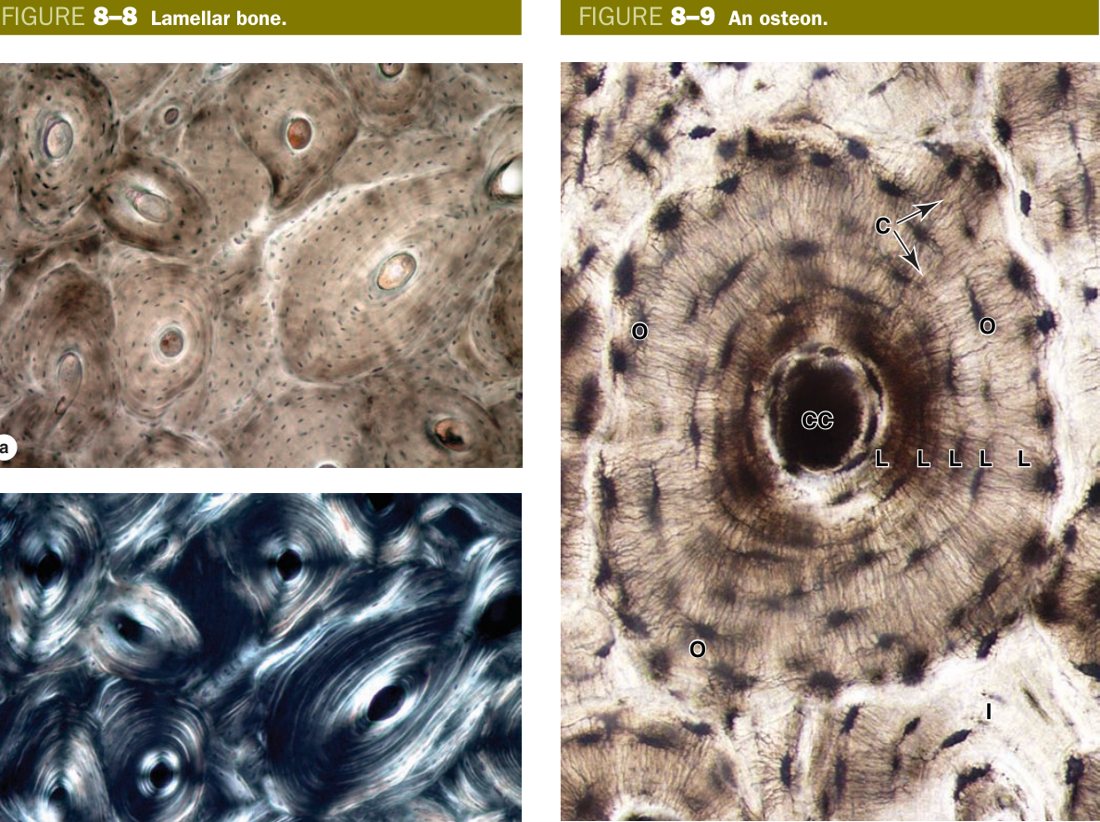
Bone matrix is not poured as a single slab. Osteoblasts secrete it in thin sheets called lamellae, each only 3–7 micrometres thick. Within a single lamella, the collagen fibres lie parallel to each other; in the next lamella, the fibres run at a different angle — almost at right angles to the first. This crossed-ply arrangement is exactly the principle behind plywood, and it is what gives bone its remarkable resistance to fractures from any direction. A blow that would split parallel-grained wood is dissipated across the cross-grain of bone.
Bone lamella: bone matrix is laid down in thin layers (3–7 µm thick) of mineralised matrix. Within one lamella the collagen fibres are parallel; the fibres of adjacent lamellae run nearly at right angles to each other — a plywood-like arrangement that gives bone its strength.
Osteogenesis imperfecta (brittle-bone disease) is the textbook collagen-I disorder — defective COL1A1/COL1A2 means weak organic scaffolding, giving children with otherwise normal mineralisation a string of fractures from trivial trauma, blue sclerae, hearing loss. Osteomalacia / rickets is the opposite failure: collagen is fine, but vitamin D deficiency means osteoid is laid down and not mineralised — soft, bendy bones.
• The inorganic crystal in bone is? → calcium-phosphate hydroxyapatite
• What is osteoid? → the freshly secreted, uncalcified organic matrix (mostly type I collagen + osteocalcin/osteonectin) before mineralisation
• What is a bone lamella, and why is the plywood arrangement important? → a 3–7 µm sheet of mineralised matrix; fibres in adjacent lamellae run at right angles, dispersing stress in every direction
• Decalcify a bone — what is left behind, and how does it feel? → the collagen organic scaffold; flexible, rubbery
• Which disease destroys collagen I and gives blue sclerae plus brittle bones? → osteogenesis imperfecta
The Four Bone Cells
Four cell types do all the work of bone, and they fall neatly into two camps. Three of them — osteoprogenitor, osteoblast and osteocyte — are stages in the life of a single mesenchymal lineage that builds bone. The fourth, the osteoclast, is a giant multinucleated cell from the blood monocyte lineage whose only job is to resorb it. The balance between these two lineages is what determines whether you gain bone, hold it, or lose it — the central drama of osteoporosis, hyperparathyroidism, and Paget disease.
The lineage chain reads: osteoprogenitor → osteoblast → osteocyte. A surface osteoprogenitor (a flat spindle stem cell on periosteum/endosteum) divides, differentiates into a cuboidal, basophilic osteoblast, and starts secreting osteoid. As osteoid mineralises around it, the osteoblast finds itself entombed inside a lacuna it has built — at this moment it stops secreting bulk matrix and is reclassified as an osteocyte. It still maintains the matrix and senses mechanical load, but it has retired from large-scale construction. Crucially, an osteoblast never becomes an osteoclast: the two come from completely different precursor lineages.
The osteoclast is something else entirely. It begins as circulating blood monocytes that recognise the surface of bone via RANK receptors. Osteoblasts present RANKL (and the cytokine M-CSF), the monocytes fuse together into a multinucleate giant (2–50 nuclei, ∼100 µm across), and the giant cell parks itself on the bone surface inside a shallow erosion pit called a Howship lacuna. Its membrane against the bone develops a deeply infolded ruffled border — the business end — which pumps H⁺ out (acidifying the underlying matrix to dissolve hydroxyapatite) and dumps lysosomal cathepsin K to digest the exposed collagen. Osteoblasts also secrete OPG, a decoy that mops up RANKL and switches osteoclasts off. The RANK/RANKL/OPG triangle is one of the most asked endocrine-bone questions you will meet.
| Cell | LM | EM | Function |
|---|---|---|---|
| Osteoprogenitor | Small, spindle, on bone surface; weakly basophilic | Few organelles | Divide & differentiate → osteoblast |
| Osteoblast | Cuboidal, on surface; strongly basophilic cytoplasm | Abundant RER, Golgi, matrix vesicles | Secrete osteoid (uncalcified matrix); when trapped → osteocyte |
| Osteocyte | Ovoid, embedded in a lacuna; processes run in canaliculi | RER, Golgi, gap junctions between processes | Maintain matrix; exchange nutrients/signals via canalicular network |
| Osteoclast | Multinucleate (2–50), giant (~100 µm), acidophilic, striated (ruffled) border | Ruffled border, many lysosomes & vacuoles | Secrete acid + lysosomal/proteolytic enzymes → resorb bone (from blood monocytes/MPS) |
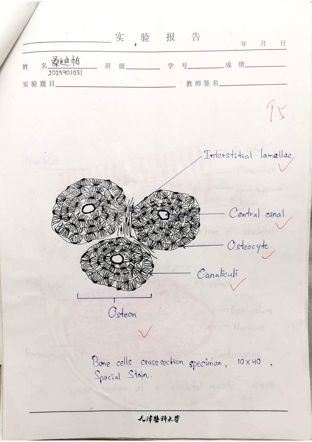
A small architectural detail that the exam loves: the osteocyte does not just sit alone in its lacuna. Long, thin cytoplasmic processes radiate out from it in all directions, each running through a tiny tunnel in the matrix called a canaliculus. Adjacent osteocytes meet, tip to tip, inside these canaliculi and form gap junctions. The whole canalicular network is therefore an interconnected web that lets nutrients, waste and mechano-signals diffuse through the otherwise impenetrable mineralised matrix. Without it, osteocytes would suffocate.
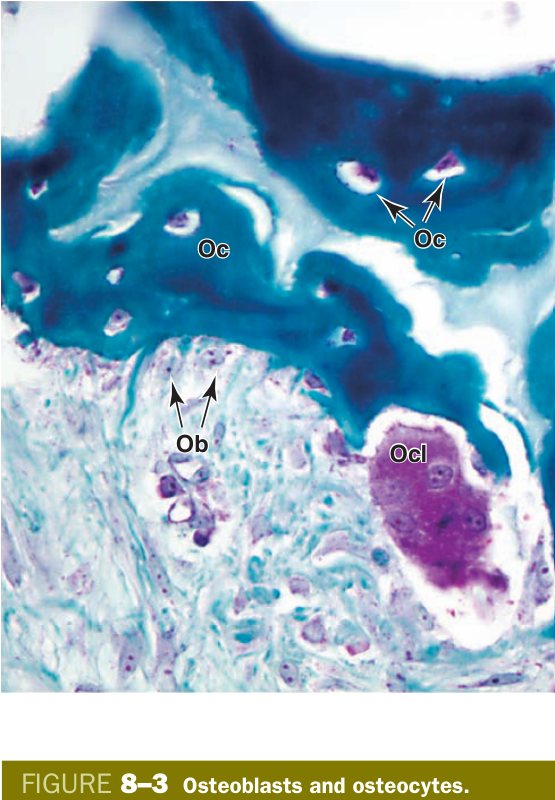
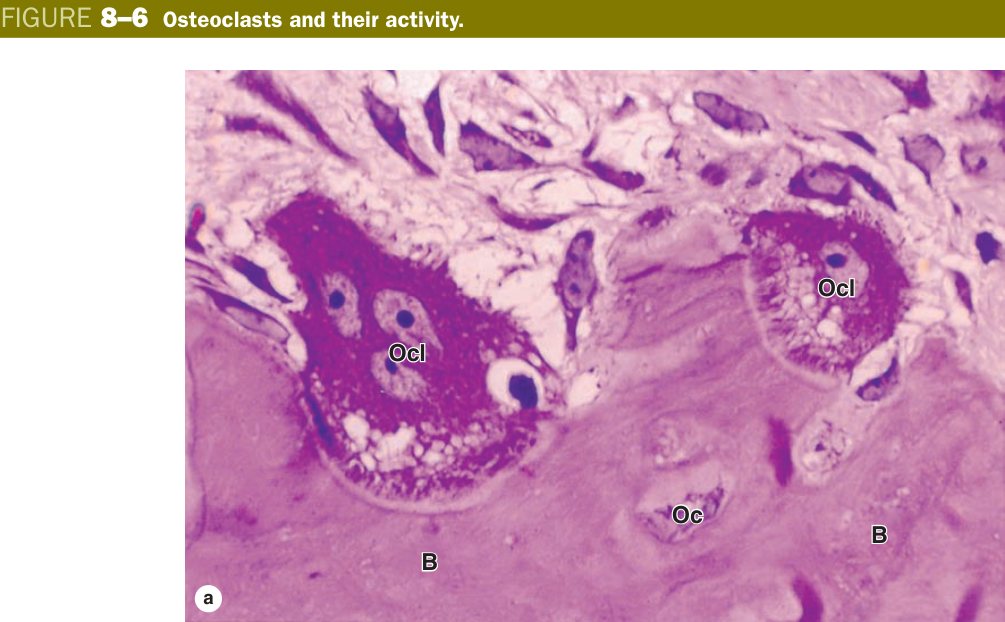
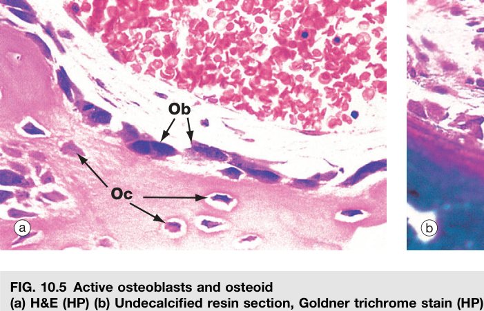
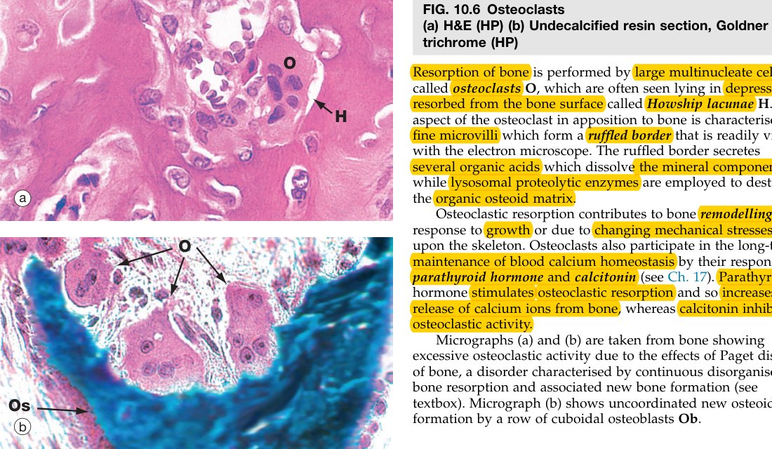
Think of bone as a city under permanent renovation. Osteoblasts are the bricklayers laying fresh osteoid; osteocytes are retired bricklayers walled into their own buildings, still keeping watch through canalicular telephone lines. Osteoclasts are demolition crews from a completely different company (the blood-monocyte lineage), hired in to chew out old buildings so new ones can go up.
“Blasts Build, Clasts Crush.” OsteoBlast = Builds bone (basophilic, one nucleus, secretes osteoid). OsteoClast = Cuts/resorbs (multinucleate giant, acidophilic, ruffled border). The osteocyte is the retired osteoblast walled into a lacuna, keeping in touch with its neighbours through gap junctions in the canaliculi. Osteoblast → osteocyte (it does not become an osteoclast).
Osteoporosis — especially post-menopausal — is too much osteoclast (the brake on RANKL falls away with oestrogen). Osteopetrosis ("marble-bone disease") is too little: osteoclasts cannot acidify the resorption pit because of a defect in carbonic anhydrase II or the proton pump — bone piles up, fills the marrow space, brittle. Paget disease is chaotic remodelling with mosaic lamellae. Bisphosphonates and denosumab (an anti-RANKL antibody) treat osteoporosis by silencing osteoclasts.
• Where does the osteoclast come from? → fusion of blood-monocyte/macrophage precursors
• What does an osteocyte sit inside, and how do its processes reach neighbours? → a lacuna; processes run through canaliculi and meet at gap junctions
• Name the signalling triad that controls osteoclast formation. → RANK (on osteoclast) · RANKL (on osteoblast) · OPG (decoy receptor)
• Why does osteopetrosis (defective carbonic anhydrase II) give dense brittle bones? → osteoclasts cannot acidify the resorption pit, so bone is never resorbed and remodelled
• What drug class silences osteoclasts to treat osteoporosis? → bisphosphonates (and the anti-RANKL antibody denosumab)
Compact Bone & the Osteon
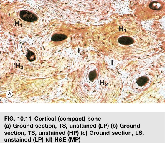
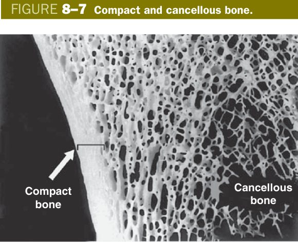
In the diaphysis (shaft) of a long bone, the dense outer rind is called compact (cortical) bone. Cut it in cross section and you find lamellae arranged in three patterns, in this order from outside to inside: outer circumferential lamellae hugging the periosteum, a thick middle layer made almost entirely of osteons (Haversian systems), with little wedges of interstitial lamellae filling the gaps between them, and finally inner circumferential lamellae hugging the marrow cavity. Each pattern tells a story: circumferential lamellae are the original surface bone, osteons are the active remodelling units, and interstitial lamellae are the leftover remnants of older osteons that have been partially eaten away by previous remodelling cycles.
| Pattern | Location / structure |
|---|---|
| Circumferential lamellae | Outer (external surface, thicker & more regular) and inner (internal/marrow surface) — encircle the whole shaft |
| Osteon (Haversian system) | Cylindrical units between the circumferential lamellae — the structural unit of compact bone |
| Interstitial lamellae | Between osteons — the remnants of old, partially-resorbed osteons |
The osteon is the irreducible functional unit of compact bone — the brick the whole wall is built from. Picture a long cylinder, a few hundred micrometres wide, oriented parallel to the long axis of the bone. Down the centre runs a Haversian (central) canal carrying a small artery, a vein, a nerve and loose connective tissue. Around the canal, bone matrix is wrapped in 4–20 concentric layers — the Haversian lamellae. Sandwiched between adjacent lamellae lie osteocytes in lacunae, each one extending its processes through canaliculi to the central canal (their nutrient supply) and to neighbouring osteocytes. This is why osteons cannot be much wider than ∼200 µm: it is the maximum distance over which the canalicular network can move nutrients before the deepest osteocytes starve.
Osteon (Haversian system): the cylindrical structural unit of compact bone, located between the inner and outer circumferential lamellae. It consists of a central Haversian canal (carrying blood vessels, nerves and loose CT) surrounded by concentric Haversian lamellae, with osteocytes in lacunae between the lamellae. Osteons run parallel to the long axis.
If Haversian canals run lengthwise down the bone, how do they get blood supply from the periosteum on the outside or the marrow on the inside? Through Volkmann's (perforating) canals — transverse channels that drill across the lamellae and link adjacent Haversian canals to each other and to the bone surfaces. The key difference: Volkmann canals do not have concentric lamellae around them. They simply punch through the existing architecture. While you are mapping vessels, also note Sharpey's (perforating) fibres — thick bundles of type I collagen anchoring the periosteum (and tendons) into the outer circumferential lamellae, like guy-ropes pegged into the cortex.
An osteon is a long pipe with a vessel down the middle and concentric rings of bone around it. Imagine a bundle of drinking straws glued together, running the length of the bone — that is the compact cortex. Volkmann canals are the cross-connections you punch between straws to share juice. Interstitial lamellae are the half-melted straws you couldn't quite remove during the last renovation.
Inside the cortical shell, the architecture relaxes. Spongy (trabecular or cancellous) bone is a three-dimensional latticework of bony struts called trabeculae, with marrow filling the spaces between them. The trabeculae are still made of lamellae and contain osteocytes in lacunae, but they have no osteons — they are too thin to need a central canal because the marrow on either side bathes them directly. Spongy bone dominates the epiphyses of long bones, the bodies of vertebrae and the diploë of flat bones, where its honeycomb construction maximises strength for minimum weight, exactly along the lines of mechanical stress.
Every bone is wrapped in two specialised connective-tissue linings. The periosteum sheaths the outside — an outer fibrous layer (vascular, with Sharpey's fibres anchoring tendons) plus an inner osteogenic layer of osteoprogenitor cells that can fire up new osteoblasts whenever appositional growth or fracture repair is needed. The endosteum is a much thinner version that lines all the internal surfaces: the marrow cavity, the trabecular spaces, the Haversian and Volkmann canals. Together, periosteum and endosteum are the bone's reserve of osteoprogenitor cells — the reason a child's snapped femur can heal back to normal architecture, because the cellular machinery for protection, growth, repair and remodelling is sitting there waiting.
- Spongy (trabecular) bone: a meshwork of trabeculae made of parallel-arranged lamellae with osteocytes; no true osteons (nourished by marrow); found in epiphyses and flat bones.
- Periosteum (outer fibrous + inner osteogenic layer) and endosteum (lines the marrow cavity/canals) — functions: protection, growth, repair and remodelling of bone.
• The structural unit of compact bone is the? → osteon (Haversian system)
• What is in a Haversian canal? → small artery, vein, nerve and loose connective tissue
• How does spongy bone differ from compact bone architecturally? → lattice of trabeculae fed directly by marrow; no osteons
• Difference between a Haversian and a Volkmann canal? → Haversian = longitudinal, surrounded by concentric lamellae; Volkmann = transverse, links Haversian canals, no concentric lamellae
• Why is the periosteum essential for fracture repair? → its inner osteogenic layer is full of osteoprogenitor cells that can become osteoblasts
• What do Sharpey's fibres do? → collagen bundles that anchor periosteum and tendons into the cortical bone
Ossification (Overview)
Bone develops in two completely different ways depending on which part of the skeleton you are looking at. Intramembranous ossification is the direct route: mesenchymal cells in a sheet of embryonic connective tissue condense, differentiate straight into osteoblasts, and start laying down osteoid right there — no cartilage template is ever made. This is how the flat bones of the skull vault, most of the clavicle, and the mandible form. Endochondral ossification is the long-bone route: the embryo first carves out a miniature hyaline-cartilage model of the future bone, then progressively replaces that cartilage with bone, starting from a primary centre in the diaphysis and later from secondary centres in the epiphyses.
| Type | Process | Where |
|---|---|---|
| Intramembranous | Mesenchyme → osteoblasts secrete osteoid directly (no cartilage model) | Flat bones of skull, clavicle, mandible |
| Endochondral | A hyaline cartilage model is progressively replaced by bone; lengthening occurs at the epiphyseal (growth) plate | Long bones, base of skull, vertebrae |
The clinically important part of endochondral ossification is what happens after birth at the epiphyseal (growth) plate — the thin disc of hyaline cartilage sandwiched between the diaphysis and each epiphysis that lengthens the bone. Read it as a moving assembly line in five zones, from epiphyseal side downward: (1) reserve / resting (quiet chondrocytes) → (2) proliferation (chondrocytes divide and stack into columns like coin piles) → (3) hypertrophy (cells balloon up) → (4) calcification (cartilage matrix mineralises and chondrocytes die) → (5) ossification (vessels invade and osteoblasts replace the calcified cartilage scaffold with woven bone). The whole plate marches steadily toward the diaphysis, so the bone gets longer while the plate itself stays the same thickness — until puberty, when sex steroids close the plate by replacing it entirely with bone.
Once the skeleton is built, it never sits still. The basic multicellular unit (BMU) of remodelling pairs an osteoclast cutting cone — chewing a tunnel through old bone — with a trailing team of osteoblasts laying down fresh concentric lamellae around a new vessel. Each pass of a BMU through cortical bone literally builds one new osteon. A similar process repairs a fracture: bleeding gives a haematoma → fibroblasts and chondroblasts move in to form a soft procallus → fibrocartilaginous callus → osteoblasts replace it with a bony hard callus → remodelling restores the original cortical architecture over months.
Intramembranous ossification = building a wall directly with bricks. Endochondral ossification = first carving a wall in soft clay, then slowly swapping every clay block for a brick while you go. The growth plate is the conveyor belt where the clay is still being made at the top and the brick swap is happening at the bottom.
The epiphyseal growth plate is the engine of longitudinal growth: growth-hormone excess before plate closure causes gigantism; after closure it causes acromegaly (the plates can no longer lengthen long bones, so periosteal apposition thickens jaw, hands and feet instead). The plate is mechanically weak — Salter–Harris fractures cross it and can disturb future growth. Achondroplasia (constitutively active FGFR3) suppresses the proliferation zone, giving short limbs but a normal-sized trunk. Rickets / osteomalacia stalls calcification of the plate so cartilage piles up and bones bow.
• Which mode forms long bones, vertebrae and the base of skull? → endochondral (hyaline cartilage model is replaced by bone)
• List the five zones of the growth plate from epiphysis to diaphysis. → reserve → proliferation → hypertrophy → calcification → ossification
• Why does growth-hormone excess after plate closure not make you taller? → the cartilage of the growth plate is gone — long bones can no longer elongate; only appositional thickening occurs (acromegaly)
• What is a BMU? → the basic multicellular unit of remodelling: an osteoclast cutting cone followed by osteoblasts depositing a new osteon
• Four stages of fracture healing in order? → haematoma → fibrocartilaginous (soft) callus → bony (hard) callus → remodelling
TMU Exam Drill
📝 Open the full TMU Question Bank — 20 MCQ + 6 terms + 5 essays →
Authentic Tianjin Medical University past-paper questions (2021 Final & the multi-section Final with answer key) mapped to this unit, in the real exam format. Click Show answer to self-test.
□ Single best answer
- A. osteoprogenitor cells
- B. osteoblasts
- C. osteocytes
- D. osteoclasts
- E. chondrocytes
□ Explain the following terms
Cartilage & Bone complete
Chondrocytes, isogenous groups, 4 bone cells & the osteon mastered. Next: Muscle.