Connective Tissue
General Characteristics & Classification
Everywhere you have looked so far in histology, the cells have been doing the heavy lifting and the matrix has been little more than a film of glue. Connective tissue flips the script. Here the cells are scattered like raisins in a pudding, and the pudding itself — the extracellular matrix — is the tissue. Whenever you see a section dominated by fibres and amorphous space with only a sprinkle of nuclei, you are looking at connective tissue.
That single design choice explains almost everything else. Because the matrix is the tissue, the cells lose any sense of polarity (no apex, no base) and the tissue is generally well vascularised — blood vessels run through the spaces between fibres to feed every cell of every organ they support. And because the matrix is built by cells, you can trace all of connective tissue back to a single embryonic ancestor: mesenchyme, the loose stellate cell-in-jelly that fills the early embryo.
Think of epithelium as a brick wall — bricks (cells) tightly mortared, no gaps. Connective tissue is a fruit salad — a few pieces of fruit (cells) floating in a lot of syrup (matrix). Same body, opposite design.
Connective tissue is a basic tissue in which a relatively small number of cells is loosely scattered in an abundant extracellular matrix. The matrix (not the cells) is the major constituent and consists of fibres + ground substance + tissue fluid. Cells show no polarity; the tissue is generally well vascularised. It derives embryologically from mesenchyme.
The four big families of connective tissue all share that “cells in matrix” plan, but the matrix is tuned for very different jobs. In connective tissue proper the matrix is soft and lets things diffuse; in cartilage it is firm and rubbery; in bone it is mineralised and rock-hard; in blood it is liquid (plasma). Same blueprint, four hardnesses.
| Class | Members |
|---|---|
| Connective tissue proper | Loose (areolar) CT, dense CT, adipose tissue, reticular tissue |
| Cartilage | Hyaline, elastic, fibrocartilage (Unit 4) |
| Bone | Compact & spongy (Unit 4) |
| Blood | A fluid connective tissue (Unit 8) |
Functionally, every connective tissue in the body is doing one of four jobs: it connects structures together, supports them mechanically, carries nutrients and waste for metabolic exchange, and houses the cellular machinery of defence and repair. Keep this quartet (connect · support · nourish · defend) in your head and every cell type in this unit will slot into one of them.
• Three components of the ECM? → fibres, ground substance, tissue fluid
• The three fibre types? → collagenous, elastic, reticular
• All connective tissues come embryologically from? → mesenchyme
• Why is connective tissue almost always well vascularised? → the loose matrix allows vessels to run through, feeding adjacent epithelia & muscles
• Four big classes? → CT proper, cartilage, bone, blood
Loose (Areolar) Connective Tissue — Overview
If you imagine wrapping every organ, every vessel, and every nerve in a soft sponge that lets blood vessels pass through it freely, that sponge is loose (areolar) connective tissue. It is the universal packing material of the body — lamina propria of every wet epithelium, the “everywhere else” tissue around muscles, the soft bed under your skin. Because it is the model CT, almost every cell and fibre we will name in this unit lives here first.
Loose CT earns its name from how it looks under the microscope: a meshwork of widely-spaced fibres with abundant ground substance and many cell types floating between them. The matrix dominates, the cells are sparse and varied, and the whole thing is highly vascular. Learn its four components and you have the syllabus skeleton for everything that follows.
- Cells — fibroblast, macrophage, plasma cell, mast cell, fat cell, undifferentiated mesenchymal cell, leukocytes (7 types).
- Fibres — collagenous, elastic, reticular.
- Ground substance — jelly-like amorphous GAG/proteoglycan matrix.
- Tissue fluid — the interstitial fluid medium.
Loose CT is the polystyrene packing peanuts that cushion every organ in the “shipping crate” of the body. Loose enough for vessels and lymphatics to weave through, strong enough that nothing rattles.
• The four components of loose CT? → cells, fibres, ground substance, tissue fluid
• How many distinct cell types live in loose CT? → seven (fibroblast, macrophage, plasma cell, mast cell, fat cell, mesenchymal cell, leukocytes)
• Why is loose CT well vascularised? → the loose meshwork freely admits capillaries & lymphatics — this is how it feeds overlying epithelium
The Cells of Loose CT
If you remember nothing else from this unit, learn these four cells: fibroblast (builder), macrophage (eater), plasma cell (antibody factory), and mast cell (alarm bell). Each appears in mid-terms as a define-and-distinguish, and together they cover “what does connective tissue actually do?” — building matrix, defending against invaders, mounting humoral immunity, and triggering allergy.
A useful staining shortcut up front: cells full of RER (protein factories) stain basophilic (blue/purple), because RNA is acidic and grabs basic dye. So fibroblasts are weakly basophilic (modest RER for matrix), plasma cells are intensely basophilic (cartwheel nucleus + huge RER for antibody), and macrophages — whose work is digestion not synthesis — are acidophilic (pink). Recognising this colour code on a slide is half the battle.
| Cell | LM appearance | EM | Function |
|---|---|---|---|
| Fibroblast | Large, stellate/spindle, branched; large pale ovoid nucleus, 1–2 nucleoli; weakly basophilic cytoplasm | Abundant RER, free ribosomes, well-developed Golgi | Synthesise fibres + ground substance |
| Macrophage | Irregular, blunt processes; round, eccentric nucleus, smaller/darker than fibroblast; acidophilic cytoplasm | Surface projections; many lysosomes, phagosomes, pinosomes | Phagocytosis, antigen presentation, secretion (lysozyme, IL-1, IFN, complement) |
| Plasma cell | Ovoid; eccentric clock-face / cartwheel nucleus; intensely basophilic cytoplasm + pale juxtanuclear “Hof” | Extensive parallel RER, ribosomes, Golgi (the pale Hof) | Produce antibodies (immunoglobulins) → humoral immunity |
| Mast cell | Large round/ovoid; small pale central nucleus; cytoplasm packed with metachromatic basophilic granules | Membrane-bound electron-dense granules | Allergic / immediate hypersensitivity (release histamine, heparin) |
| Fat cell (adipocyte) | Large; single big lipid droplet; flattened peripheral nucleus; thin cytoplasm rim (“signet ring”) | One large lipid droplet, thin organelle rim | Synthesise & store lipid |
| Undiff. mesenchymal cell | Small, spindle, along vessels | Few organelles | Multipotent reserve cell for repair |
| Leukocytes | Migrate from blood (lymphocytes, eosinophils, neutrophils) | — | Defence / inflammation |
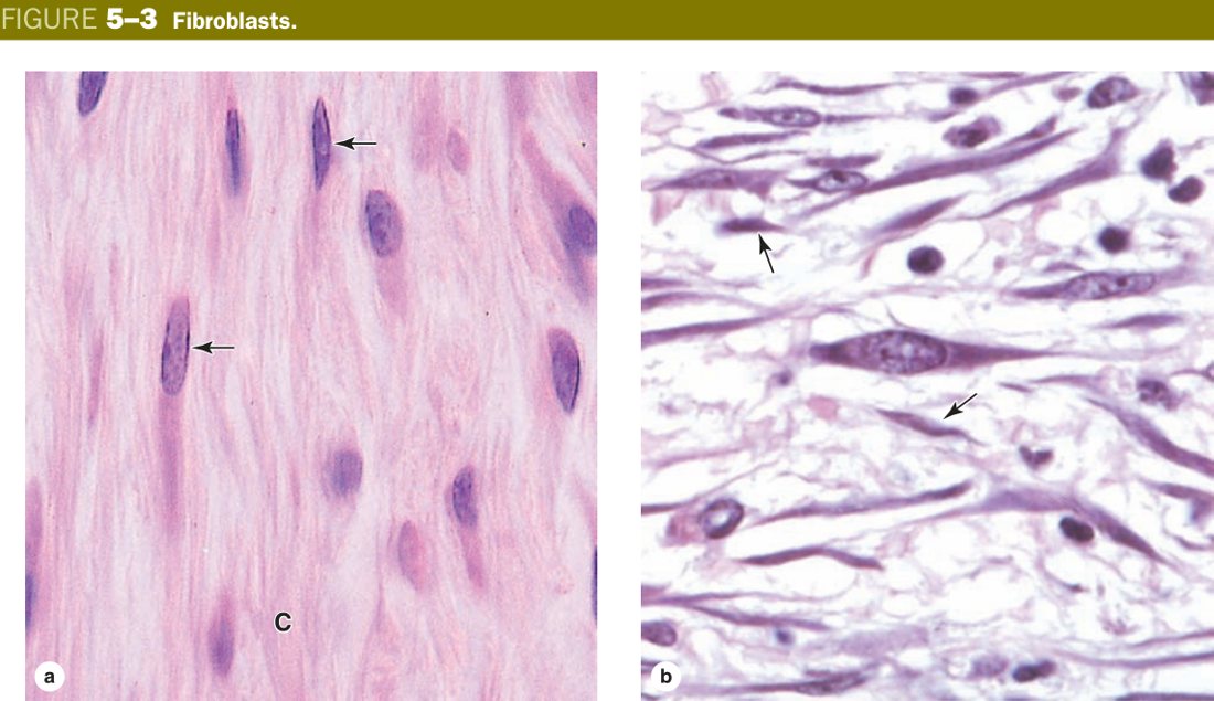
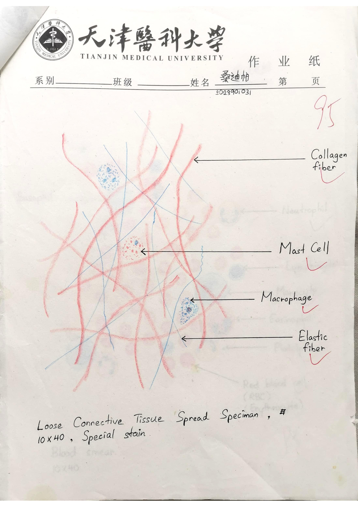
The fibroblast is the workhorse of connective tissue — it builds almost the entire matrix you will ever study. Spread across a smear of loose CT, fibroblasts look like flat starfish with branching processes; in section, you mostly see their pale, oval nuclei. The cytoplasm is faintly basophilic because the cell is stuffed with rough endoplasmic reticulum churning out procollagen, elastin and proteoglycan precursors for export. In a sense, every collagen fibre, every elastic fibre and every gram of ground substance in your body has, at some point, passed through a fibroblast's RER.
When the building is done and the matrix is stable, the same cell quietens into a fibrocyte: smaller, spindle, fewer processes, scant cytoplasm, sparse RER. Fibrocytes still maintain matrix at a low rate. After an injury they re-activate — the fibrocyte plumps up, RER expands, cytoplasm becomes basophilic again, and the cell starts laying down repair collagen. So “fibroblast vs fibrocyte” is really just “active vs resting” states of one cell type.
Fibroblast = the bricklayer on the building site; fibrocyte = the same worker dozing in a folding chair once the wall is up. Cut yourself and the chair empties.
Fibroblast: the most numerous, principal cell of CT. LM — large, flat, stellate/spindle with branching processes; large, ovoid, pale-staining nucleus with 1–2 prominent nucleoli; weakly basophilic cytoplasm. EM — abundant RER, free ribosomes and a well-developed Golgi. Function — synthesises the fibres and ground substance of the matrix. (The inactive form = fibrocyte: spindle, fewer processes, smaller darker nucleus, acidophilic cytoplasm, sparse RER.)
Defective fibroblast collagen causes a family of inherited disorders. Ehlers–Danlos syndrome (mutations in collagen V or III) gives hyperextensible skin and hypermobile joints. Osteogenesis imperfecta (collagen I) gives brittle bones and blue sclerae. Scurvy (vitamin C deficiency) blocks prolyl/lysyl hydroxylation in the fibroblast's RER — collagen cannot triple-helix properly, wounds re-open and old scars fall apart.
• Cytoplasm of an active fibroblast is acidophilic or basophilic, and why? → weakly basophilic, because abundant RER (RNA is acidic, takes up basic dye)
• Two products of the fibroblast? → fibres + ground substance
• What is a fibrocyte? → the inactive, spindle, scant-cytoplasm form of the same cell
• A collagen-I gene mutation causes which clinical disorder? → osteogenesis imperfecta (brittle bone disease)
• Why does scurvy break collagen? → vitamin C is needed for prolyl/lysyl hydroxylase, without which the triple helix is unstable
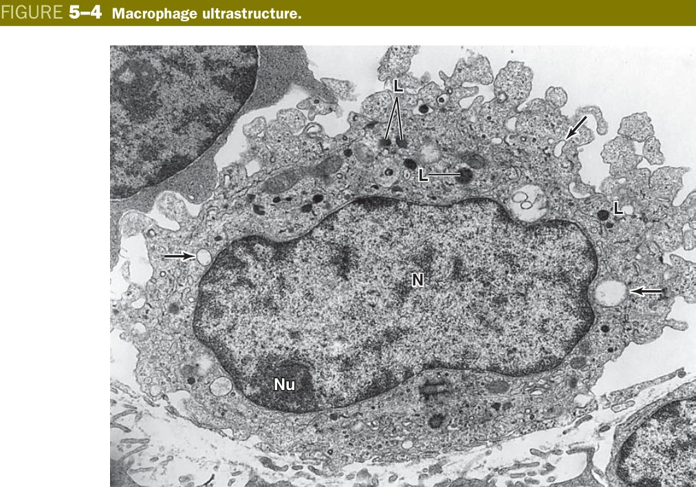
Where the fibroblast builds, the macrophage eats. A blood monocyte squeezes out of a venule, settles in the tissue, and turns into a macrophage — an irregular cell with blunt pseudopods sampling its surroundings. Under the EM its cytoplasm is a warehouse of lysosomes, phagosomes and pinocytotic vesicles, because the whole job description is digestion: ingest bacteria, dead cells, dust, debris, and present the leftover peptides to lymphocytes as antigen.
On a routine H&E the macrophage looks small, dark and pink (acidophilic), with an eccentric nucleus that is smaller than a fibroblast's. The giveaway is engulfed material in the cytoplasm — carbon, haemosiderin, lipid — or, in special-stain experiments, granules of vital dye that the macrophage has greedily taken up. That “dirty cytoplasm” is the visual signature.
Macrophages are the body's sanitation crew and intelligence agents in one. They clean up rubbish, and then post photos of the rubbish (antigens on MHC-II) on a noticeboard so the immune system can identify the culprit.
Macrophage: a phagocytic CT cell derived from blood monocytes. LM — irregular outline with short blunt processes; rounded, eccentric nucleus that is smaller and darker than the fibroblast's; acidophilic cytoplasm (often containing engulfed material/vital-dye granules). EM — irregular surface with many projections; rich in lysosomes, phagosomes and pinosomes. Function — chemotaxis, phagocytosis (specific receptor-mediated against bacteria/viruses + non-specific of dust/debris), antigen presentation & immune regulation, and secretion of lysozyme, complement, IL-1 and interferon.
The same monocyte lineage settles in different organs under different local names, and together they form the mononuclear phagocyte system (MPS) — Kupffer cells in liver, microglia in CNS, osteoclasts in bone (fused multinuclear macrophages), alveolar/“dust” cells in lung, Langerhans cells in skin epidermis. Same cell, different addresses.
In tuberculosis, macrophages engulf Mycobacterium tuberculosis but cannot digest it; they aggregate into a granuloma with central caseation, a hallmark histological pattern. In atherosclerosis, macrophages binge on oxidised LDL and become “foam cells” in the arterial intima — literally cytoplasm full of lipid droplets.
• EM hallmark? → abundant lysosomes, phagosomes & pinosomes
• H&E cytoplasm of macrophage vs fibroblast? → macrophage is acidophilic (pink), fibroblast weakly basophilic
• Tissue macrophages of liver / CNS / bone / lung / skin? → Kupffer / microglia / osteoclast / alveolar / Langerhans
• Two macrophage-driven diseases? → tuberculosis granuloma, atherosclerotic foam cell
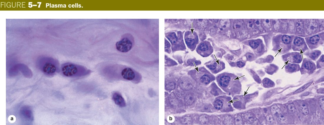
The plasma cell is one of those rare histology cells you can identify with a single glance, because nothing else looks like it. The nucleus sits eccentrically and its heterochromatin is clumped into spokes radiating from the centre — the famous “cartwheel” or “clock-face” pattern. The cytoplasm is intensely basophilic except for a single pale crescent next to the nucleus, the Hof, which is the Golgi where antibody is packaged.
All of that morphology is in service of one job: making antibody. A plasma cell is a B lymphocyte that has met its antigen, exited the lymph node, and re-tooled into a full-time immunoglobulin factory. Hence the parallel arrays of RER (visible on EM as endless stacks of cisterns), the prominent Golgi, and the strong basophilia — all the textbook features of a protein-export specialist. You will see plasma cells in any tissue mounting a chronic antibody response: lamina propria of gut, the medullary cords of lymph nodes, sites of chronic inflammation.
If the fibroblast is a builder secreting collagen scaffolding, the plasma cell is a printing press churning out the same antibody book by the million. Both have huge RER — but the plasma cell's product is shipped into blood, not laid down beside the cell.
Plasma cell: an antibody-secreting cell derived from B-lymphocytes. LM — ovoid/round; eccentric nucleus with coarse heterochromatin in a “cartwheel / clock-face” pattern; intensely basophilic cytoplasm with a pale juxtanuclear area (the Golgi/“Hof”). EM — extensive parallel arrays of RER, free ribosomes, well-developed Golgi. Function — produces antibodies (immunoglobulins), taking part in humoral immunity.
Malignant proliferation of a single plasma-cell clone is multiple myeloma. The pathology mirrors the cell biology: too many plasma cells crowding marrow cause bone pain & lytic lesions; their monoclonal antibody appears as an M-spike on serum electrophoresis; light chains overload the kidney (myeloma kidney). Conversely, a tissue stuffed with reactive plasma cells signals chronic inflammation — the immune system has settled in to fight a persistent antigen.
• Nuclear pattern? → eccentric “cartwheel / clock-face” heterochromatin
• What is the pale juxtanuclear “Hof”? → the prominent Golgi packing antibody
• Why is the cytoplasm so strongly basophilic? → densely packed parallel RER for antibody synthesis
• Plasma-cell malignancy? → multiple myeloma (M-spike, lytic bone lesions, light-chain renal failure)
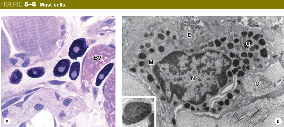
The mast cell is the body's alarm button. It sits along small blood vessels in loose CT, its cytoplasm packed with coarse granules that contain pre-made histamine, heparin and eosinophil chemotactic factor. Specific IgE made by plasma cells (after a first exposure to allergen) coats the mast cell's surface via FcεRI receptors. On second exposure the allergen cross-links two adjacent IgE molecules, the receptors aggregate, and within seconds the cell degranulates — releasing its mediators in a single explosive burst.
On a slide stained with H&E the mast cell is unremarkable, but stained with toluidine blue (a basic blue dye) the granules turn a startling purple-red. This colour-shift is metachromasia, and it happens because the densely sulphated heparin inside the granules forces toluidine-blue molecules to stack head-to-tail, which shifts their absorption spectrum. The same trick identifies mast cells in every tissue you examine.
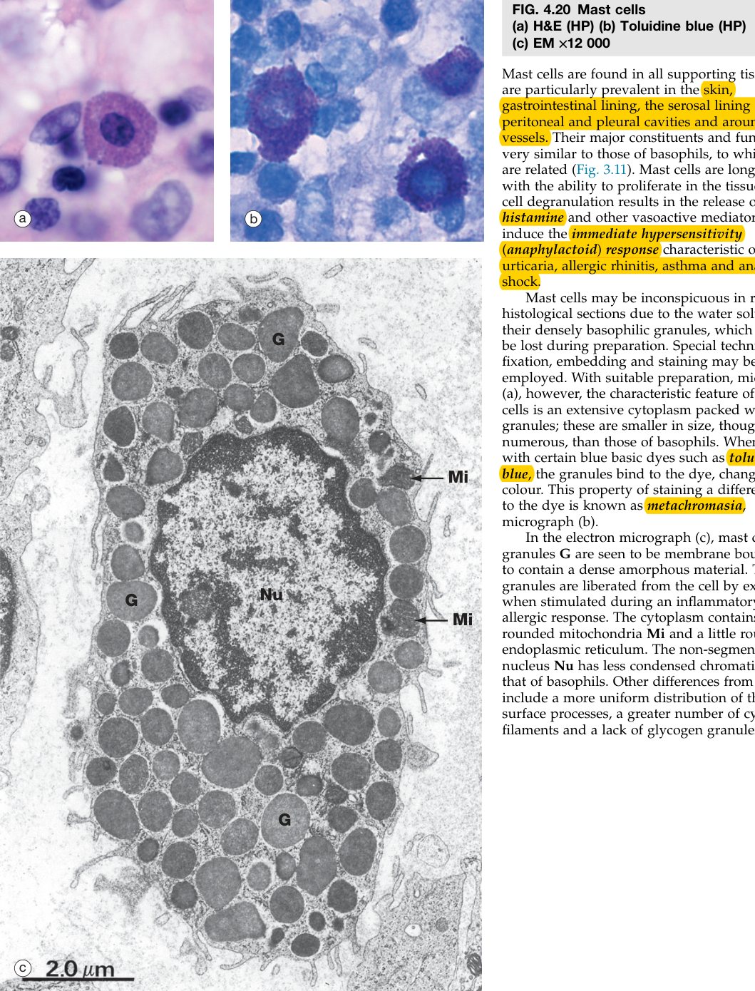
Mast cells are sentries with a satchel of grenades. The IgE on their surface is the trip-wire; the allergen is the boot that catches the wire. One step on the wire and every grenade goes off at once — sneezing, wheal, anaphylaxis.
Mast cell: a large round/ovoid CT cell found along small blood vessels. LM — small, pale, centrally-located nucleus; cytoplasm packed with coarse basophilic granules that are metachromatic (stain purple-red with toluidine blue). EM — membrane-bound electron-dense granules. Granule/cytoplasm contents — histamine, heparin, eosinophil chemotactic factor (granules); leukotrienes (cytoplasm). Function — mediates allergic / immediate (type I) hypersensitivity reactions.
Mast cells and blood basophils share the same mediators (histamine + heparin) and both drive allergy — the difference is location: mast cells live in tissue, basophils circulate in blood. Granules are metachromatic — the classic toluidine-blue example.
Massive simultaneous mast-cell degranulation across many tissues is anaphylaxis: histamine causes vasodilation & hypotension, bronchoconstriction, urticaria and laryngeal oedema — treated with intramuscular adrenaline, which reverses each of those effects. Lesser degrees of the same biology produce asthma, allergic rhinitis (“hay fever”) and urticaria. Heparin from the same granules is an endogenous anticoagulant.
• Two major mediators stored in the granules? → histamine + heparin
• Stain that demonstrates metachromasia? → toluidine blue (granules turn purple-red)
• Why does metachromasia happen here? → heparin is a heavily sulphated GAG that stacks dye molecules and shifts their colour
• Surface receptor that triggers degranulation? → FcεRI bound to IgE, cross-linked by allergen
• First-line drug for anaphylaxis? → intramuscular adrenaline
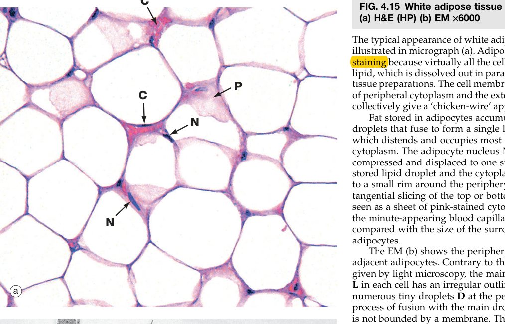
A mature white adipocyte is a balloon of stored lipid. A single huge fat droplet fills almost the entire cell, pushing the cytoplasm and the now-flattened nucleus to the periphery. In routine processing the lipid is dissolved away by the alcohols and xylene, so the cell appears as a clear “empty” ring with a sliver of cytoplasm at the rim — the classic signet-ring shape. Beyond storing energy and cushioning organs, adipocytes are now recognised as an endocrine organ secreting leptin and adiponectin.
Two other minor cell types complete the loose-CT cast. The undifferentiated mesenchymal cell is a small spindle reserve cell that sits along capillaries and can re-differentiate to repair injury — this is one of the body's pools of multipotent adult stem cells. The leukocytes (lymphocytes, eosinophils, neutrophils) are not true residents at all; they wander out of vessels during inflammation, do their job, and either die there or move on. Their abundance in a tissue is a snapshot of how much immune activity is happening.
- Fat cell (adipocyte): large, round/polygonal, occurring singly or in groups; a single large lipid droplet pushes the cytoplasm and the flattened nucleus to the periphery (signet-ring). Lipid dissolves in processing → appears empty. Function: synthesis & storage of lipid.
- Undifferentiated mesenchymal cells: multipotent reserve cells (often perivascular).
- Leukocytes: wander in from blood during defence/inflammation (lymphocytes, eosinophils, neutrophils).
• Where does the nucleus of a white adipocyte sit? → peripheral and flattened, squashed by the single fat droplet
• What is the multipotent reserve cell of adult loose CT called? → the undifferentiated (perivascular) mesenchymal cell
• Why does leukocyte number in a tissue fluctuate? → they migrate in from blood during inflammation; not permanent residents
The Fibres
Connective tissue has three fibre types and each solves a different mechanical problem. Collagen resists pulling (tensile strength). Elastin recoils after stretch. Reticular fibres make a fine supporting net for delicate organs. Match the fibre to the job and you know where to find it: tendons are full of collagen, the aorta is full of elastin, and lymph node stroma is full of reticular fibres.
A second clue is the stain. Collagen takes up eosin and stains pink on routine H&E. Elastic fibres are nearly invisible on H&E and need a special stain (orcein turns them brown, aldehyde-fuchsin purple). Reticular fibres are argyrophilic — they bind and reduce silver salts and so appear black on a silver-impregnation stain. If your slide is black-on-pale, you are looking at reticulin.
Three fibres = three engineering jobs. Collagen is the rope (won't break under tension). Elastin is the rubber band (snaps back). Reticular is the safety-net mesh (holds soft organs together without crushing them).
| Fibre | LM / stain | EM & component | Property / where |
|---|---|---|---|
| Collagenous (white) | Most numerous; single or in bundles, straight/wavy, branch into networks; pink (acidophilic) on H&E | Closely-packed fibrils with 64 nm periodic banding; type I (& III) collagen | Great tensile strength (resists pulling) |
| Elastic (yellow) | Thinner, fewer, solitary, branch & anastomose; H&E faint pink/refractile; orcein → brown, aldehyde-fuchsin → purple | Core of elastin + peripheral fibrillin microfibrils | Elasticity (rubber-like recoil) — arteries, lung, skin |
| Reticular | Very thin (0.2–1.0 µm), delicate network; invisible on H&E; silver → black (argyrophilic), also PAS+ | 64 nm banding; type III collagen + carbohydrate | Supporting framework (stroma) around vessels, cells; reticular lamina of BM |
A useful note on collagen types you will keep meeting: Type I is the workhorse of tendon, bone, skin and capsules. Type II is unique to hyaline and elastic cartilage (Unit 4). Type III is reticulin — the thin fibres of lymphoid stroma and early wound matrix. Type IV is a special mesh that forms only in basement membranes (so anything that says “basal lamina” means collagen IV). When you read a clinical case about a collagen gene, the disease usually tells you the type: Ehlers-Danlos = III/V, osteogenesis imperfecta = I, Alport syndrome = IV.
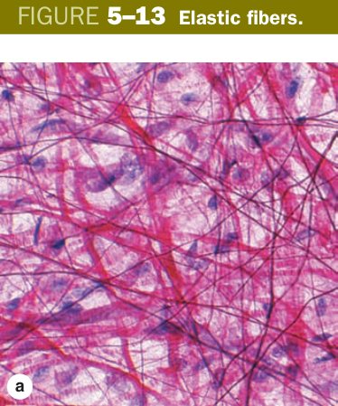
Elastic fibres are worth a closer look because they are a two-part build. A core of amorphous elastin (a rubber-like protein) is laid down on a scaffold of fibrillin-1 microfibrils secreted first by the fibroblast. The fibrillin holds elastin in the right place while it cross-links into its final extensible polymer. Lose fibrillin (the FBN1 mutation in Marfan syndrome) and the elastic fibres of the aorta and zonule of the lens are built sloppily — hence aortic-root dissection, lens dislocation and the tall arachnodactyly phenotype.
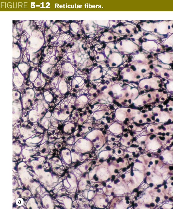
Reticular fibres are argyrophilic = silver-loving: they bind and reduce silver salts to appear as fine black threads, because their high carbohydrate content reduces the silver. They are essentially fine type III collagen and form the delicate stroma of haemopoietic and lymphoid organs, liver and endocrine glands.
Scurvy (vitamin C deficiency) impairs collagen hydroxylation → weak collagen → poor wound healing, bleeding gums, loose teeth. Ehlers–Danlos (collagen defect) → hyperextensible skin, hypermobile joints. Marfan syndrome (fibrillin-1 mutation → defective elastic fibres) → aortic-root aneurysm, lens dislocation, tall arachnodactyly.
• Collagen type in tendon & bone? → Type I
• Collagen type in hyaline cartilage? → Type II
• Collagen type in reticular fibres? → Type III
• Collagen type in basement membrane? → Type IV
• Two molecular components of an elastic fibre? → elastin core + fibrillin-1 microfibrils
• FBN1 mutation causes which syndrome? → Marfan (aortic dissection, lens dislocation)
• Why do reticular fibres stain black with silver? → they are argyrophilic — their carbohydrate reduces silver salts
Ground Substance & Tissue Fluid
Between the fibres and around the cells sits a viscous, transparent gel that you cannot see directly on a normal H&E: the ground substance. It is built from three molecular families — glycosaminoglycans (GAGs, long sulphated sugar chains), proteoglycans (GAGs bolted onto a core protein, resembling bottle-brushes), and glycoproteins (such as fibronectin and laminin) that glue cells to fibres.
The dominant GAG is hyaluronan (hyaluronic acid), an unusually long, unbranched, unsulphated polymer that acts as the backbone onto which proteoglycans hook. The whole assembly traps water and forms a hydrated gel through which nutrients, waste and signalling molecules can diffuse but bacteria cannot easily push. That is what is meant by the “molecular sieve” — the GAG mesh is a size filter and a physical barrier in one. Some bacteria, notably streptococci and clostridia, secrete hyaluronidase to dissolve the mesh and spread — the molecular reason “flesh-eating” cellulitis travels so fast.
Other GAGs you should be able to name: chondroitin sulphate and dermatan sulphate in cartilage, skin and bone; heparan sulphate in basement membranes and on cell surfaces; keratan sulphate in cartilage and cornea. The intracellular cousin heparin (highly sulphated, in mast-cell granules) is the one we have already met — same chemistry, different location and pharmacology.
The ground substance is the gelatin in a fruit jelly — it sets the whole tissue, but lets dissolved sugar and flavours diffuse through. Slice it and water runs out; the gel is mostly trapped water held in place by sugar chains.
Bathing the cells inside this gel is tissue fluid (interstitial fluid). It is filtered out of plasma at the arterial end of capillaries, where hydrostatic pressure exceeds osmotic pressure, and most of it is re-absorbed at the venous end, where the balance is reversed. The small surplus (~10%) drains away through lymphatics. This is the daily exchange that feeds every cell of every tissue. Disturb it — raised venous pressure (heart failure), lost plasma protein (nephrotic syndrome, liver failure) or blocked lymphatics (filariasis) — and fluid accumulates as oedema.
- Ground substance: a jelly-like, amorphous, homogeneous gel filling the space between cells and fibres. Composed of glycosaminoglycans (GAGs) — chiefly hyaluronic acid (forms the framework) — plus proteoglycans and glycoproteins.
- Molecular sieve / barrier: the dense GAG meshwork acts as a molecular sieve regulating diffusion and as a physical barrier hindering the spread of bacteria (some bacteria secrete hyaluronidase to break it down and invade).
- Tissue fluid: filtered from plasma at the arterial end of capillaries and largely reabsorbed at the venous end; it is the medium for metabolite exchange between blood and cells. Imbalance → oedema (excess) or dehydration (deficit).
• Backbone GAG of the molecular sieve? → hyaluronic acid
• Enzyme bacteria use to spread through the matrix? → hyaluronidase
• Two adhesive glycoproteins of the matrix? → fibronectin, laminin
• Where is tissue fluid filtered out, and where re-absorbed? → arterial end of capillary → venous end (excess returns via lymphatics)
• Four mechanisms that produce oedema? → raised venous pressure, low plasma protein, increased vessel permeability, lymphatic obstruction
Dense Connective Tissue
If loose CT is a sponge with cells scattered through it, dense CT is the opposite: far more fibres, far fewer cells. The fibres dominate the picture and the few fibroblasts squeeze between bundles as thin spindle nuclei. The trade-off is loss of metabolic flexibility for a huge gain in mechanical strength — which is why dense CT is the tissue of tendons, ligaments and the deep dermis, not of the soft spaces between organs.
Within dense CT, the key question is direction of pull. If the load always pulls in one direction (a tendon between muscle and bone, a ligament between two bones), the collagen is laid down in parallel bundles aligned with that load — dense regular CT. If the load can come from any direction (dermis stretched in every plane, an organ capsule under pressure from inside), the collagen is woven in all directions to form a 3-D mesh — dense irregular CT.
Regular = a single-direction climbing rope. Irregular = a basket woven in every direction. Tendons need ropes; skin needs baskets.
| Type | Structure | Distribution |
|---|---|---|
| Dense regular CT | Parallel, closely-packed collagen bundles; flattened fibroblasts squeezed in rows between bundles | Tendons, ligaments, aponeuroses, cornea |
| Dense irregular CT | Collagen bundles interwoven in all directions → 3-D network (resists multidirectional stress) | Dermis, capsules of organs, submucosa |
| Elastic tissue | Elastic fibres in parallel bundles or membranes | Ligamentum nuchae & flavum, walls of large (elastic) arteries |
A special case is dense elastic tissue, where parallel sheets of elastic fibres rather than collagen carry the load — the ligamentum nuchae and flavum of the spine, and the elastic laminae of the aorta and other large arteries. The elastic laminae let the aorta inflate during systole and then recoil to push blood forward during diastole; if they degenerate (as in Marfan or long-standing hypertension), an aneurysm or dissection follows.
• Dense regular vs irregular — one-line difference? → regular = parallel bundles, single-direction load (tendon); irregular = woven mesh, multi-direction load (dermis)
• Example of dense regular CT besides tendon? → ligament, aponeurosis, cornea
• Where do you find dense elastic tissue? → ligamentum nuchae & flavum, elastic laminae of large arteries
• Functional consequence of elastic-lamina failure in the aorta? → aneurysm and dissection
Adipose & Reticular Tissue
Adipose tissue is connective tissue whose cells (adipocytes) so dominate the picture that the matrix looks like a thin lacework between fat globules. There are two flavours and they look completely different under the microscope because they do different jobs. White adipose tissue stores energy and insulates; its cells hold one big lipid droplet (unilocular), the nucleus is shoved peripherally, and mitochondria are scarce. Brown adipose tissue burns energy to make heat; its cells hold many small droplets (multilocular), the nucleus is central, and the cytoplasm is crammed with large iron-rich mitochondria — those mitochondria give the tissue its brown colour and its function.
The trick that makes brown fat thermogenic is a special inner-mitochondrial-membrane protein called uncoupling protein 1 (UCP-1, thermogenin), which lets protons leak back across the membrane without driving ATP synthase. The energy of substrate oxidation is released as heat instead of stored as ATP. This is why brown fat is so prominent in newborns (concentrated in the interscapular and perirenal pads) — babies cannot shiver effectively and rely on non-shivering thermogenesis to stay warm.
White fat is the body's pantry — one big jar of butter, low metabolic activity. Brown fat is the body's furnace — many small fuel pellets fed into many burners (mitochondria) and the heat is dumped, not banked.
| White adipose tissue | Brown adipose tissue | |
|---|---|---|
| Droplets | Single large lipid droplet (unilocular) | Multiple small droplets (multilocular) |
| Nucleus | Flattened, peripheral | Central, round |
| Mitochondria | Few | Numerous, large (rich in cytochrome → brown); rich capillaries |
| Distribution | Subcutaneous tissue, mesentery | Neonates (interscapular) |
| Function | Energy reservoir, shock-absorber, thermal insulation | Heat production (thermogenesis) |
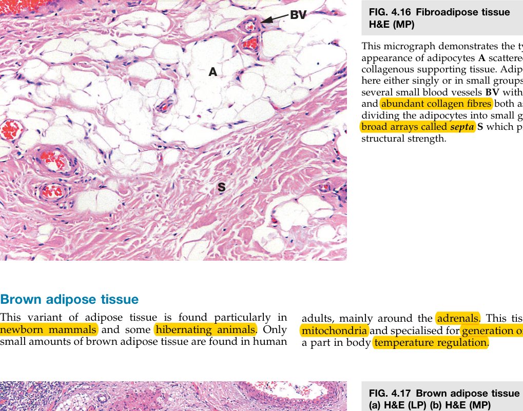
A second specialised CT proper is reticular tissue. It looks nothing like adipose: stellate reticular cells sit on a delicate, three-dimensional meshwork of reticular fibres (type III collagen, argyrophilic) and ground substance fills the gaps. The result is a soft sponge whose holes are exactly the right size for blood-forming and immune cells to sit in. This is why reticular tissue forms the supporting stroma of bone marrow, lymph node and spleen — revisited in Units 4 & 8.
A third minor specialised CT proper is mucous (mucoid) connective tissue, a jelly-like matrix dominated by hyaluronan with widely-spaced fibroblasts. Its classic location is Wharton's jelly of the umbilical cord, where it cushions the umbilical vessels. It is otherwise mainly a tissue of the embryo — you do not see much of it in the adult.
• Mitochondrial protein that makes brown fat thermogenic? → UCP-1 (thermogenin)
• Where is brown fat most prominent in humans? → the newborn (interscapular, perirenal pads)
• Reticular tissue is built around which collagen type? → Type III, argyrophilic
• Three organs whose stroma is reticular tissue? → bone marrow, lymph node, spleen
• What is Wharton's jelly? → mucoid CT (hyaluronan-rich) of the umbilical cord cushioning the umbilical vessels
TMU Exam Drill
📝 Open the full TMU Question Bank — 20 MCQ + 6 terms + 5 essays →
Authentic Tianjin Medical University past-paper questions (2021 Final & the multi-section Final with answer key) mapped to this unit, in the real exam format. Click Show answer to self-test.
□ Single best answer
- A. The cells are large and have many processes.
- B. The nucleus is large, oval and light-stained.
- C. The cytoplasm is strongly acidophilic.
- D. Abundant RER, free ribosomes and developed Golgi.
- E. Fibroblasts synthesize fibers and ground substance.
- A. Round or oval in shape.
- B. Nucleus round and eccentric (cartwheel/clock-face).
- C. Cytoplasm strongly basophilic.
- D. Formed from monocytes in blood.
- E. Secretes antibodies.
Connective tissue complete
7 cells + 3 fibres + ground substance mastered. Next: the specialised CTs — Cartilage & Bone.