Epithelial Tissue
General Features of Epithelium
Run your tongue across the roof of your mouth and you are touching epithelium. Look at the inner pink of your eyelid — epithelium. Every surface where your body meets the outside world, or where one inside compartment meets another, is sealed by a sheet of cells packed shoulder-to-shoulder with almost no space between them. That is the defining trick of this tissue: cells dominate, matrix is minimal. Connective tissue is the opposite — sparse cells floating in a sea of matrix. Hold that contrast in your head and half the exam answers in this unit fall out by themselves.
Why do bodies need such a tissue at all? Because surfaces have to do three contradictory jobs at once — cover (keep the inside in and the outside out), exchange (let oxygen, water and nutrients cross selectively), and secrete (release enzymes, mucus, hormones). A leaky sheet would betray job one; a thick sheet would betray job two. Evolution's answer is a single layer (or a stacked stack) of polarised cells anchored on a basement membrane, kept tight by junctions, and renewed constantly from a mitotic base. Everything else in this unit — classification, junctions, glands — is just engineering detail around that one idea.
Epithelium is a basic tissue composed of closely aggregated cells with very little extracellular matrix, arranged in continuous sheets (or cords/follicles) that cover body surfaces, line cavities and tubes, and form the parenchyma of glands.
Picture a brick wall — the bricks are cells, the thin film of mortar is the tiny ECM. Now picture a fruit salad — a few pieces of fruit floating in a lot of syrup. The wall is epithelium; the fruit salad is connective tissue. Same body, opposite design philosophies.
Now the six features that examiners love. They are simply the consequences of being a cellular sheet on a surface — do not memorise them as a flat list, derive them. If you are a tightly-packed sheet, you have no room for vessels (avascular). If you sit on a surface, you must have a top and a bottom (polarity). If you have a bottom, something has to anchor you to whatever is below (basement membrane). If you take wear at the surface, you must be able to replace yourself (high regeneration). If you guard the body's borders, you should warn the brain (rich nerve endings).
These six features are also the “compare epithelium with connective tissue” essay (mid-term, 18 marks):
| Feature | Epithelium | (contrast: Connective tissue) |
|---|---|---|
| Cellularity / ECM | Cells closely packed; minimal ECM; form cellular sheets | Few cells; abundant ECM |
| Polarity | Distinct free (apical) surface, lateral surfaces, and basal surface resting on a basement membrane | No polarity; cells scattered in matrix |
| Vascularity | Avascular — no blood vessels; nourished by diffusion from underlying CT | Highly vascular |
| Innervation | Rich in free nerve endings (sensitive) | Present |
| Basement membrane | Always rests on a basement membrane | Absent |
| Regeneration | High capacity for renewal (continuous mitosis) | Variable |
The avascular point deserves a second look, because students get it wrong every year. If epithelium has no blood vessels, how do its cells get oxygen? By diffusion across the basement membrane from capillaries in the underlying connective tissue. That is also why epithelium can never be too thick — the deepest cell still has to be within diffusion range. When you see a thick epithelium (epidermis, oesophagus), look closely: the superficial layers are dying or already dead. They got too far from the food supply.
Epithelium does not do one job; it does a family of jobs. The textbook splits the tissue itself by function into four groups — covering, glandular, sensory, contractile (myoepithelial). The first two are the giants of this unit; the other two are footnotes for later. Read the table and notice that the same kind of cell, by rearranging itself, can either line a surface or sink into the connective tissue to form a gland. Glands are not a different tissue; they are epithelium that has dug in.
| Class | Component | Main role |
|---|---|---|
| Covering epithelium | Sheets of cells covering surfaces / lining lumina | Protection, absorption, secretion, transport |
| Glandular epithelium | Glandular (secretory) cells; forms glands | Secretion (exocrine + endocrine) |
| Sensory epithelium | Specialised receptor cells | Sensory reception (taste, smell, hearing) |
| Myoepithelium | Contractile epithelial cells around secretory units | Contractility (expel secretion) |
Overall epithelial functions: protection · secretion · absorption · excretion · sensory reception · contractility.
• Three surfaces of an epithelial cell? → free (apical), lateral, basal
• Why is epithelium avascular? → cells too closely packed; nourished by diffusion from CT capillaries across the basement membrane
• Four functional classes? → covering, glandular, sensory, myoepithelial
• Which feature does CT lack that every epithelium has? → basement membrane + polarity
Covering Epithelium — Classification
There are eight covering epithelia and you have to know each one by name, by section appearance, by location and by function. That sounds brutal until you realise the naming system itself does most of the work for you. The name is a two-word recipe: (1) how many layers — simple (one) or stratified (two or more), and (2) what shape the surface cells are — flat (squamous), cube (cuboidal), or tall (columnar). Add two oddballs — pseudostratified (looks layered but is not) and transitional (changes shape with stretch) — and you have the whole list.
Why does any of this matter at the microscope? Because shape betrays function. Flat cells are thin — perfect for diffusion. Cube and column cells are thick, full of organelles — perfect for secretion and absorption. Many layers of flat cells stacked dead at the top are perfect for protection against abrasion. The body's choice of epithelium at any site is a clue to what the site has to do.
Named by (1) number of cell layers and (2) shape of the surface cells. Plus two special types: pseudostratified and transitional.
| Type | Distribution (learn exactly) | Main function |
|---|---|---|
| Simple squamous | Endothelium (heart/blood/lymphatic lining), mesothelium (pleura, peritoneum, pericardium/epicardium), alveoli, parietal layer of Bowman's capsule | Lubrication, diffusion, transport |
| Simple cuboidal | Renal tubules, thyroid follicles, small ducts, surface of ovary | Secretion, absorption |
| Simple columnar | Stomach → intestine (GIT), gall bladder, uterus | Secretion, absorption |
| Pseudostratified ciliated columnar | Respiratory passages (trachea, bronchi) | Protection, mucociliary transport |
| Stratified squamous — keratinized | Epidermis of skin | Protection, prevents water loss |
| Stratified squamous — non-keratinized | Mouth, pharynx, oesophagus, vagina, cornea | Protection (moist surfaces) |
| Stratified columnar | Conjunctiva, parts of male urethra, large gland ducts | Protection |
| Transitional (urothelium) | Renal calyces, ureter, urinary bladder, upper urethra | Protection + distension |
Diffusion surfaces are thin (simple squamous): alveoli, capillary endothelium, Bowman's capsule. Wet-but-abraded surfaces are non-keratinized stratified squamous: the food path mouth→pharynx→oesophagus + vagina + cornea. Dry abraded surface is keratinized: skin. Stretchy urine surfaces are transitional: the urinary tract (calyx→bladder→upper urethra).
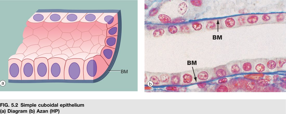
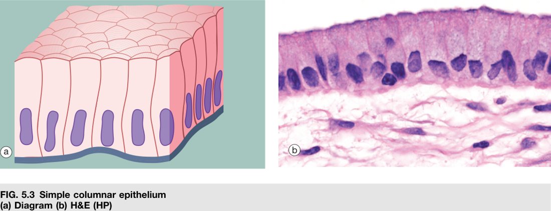
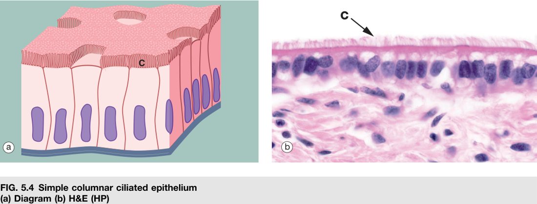
Wheater's atlas — the covering epithelia at a glance (each plate: labelled diagram + matching H&E micrograph):
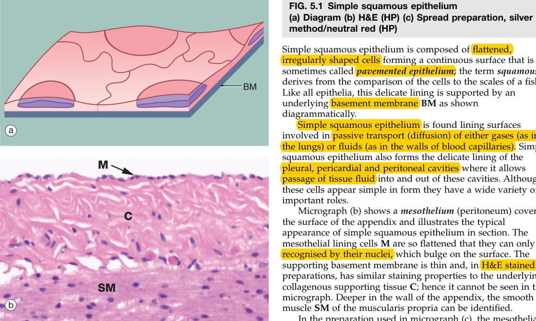
When the body's only job at a surface is to let things across, it picks the thinnest cell it can build — a flat, single-layered pancake. That is simple squamous epithelium. Stand it on its edge in a section and you see a fine pink line with the occasional bulge where a nucleus sits; flatten it surface-on and you see irregular tiles with jigsaw borders, like crazy-paving. Each cell is so thin that a red blood cell crossing an alveolar capillary is only microns from the air it must collect.
Two named varieties of this same epithelium have their own exam-worthy labels — endothelium when it lines the lumen of the cardiovascular and lymphatic system, mesothelium when it lines the great serous cavities (pleura, peritoneum, pericardium). Examiners love these two terms because they trip up the student who has memorised “simple squamous = thin” without learning the proper noun for the location. Get them cold.
- Surface view: irregular cells with serrated (jigsaw) borders fitted together (silver-stained).
- Vertical section: a single layer of flattened cells with a flattened, ellipsoid nucleus that bulges at the cell centre; cytoplasm extremely thin.
Endothelium = the simple squamous epithelium lining the inner surface of the cardiovascular and lymphatic systems (heart, blood & lymph vessels). Mesothelium = the simple squamous epithelium lining the serous cavities — covering the pleura, peritoneum and pericardium (incl. the epicardium).
The thinness of simple squamous epithelium enables rapid gas exchange at the alveolus and filtration at the glomerulus. Malignant transformation of mesothelium → mesothelioma (asbestos exposure). Damaged endothelium initiates atherosclerosis and thrombosis.
• Lines heart, blood & lymph vessels → ? → endothelium
• Lines pleura, peritoneum, pericardium → ? → mesothelium
• Why is this epithelium chosen at the alveolus and glomerulus? → thinness allows rapid diffusion / filtration
• Asbestos-linked malignancy of serous lining? → mesothelioma
Take that flat pancake and pile in more cytoplasm and organelles until the cell is roughly as tall as it is wide — you now have a cube. Why a cube? Because the cell is no longer just letting things drift across; it has work to do. It is pumping sodium, reabsorbing glucose, secreting thyroglobulin. That work needs mitochondria, ribosomes and a respectable Golgi, and they all need room.
The give-away on a slide is the nucleus: a single, round, centrally-placed nucleus in each cube. Find that pattern and you are nearly always looking at a renal tubule or a thyroid follicle, with small ducts as the third common haunt. Compare and contrast with simple columnar in the next subtopic — the nucleus position alone usually decides the answer.
- Surface view: polygonal cells. Vertical: single row of cube-shaped cells; spherical, centrally-placed nucleus.
- Distribution: renal tubules, thyroid follicles, small ducts. Function: secretion & absorption.
• Three classic locations? → renal tubules, thyroid follicles, small ducts
• Function? → secretion + absorption
• Nucleus cue that separates cuboidal from columnar? → central round (cuboidal) vs basal oval (columnar)
Stretch that cube vertically and you get a column. The extra height is not for decoration — it is more cytoplasm for more secretory and absorptive machinery, plus a long apical border on which to plant microvilli. This is why the entire luminal length of the gut from stomach to rectum chose simple columnar: there is a lot to absorb and a lot to secrete, and you want to do both per square millimetre.
The histology trick is the nucleus — oval, lined up in the basal third of every cell, like a row of soldiers along the bottom. Above the nuclei, you often see goblet cells (pale, mucus-filled balloons) and an apical brush border made of microvilli. Brush border = absorptive turbo-boost; goblet cells = lubrication and protection. Together they are the signature of the small intestine.
- Vertical: single row of tall column cells; oval nucleus in the basal third, all aligned at the same level.
- Often bears apical microvilli (striated/brush border) and interspersed goblet cells (intestine).
- Distribution: GIT (stomach→intestine), gall bladder, uterus. Function: secretion & absorption.
• Apical specialisation that magnifies absorption? → microvilli (brush/striated border)
• Mucus-secreting cell interspersed in this epithelium? → goblet cell
• Major locations? → GIT (stomach → rectum), gall bladder, uterus
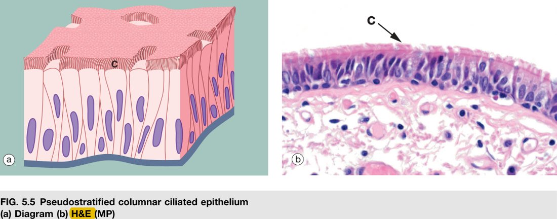
Here is the trap. You look down the microscope at the trachea and see nuclei scattered at different heights — some near the base, some halfway up, some near the surface. Your first instinct is “stratified, several layers.” Wrong. Every cell here actually touches the basement membrane; some are short and never reach the lumen, others are tall and do. The nuclei sit wherever the cell's widest part happens to be, which gives the illusion (“pseudo”) of layers. It is one layer of cells of unequal height, not multiple layers.
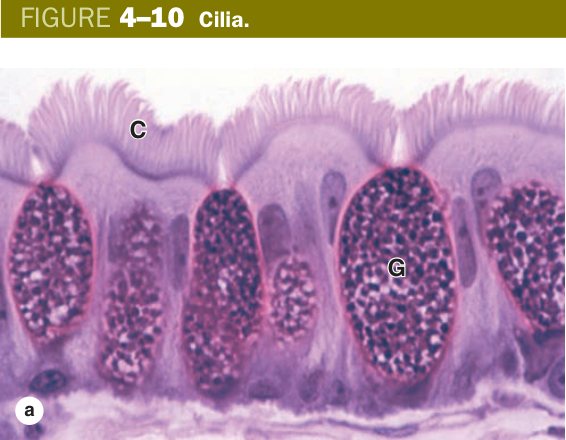
The functional cast is mixed: tall ciliated columnar cells dominate, goblet cells pump out mucus, and short basal cells sit on the floor as a stem-cell reserve. The result is the mucociliary escalator — mucus traps dust and bugs, cilia sweep the loaded mucus upward toward the throat to be swallowed. Smoking paralyses the cilia, the escalator stops, and the airways drown in their own secretions. That is the structural story behind the smoker's cough.
- Cell types: ciliated columnar cells, goblet cells (mucus), short spindle (basal) cells, conical (basal) cells.
- Distribution: respiratory passages (trachea, bronchi). Function: protection + mucociliary transport (cilia sweep trapped particles + mucus upward).
• The four cell types? → ciliated columnar, goblet, short spindle (basal), conical
• The cleanest distinguishing rule from true stratified? → every cell touches the basement membrane
• Classic site & function? → trachea/bronchi; mucociliary clearance
• What does smoking do to it? → paralyses cilia → mucus stagnates → chronic cough/bronchitis
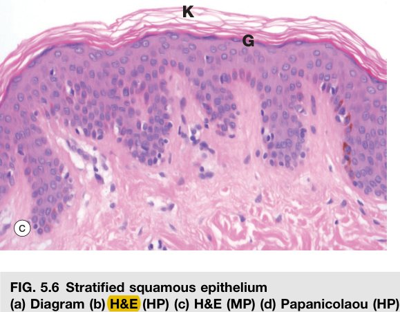
Now the body needs a tougher surface — something that can shrug off rubbing, chewing, scraping, walking. The answer is to stack many layers and let the top layers be sacrificial. New cells are born at the bottom by mitosis, climb upward as fresh ones push them along, flatten as they go, and finally die and shed from the top. The whole epidermis turns over every 4–6 weeks.
Two flavours: keratinised, where the surface cells die hard — they fill with keratin, lose their nuclei, and become a dry waterproof crust (skin); and non-keratinised, where the surface cells stay nucleated and moist (mouth, oesophagus, vagina, cornea). The choice depends on the local climate: dry external surfaces need waterproofing, internal wet surfaces do not. Either way, the engine is the basal layer — the cuboidal/low-columnar cells sitting on the basement membrane, dividing constantly. Wound healing, regeneration, and (sadly) basal cell carcinoma all start here.
Many layers; named for the flat surface cells. From base up: basal layer (single row of cuboidal/low-columnar, mitotically active) → several polyhedral layers → flattened/spindle layers → flat surface cells.
| Sub-type | Surface cells | Distribution |
|---|---|---|
| Keratinized | Dead, anucleate, filled with keratin | Epidermis of skin |
| Non-keratinized | Nucleated, little keratin (moist) | Mouth, pharynx, oesophagus, vagina, cornea |
The mitotic basal layer drives continuous renewal and wound healing; it is also the origin of basal cell carcinoma. Chronic acid reflux can convert oesophageal non-keratinized stratified squamous to intestinal columnar epithelium (Barrett's metaplasia → adenocarcinoma risk).
• Why does the surface flatten as cells rise? → they migrate, dehydrate, and accumulate keratin/become spindle-shaped
• Keratinised vs non-keratinised — locations? → epidermis vs mouth/pharynx/oesophagus/vagina/cornea
• Function justification for many layers? → protection against abrasion + (with keratin) waterproofing
• Barrett's oesophagus is a switch from ___ to ___? → non-keratinised stratified squamous → simple columnar (intestinal type)
A rare type — the body almost never wants tall columnar cells in layers. Where it does appear, only the surface layer is columnar; the deeper layers are polygonal and short. Think of it as a stratified epithelium that put a row of columnar cells on top as the working face. You will see it lining the conjunctiva, parts of the male urethra, and the larger excretory ducts of glands. Its job is protection in places that need slightly more substance than a single columnar row could provide.
- Deep layer(s) of polygonal cells; surface layer of columnar cells. Relatively rare.
- Distribution: conjunctiva, parts of the male urethra, large excretory ducts. Function: protection.
• Three classic sites? → conjunctiva, parts of male urethra, large gland excretory ducts
• Main function? → protection
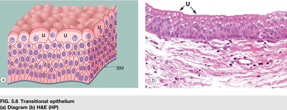
The bladder has an engineering problem the rest of the body does not: it has to triple in volume several times a day and then shrink back, while keeping a fiercely cytotoxic urine on the inside and the body fluids on the outside. No fixed-shape epithelium can do that. The answer is transitional epithelium — a stratified epithelium whose surface cells (the famous “umbrella cells”) are large, dome-shaped, often binucleate, and have a thick apical plaque membrane built specifically to resist urine.
When the bladder is empty, the cells pile up — you count 6–8 apparent layers, with the umbrella cells bulging into the lumen. Fill the bladder, the wall stretches, and those same cells slide over each other and flatten until you can only see 2–3 layers. It is the same epithelium changing shape, not changing cell number. That single sentence is worth a mark every time the question appears.
| State | Appearance |
|---|---|
| Contracted (empty bladder) | More cell layers; rounded dome-shaped surface cells |
| Distended (full bladder) | Fewer layers; surface cells flattened/stretched |
- Distribution: renal calyces, ureter, urinary bladder, upper urethra. Function: protection + accommodates volume changes; barrier against hypertonic, cytotoxic urine.
Transitional epithelium is the origin of urothelial (transitional cell) carcinoma — the commonest bladder cancer; classic risk factors are smoking and aniline-dye exposure; painless haematuria is the hallmark.
• Empty bladder = how many layers look? → more (~6–8); cells dome up
• Full bladder? → fewer (~2–3); cells flatten and slide
• Locations? → renal calyces, ureter, bladder, upper urethra
• Cancer of origin? → urothelial (transitional cell) carcinoma — bladder; smoking/aniline dyes
Specializations of the Free (Apical) Surface
The free surface is where the cell meets the lumen — air, urine, gut contents, a duct. Whatever the cell needs to do to or with that lumen, it has to do from this surface. So evolution has built three specialisations into the apical membrane, each with a different cytoskeletal core and a different job. Learn them as a trio — microvillus, cilium, stereocilium — because exam questions almost always ask you to tell them apart.
The trick is the core. Microvilli have an actin core. Cilia have a microtubule core (9+2). Stereocilia are confusingly named — despite the word “cilia”, they are actually very long microvilli with an actin core, not motile. Get the core right and every other property (motility, length, function) follows automatically.
| Structure | Core (EM) | LM appearance | Motility | Function / site |
|---|---|---|---|---|
| Microvillus | Bundle of actin filaments | Striated / brush border | Non-motile | ↑ absorptive surface area — small intestine, PCT of kidney |
| Cilium | 9 + 2 microtubule axoneme + basal body | Fine hair-like, longer than microvilli | Motile (directional beat) | Sweep mucus/ova — respiratory tract, uterine tube |
| Stereocilium | Actin (very long microvilli) | Long, often branched/clumped | Non-motile | Absorption / sensory — epididymis, hair cells of inner ear |
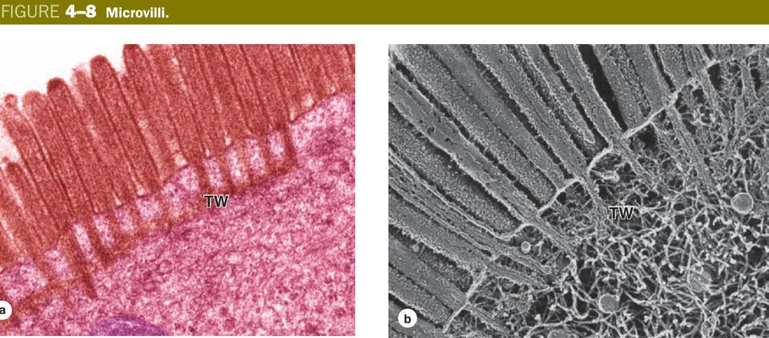
Why microvilli? Because a flat membrane has a fixed surface area, and absorption is rate-limited by area. By projecting hundreds of finger-like extensions, an enterocyte multiplies its apical surface roughly thirty-fold. Each microvillus contains a bundle of parallel actin filaments anchored into the cytoplasmic terminal web. They do not move; they just are — standing forests increasing area for sodium-glucose cotransporters, peptidases, and water uptake.
Why cilia? Because some surfaces need to move the lumen contents, not absorb them. The cilium is built like a tiny oar: nine peripheral doublets of microtubules arranged around a central pair (the 9+2 axoneme), anchored to a basal body just under the membrane, powered by dynein arms that walk one doublet against its neighbour. The result is a directional beating wave — mucus rides upward in the trachea, the ovum drifts toward the uterus in the fallopian tube. Without dynein, the cilia freeze; with frozen cilia, the airways and tubes silt up.
Microvillus: delicate finger-like projection of the apical cell membrane and cytoplasm containing a core of fine actin filaments; seen by LM as a striated/brush border; increases free surface area for absorption. Cilium: longer, motile projection of membrane and cytoplasm containing a 9+2 arrangement of microtubules arising from a basal body; its directional beat sweeps surface substances along.
Microvilli are shag carpet — lots of little fibres standing still to soak things up. Cilia are rowing oars — longer, fewer, and beating in coordinated strokes to push the lumen contents along. Stereocilia are fishing rods — very long shag fibres, still passive, sometimes used as antennae (inner-ear hair cells).
Primary ciliary dyskinesia (Kartagener syndrome): defective dynein arms → immotile cilia → chronic sinusitis, bronchiectasis, infertility, and situs inversus. Loss of intestinal microvilli (brush border) in coeliac disease → malabsorption.
• Core of a motile cilium? → 9+2 microtubule axoneme + basal body; dynein arms power the beat
• Stereocilium — cilium or microvillus? → very long microvillus (actin), non-motile, despite the name
• Brush/striated border on LM = ? → densely packed microvilli of enterocytes / PCT cells
• Kartagener triad? → bronchiectasis + sinusitis + infertility (± situs inversus) from dynein defect
Lateral Surface — Cell Junctions
A sheet of cells is only as good as the joins between them. Leave gaps and water leaks. Leave them slippery and they slide apart under stress. Leave them isolated and they cannot talk to each other to coordinate a response. So epithelial cells use a toolkit of four junction types along their lateral surfaces — each one engineered for a different problem.
Start at the top and read down. Right at the apex, a tight junction (zonula occludens) fuses adjacent membranes into a continuous belt; nothing slips between the cells, and the apical and basolateral membrane domains stay segregated. Just below it, an intermediate junction (zonula adherens) grips neighbour to neighbour through cadherins linked to actin — mechanical adhesion. Deeper still, scattered as plaques, are the desmosomes (macula adherens): spot welds linking keratin intermediate filaments across the gap, the strong anchors that keep epidermal cells from being torn apart by friction. And anywhere along the side, gap junctions punch communicating channels between cells, letting ions and small metabolites flow directly. When tight + adherens + desmosome sit together near the apex, the trio is called the junctional complex — a recurring 4-mark term.
| Junction | Other name | Linker / molecule | Function |
|---|---|---|---|
| Tight junction | Zonula occludens | Belt; membranes fused at points (claudin, occludin) | Barrier — prevents bacteria/macromolecules passing between cells & stops outflow of tissue fluid; seals the apex |
| Intermediate junction | Zonula adherens (adherens) | Belt; cells separated by narrow space + actin filaments (cadherin) | Adhesion, maintains cell shape, transmits contractile force |
| Desmosome | Macula adherens | Plate (spot); space with a mid-line + keratin intermediate filaments (desmoglein/desmocollin) | Strong spot adhesion — firmly anchors one cell to another |
| Gap junction | Nexus / communicating | Connexons (connexin) of adjacent cells aligned | Communication — passage of ions/small molecules; couples cells electrically |
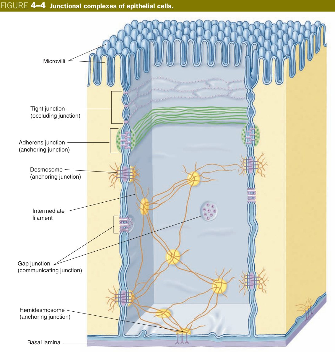
Junctional complex: when two or more types of cell junction coexist in the same region of the lateral surface (typically tight junction + intermediate junction + desmosome, in that apex-to-base order), the collection is termed a junctional complex.
Zonula = belt (encircles cell): tight + adherens. Macula = spot: desmosome. Apex→base order: “Tight, then Adhere, then Desmo, with Gaps anywhere” (TJ → ZA → desmosome → gap junction). Filament cue: adherens = actin; desmosome = keratin (intermediate filaments).
Think of two adjacent cells as two boats moored side by side. The tight junction is plastic wrap stretched between them at the deck level — nothing sloshes between. The adherens junction is a rope belt holding the rails together. The desmosome is a row of carabiners spot-welding the hulls. The gap junction is a pipe between the engine rooms so each crew can talk to the other.
Tight junctions form the blood–brain and blood–testis barriers. Autoantibodies against the desmosomal protein desmoglein-3 cause pemphigus vulgaris (intra-epidermal blisters); antibodies against hemidesmosomal antigens cause bullous pemphigoid (sub-epidermal blisters). Gap junctions synchronise cardiac muscle (intercalated discs).
• Which junction seals the paracellular space? → tight junction; molecules: claudin, occludin
• Filament linked to adherens vs desmosome? → actin (adherens) vs keratin intermediate filaments (desmosome)
• Which junction allows ions/small molecules between cells? → gap junction (connexons of connexin)
• Antibodies against desmoglein-3 → ? → pemphigus vulgaris (intra-epidermal blisters)
• What is a “junctional complex”? → two or more junction types coexisting in the same region of the lateral surface
Specializations of the Basal Surface
Every epithelium sits on a thin extracellular mat that separates it from the connective tissue below. That mat is the basement membrane, and despite its modesty on H&E (a faint pink line, easily missed), it is one of the busiest structures in the body. It anchors the epithelium, filters what passes between epithelium and CT, and instructs epithelial cells how to migrate and differentiate during repair. Stain it with silver and it blackens; stain it with PAS and it turns magenta — both tricks exploit its rich carbohydrate content (type IV collagen, laminin, perlecan, entactin).
By electron microscopy, the basement membrane resolves into two layers. The upper basal lamina (lamina lucida + lamina densa) is laid down by the epithelial cells themselves. The lower reticular lamina (reticular/type III collagen fibres) is made by fibroblasts of the underlying connective tissue. Two tissues collaborating to build one membrane — remember the division of labour, examiners ask it directly.
Basement membrane: a thin extracellular sheet between epithelium and the underlying connective tissue. LM: a pink line on H&E, black with silver, PAS-positive (magenta). EM: a basal lamina (lamina lucida + lamina densa, produced by the epithelial cells) plus a reticular lamina (reticular fibres, produced by fibroblasts of the connective tissue).
- Functions: structural support & attachment of epithelium; a selective (semi-permeable) molecular filter; and a scaffold that guides epithelial migration, proliferation and differentiation during repair.
In the glomerulus, the basement membrane is the key filtration barrier: anti-GBM antibodies cause Goodpasture syndrome; chronic hyperglycaemia thickens it in diabetic nephropathy; a defective type IV collagen causes Alport syndrome.
• Who makes which layer? → epithelial cells make the basal lamina; fibroblasts make the reticular lamina
• Why is it PAS-positive? → rich in glycoproteins (type IV collagen, laminin, perlecan)
• Three functions? → support/attachment; selective filtration; scaffold for migration & differentiation in repair
• Glomerular BM disease examples? → Goodpasture, diabetic nephropathy, Alport
If the apex needs surface area for absorption, the base sometimes needs surface area for active transport. The trick is the same but reversed — instead of pushing membrane outward (microvilli), the basal membrane folds inward deep into the cytoplasm. Wedged between the folds are mitochondria, packed like batteries against the ion pumps that have to be supplied with ATP. This is the structural signature of the renal proximal and distal convoluted tubules and of the striated ducts of salivary glands — cells that move huge volumes of sodium and water with high efficiency.
Anchoring the cell to the basement membrane is a different job, done by the hemidesmosome — literally “half a desmosome.” A desmosome has two cytoplasmic plaques (one in each neighbouring cell) connected through the gap; a hemidesmosome has only the cell-side plaque, with integrins reaching down into the basal lamina. Keratin filaments anchor into the plaque from inside; the integrins grip from outside. Result: the cell cannot be lifted off the basement membrane by ordinary shearing. Lose these and you get a blistering disease (bullous pemphigoid).
Plasma membrane infolding: the basal cell membrane folds deeply into the cytoplasm, with numerous mitochondria packed between the folds. This increases basal surface area and supplies ATP for active transport of water and ions. Seen in renal PCT/DCT and striated ducts of salivary glands.
- Hemidesmosome: “half a desmosome” — a plaque on the basal membrane that anchors the epithelial cell to the underlying basement membrane (keratin filaments insert into one plaque only).
• Two classic sites of basal infoldings? → renal PCT/DCT and striated ducts of salivary glands
• Hemidesmosome connects what to what? → basal pole of epithelial cell to the basal lamina (via integrins)
• Filament that inserts into a hemidesmosome plaque? → keratin intermediate filaments
• Antibody against hemidesmosomal antigens → ? → bullous pemphigoid (sub-epidermal blister)
Glandular Epithelium & Glands
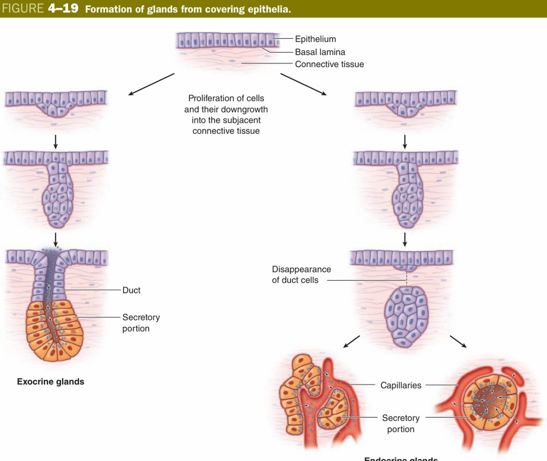
Glands are not a separate tissue; they are epithelium that has grown into the underlying connective tissue and specialised for secretion. Picture a flat sheet of epithelial cells; now imagine a small region of it sinking down into the CT and forming a tubule. If the tubule keeps its connection to the surface as a duct, the deep cells secrete into the duct and the secretion arrives back at the surface — an exocrine gland. If the connection withers away and the deep cells start secreting into the surrounding capillaries instead, you have an endocrine gland. One developmental origin, two destinies.
That is why the obvious next question — “what is the difference between exocrine and endocrine?” — has the answer “duct or no duct.” Exocrine glands keep their duct and dump enzymes, mucus, sweat, milk onto an epithelial surface. Endocrine glands lose their duct and dump hormones straight into the blood — which is why they sit in a rich capillary bed.
| Exocrine gland | Endocrine gland | |
|---|---|---|
| Duct | Has a duct — secretes onto a surface/lumen | Ductless — secretes into blood |
| Product | Enzymes, mucus, sweat, etc. | Hormones |
| Vascularity | Ordinary | Rich capillary network |
| Example | Salivary, pancreas (acini), sweat | Thyroid, adrenal, pancreatic islets |
Exocrine glands are then sub-classified along four independent axes — treat each axis as a separate question and the apparently complicated nomenclature collapses. Duct branching: simple (unbranched) vs compound (branched). Secretory unit shape: tubular vs acinar (round, also called alveolar) vs tubuloacinar (both). Nature of product: serous (watery, enzyme-rich, basophilic, e.g. parotid, pancreas) vs mucous (viscous, pale, e.g. goblet cells) vs mixed/seromucous (submandibular, with the famous serous demilunes capping mucous tubules).
The fourth axis — mode of secretion — is the most exam-favoured. It asks: how much of the cell goes out with the secretion? Merocrine: nothing — just exocytosis of vesicles, cell intact. This is the default, used by almost every gland. Apocrine: a thin rim of apical cytoplasm pinches off with the product — classically the lipid droplets of the lactating mammary gland. Holocrine: the whole cell dies and becomes the secretion — the sebaceous gland is the textbook example; new cells are continually born at the base to replace those that died at the top.
Exocrine glands are classified by:
- Duct branching: simple (unbranched duct) vs compound (branched duct).
- Secretory unit shape: tubular, acinar/alveolar, or tubuloacinar.
- Mode of secretion: merocrine (exocytosis, cell intact — most glands), apocrine (apical cytoplasm pinched off — lactating breast lipid), holocrine (whole cell disintegrates — sebaceous gland).
- Nature of product: serous (watery, enzyme-rich, basophilic cells — parotid, pancreas), mucous (viscous, pale cells — goblet cells), or mixed/seromucous (submandibular, with serous demilunes).
- Myoepithelial cells: contractile, basket-like epithelial cells around secretory units/ducts that squeeze out secretion (salivary, mammary, sweat glands).
Merocrine = the postman drops the parcel and walks away. Apocrine = the postman tears a corner off his jacket and leaves it with the parcel. Holocrine = the postman explodes and his remains are the parcel.
• Four axes of exocrine classification? → duct branching, unit shape, product, mode of secretion
• Merocrine / apocrine / holocrine examples? → pancreas / lactating mammary lipid / sebaceous
• Serous vs mucous cytology? → basophilic, dark, round basal nucleus (serous) vs pale, flattened basal nucleus (mucous)
• Myoepithelial cells — where and why? → basket cells around salivary/mammary/sweat acini; contract to expel secretion
• A goblet cell counts as what kind of gland? → unicellular exocrine (mucous) gland
TMU Exam Drill
📝 Open the full TMU Question Bank — 20 MCQ + 6 terms + 5 essays →
Authentic Tianjin Medical University past-paper questions (2021 Final & the multi-section Final with answer key) mapped to this unit, in the real exam format. Click Show answer to self-test.
□ Single best answer
- A. basement membrane
- B. tight junction
- C. microvillus
- D. desmosome
- E. basal infolding
Epithelium complete
Free-surface, lateral & basal specializations + all 8 covering types mastered. Next: Connective Tissue.