Integration of Metabolism
The organising question ★★★
Every unit from 10 to 21 has described a pathway. This one asks a different kind of question: at any given moment, which of those pathways is running, in which organ, and why? Nothing new is introduced here. What is introduced is the logic that decides between them.
The first priority of metabolism is to provide sufficient glucose to the brain and red blood cells.
The blood glucose level must be maintained above 2.2 mmol/L. Below that, consciousness fails within minutes.
Everything else — glycogen, gluconeogenesis, lipolysis, ketogenesis, muscle proteolysis — is machinery for defending that number.
Two facts about the brain make it the tissue around which all of metabolism is organised.
Glucose is the exclusive fuel for brain tissue, except in periods of extreme starvation, and brain metabolism is entirely aerobic. It cannot use fatty acids at all — they travel bound to albumin and cannot cross the blood-brain barrier (Unit 18). It has no meaningful glycogen store of its own.
The erythrocyte is even more restricted: no mitochondria at all, so it is obligately glycolytic and its dependence on glucose is absolute in every state, including starvation.
So the body is permanently supplying a large, non-negotiable glucose demand from a store — liver glycogen — that lasts less than a day. Everything that follows in this unit is the consequence of that arithmetic.
Note that it is the RATIO, not either hormone alone, that the tissues read.
High insulin/glucagon — after a meal — means fuel storage is taking place: glycogenesis, lipogenesis, protein synthesis.
Low insulin/glucagon — fasting, starvation, or untreated diabetes — means fuel mobilisation: glycogenolysis, gluconeogenesis, lipolysis, ketogenesis, proteolysis.
“When glucagon speaks, the liver listens.”
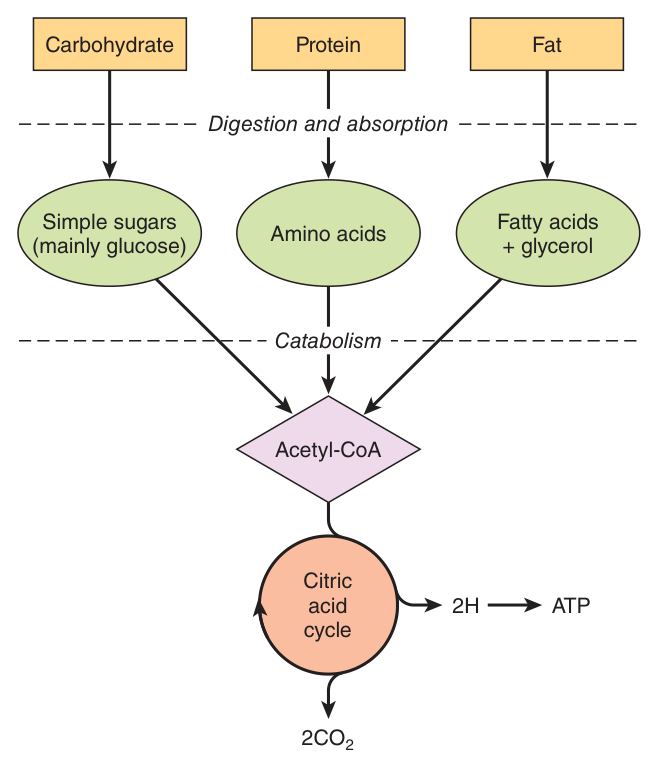
Fatty acids are more energy dense than glucose and produce ATP more efficiently, entering the citric acid cycle at the level of acetyl-CoA. Fatty acids spare blood glucose.
Glucose can also be a primary source of energy to the muscles for short periods, but it will soon be depleted, as will glycogen. When the blood glucose drops below the normal range, the liver kicks in through glycogenolysis and gluconeogenesis.
- The first priority of metabolism? → Sufficient glucose for the brain and red blood cells
- The blood glucose floor? → Above 2.2 mmol/L
- Why can the brain not use fatty acids? → They are albumin-bound and cannot cross the blood-brain barrier
- Why is the erythrocyte obligately glycolytic? → It has no mitochondria
- What is the master signal? → The insulin/glucagon RATIO
The organ profiles ★★★
Each organ has a characteristic enzyme complement, and that complement — not any conscious coordination — is what makes it behave the way it does. Learn the table as a set of capabilities, and the behaviour in every metabolic state becomes deducible.
| Organ | Preferred fuel | Characteristic enzymes | What it exports |
|---|---|---|---|
| Liver | Glucose, fatty acids, amino acids, alcohol | Glucokinase, glucose-6-phosphatase, the urea cycle, HMG-CoA synthase, glycerol kinase | Glucose, VLDL, ketone bodies, urea, albumin — the altruistic organ |
| Brain | Glucose; ketone bodies in prolonged starvation | Fully aerobic; no fatty acid oxidation | Nothing |
| Skeletal muscle | Fat at rest; glucose in short bursts | Hexokinase, NO glucose-6-phosphatase; branched-chain α-ketoacid dehydrogenase | Lactate and alanine — but keeps its glycogen to itself |
| Adipose tissue | Glucose, fatty acids | Lipoprotein lipase, hormone-sensitive lipase; NO glycerol kinase | Free fatty acids and glycerol |
| Erythrocyte | Glucose only | No mitochondria — glycolysis only | Lactate |
| Heart | Fatty acids and ketone bodies | Rich in mitochondria | Nothing |
| Kidney | Fatty acids | Glucose-6-phosphatase; renal glutaminase | Glucose (in prolonged starvation) and ammonia |
Look at the negatives in that table. Each one is a design decision with visible consequences.
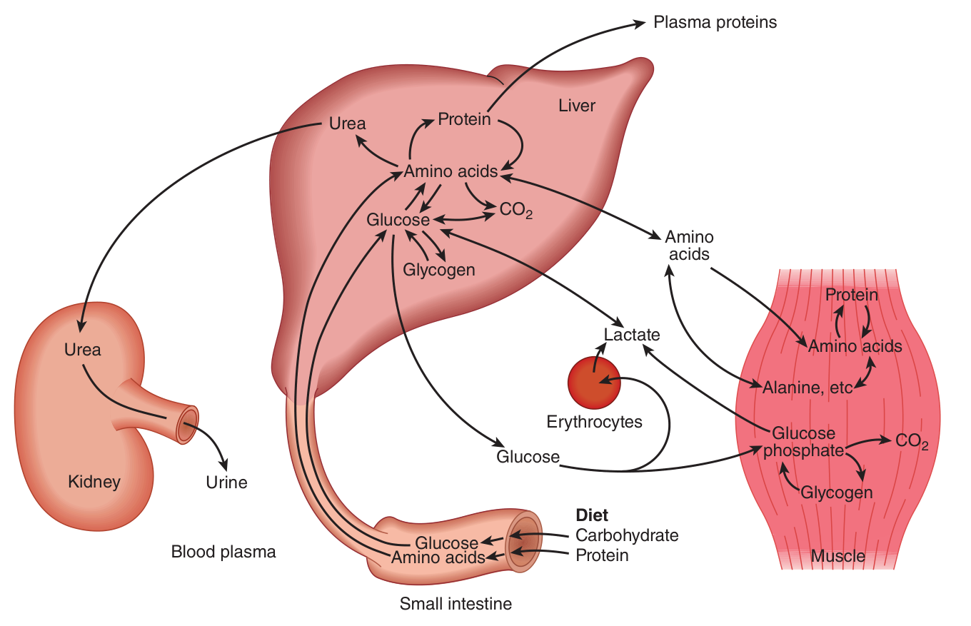
Muscle has no glucose-6-phosphatase. So muscle glycogen can never leave the muscle as glucose — it is a private store, for that muscle's own use. Skeletal muscle donates amino acids for gluconeogenesis in the liver, but it keeps its glycogen to itself. Muscle carbon reaches the blood only as lactate (Cori cycle) or alanine (glucose-alanine cycle) — and both require the liver to convert them back.
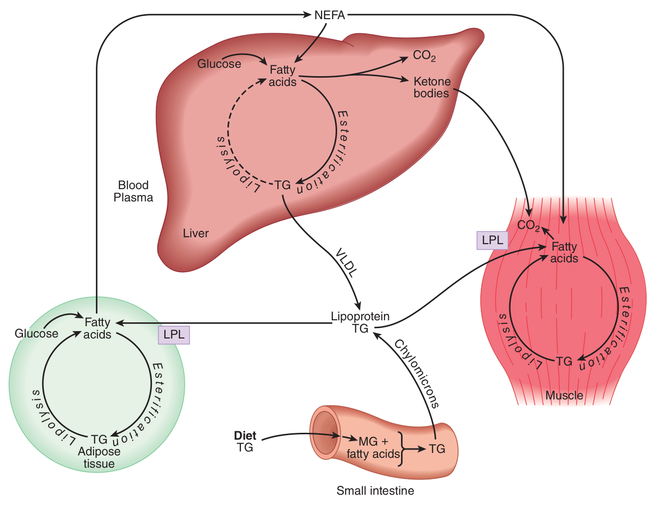
Adipose tissue has no glycerol kinase. So the glycerol released by lipolysis cannot be reused by the adipocyte and passes to the liver. It follows that fat storage requires a supply of glucose, to make glycerol-3-phosphate by glycolysis — and therefore requires insulin. This is why lipolysis runs unopposed in diabetes.
The liver lacks succinyl-CoA-acetoacetate CoA transferase. So it exports the ketone bodies it makes rather than burning them (Unit 17).
Ask what an organ cannot do, and its role in the whole becomes obvious.
- Why can muscle glycogen not raise blood glucose? → Muscle lacks glucose-6-phosphatase
- Why can the adipocyte not reuse its own glycerol? → It lacks glycerol kinase
- What does the heart prefer? → Fatty acids and ketone bodies
- Which two organs possess glucose-6-phosphatase? → Liver and kidney
- What does muscle export instead of glucose? → Lactate and alanine
The well-fed state ★★★
The ratio of insulin to glucagon is high following a meal, indicating that fuel storage is taking place.
Glucokinase in liver is adapted to trap large influxes of glucose from the hepatic portal vein and to build up glycogen stores. Two properties adapt it:
1 · A high Km. The Km of glucokinase for glucose is 10 mM, compared with 0.1 mM for hexokinase, so it phosphorylates glucose only at concentrations well above the fasting level of 5 mM — like those seen in the hepatic portal vein after a meal, about 22 mM.
2 · No product inhibition. Unlike hexokinase, glucokinase is not inhibited by glucose 6-phosphate, so it keeps working however much has already been trapped.
This is Unit 6's Km doing physiology, and it is one of the most satisfying examples in the subject.
Hexokinase, with a Km of 0.1 mM, is saturated at every physiological glucose concentration. That is exactly what a peripheral tissue wants: take up glucose at a constant rate whatever the supply, because you need it to survive.
But the liver's job is the opposite. It is not feeding itself; it is buffering the blood. It must take up a great deal of glucose when there is plenty and none at all when there is little — otherwise it would compete with the brain. A high-Km, non-inhibited enzyme does precisely that: its rate rises steeply with concentration and falls to nearly nothing at fasting levels.
The enzyme's kinetics ARE its physiological role. Recall too that in the pancreatic β cell, the same enzyme acts as the glucose sensor that triggers insulin release — the same kinetics used to measure rather than to store.
| Tissue | What happens in the fed state |
|---|---|
| Liver | Glucokinase traps glucose. High insulin/glucagon increases the activity of glycogen synthase and pyruvate dehydrogenase, so glucose goes to glycogen and the surplus to acetyl-CoA → fatty acids → VLDL. Gluconeogenesis and ketogenesis are off. |
| Muscle | Insulin increases muscle glycogen synthase activity and increases protein synthesis. GLUT4 moves to the membrane. Except for short elevations in energy demand, the preferential fuel for muscle is fat. |
| Adipose tissue | Insulin induces lipoprotein lipase, so chylomicron and VLDL triacylglycerol is stripped and stored; insulin inhibits hormone-sensitive lipase, so nothing leaves. Glucose uptake supplies glycerol-3-phosphate for esterification. |
| Brain | Burns glucose, as always. Its uptake is insulin-independent. |
- K_m of glucokinase vs hexokinase? → 10 mM vs 0.1 mM
- Portal vein glucose after a meal? → About 22 mM, against a fasting level of 5 mM
- Which enzyme is NOT inhibited by glucose 6-phosphate? → Glucokinase
- Insulin does what to hormone-sensitive lipase? → Inhibits it — insulin is the principal antilipolytic hormone
- Insulin does what to lipoprotein lipase in adipose tissue? → Induces it
Fasting ★★★
Fasting begins about 3 to 32 or 36 hours after a meal. The ratio of insulin to glucagon falls, because the blood sugar concentration falls.
| Site | The switch |
|---|---|
| Glycogen | Glucagon activates glycogen phosphorylase; glycogen synthase is inhibited. The same cAMP cascade does both — reciprocal regulation by one phosphorylation event (Unit 12). |
| Gluconeogenesis | Glucagon lowers fructose 2,6-bisphosphate, removing the inhibitor of fructose 1,6-bisphosphatase — so gluconeogenesis proceeds (Unit 13). |
| Adipose tissue | Falling insulin releases hormone-sensitive lipase: fasting results in the progressive release of alanine from muscle and mobilization of free fatty acids and glycerol. |
The free fatty acids arriving at the liver do four things at once — and this is the most examinable sequence in the unit:
1 · FFA oxidation raises acetyl-CoA, which raises citrate, which stimulates fructose 1,6-bisphosphatase.
2 · Acetyl-CoA stimulates pyruvate carboxylase → oxaloacetate → PEP → gluconeogenesis.
3 · Acetyl-CoA increases the activity of the citric acid cycle, raising ATP, which inhibits glycolysis.
4 · The NADH and ATP produced pay for gluconeogenesis, which costs 6 ATP equivalents per glucose.
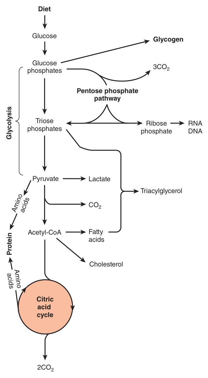
Notice the elegance of that list. Fatty acids are not glucogenic — pyruvate dehydrogenase is irreversible, so not one carbon of a fatty acid becomes glucose (Unit 17). And yet fatty acid oxidation is what makes gluconeogenesis possible.
It contributes in three ways without contributing carbon: it signals (acetyl-CoA activating pyruvate carboxylase), it funds (ATP and NADH), and it spares (every tissue burning fat is a tissue not burning glucose).
The fat is the fuel; the amino acids and glycerol are the carbon. If you can state that division of labour, you have understood the fasting liver.
Brain tissue continues to use its sole source of energy during fasting — glucose.
Skeletal muscle donates amino acids for gluconeogenesis in the liver, but it keeps its glycogen to itself. Note the detail: the branched-chain amino acids are not released into the bloodstream but are transaminated to α-ketoacids and burned locally (Unit 20 — the branched-chain α-ketoacid dehydrogenase complex is absent from the liver); all other amino acids are transported to the liver for transamination and use in gluconeogenesis.
- What removes the inhibitor of fructose 1,6-bisphosphatase? → Glucagon lowering fructose 2,6-bisphosphate
- Acetyl-CoA from β-oxidation activates which gluconeogenic enzyme? → Pyruvate carboxylase
- Which amino acids are NOT released from muscle? → The branched-chain ones — transaminated and burned locally
- Why does fasting muscle not release glucose? → No glucose-6-phosphatase
Starvation ★★★
The metabolic adaptations described in starvation show an attempt by the body to conserve blood glucose and to spare PROTEIN from continual degradation.
Protein-sparing is the point. There is no protein store — every gram degraded is a gram of functioning tissue lost.
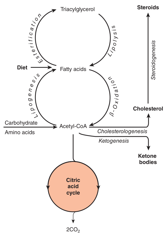
One of the key symptoms of starvation and late fasting is the development of ketosis. Gluconeogenesis slows down; free fatty acids continue to be mobilized from adipose tissue and the oxidation of fatty acids continues unabated.
Amino acids → pyruvate → oxaloacetate; the oxaloacetate is drawn into gluconeogenesis and is therefore unavailable to condense with acetyl-CoA, so acetyl-CoA is converted to ketone bodies instead.
Skeletal muscle eventually runs out of glycogen. As starvation progresses, muscle increases its reliance on free fatty acids and spares ketones for use by the brain. Starvation causes the brain to defer to the red blood cells in the consumption of glucose.
| Fuel exchange (grams per 24 h) | Day 3 | Day 40 |
|---|---|---|
| Brain — glucose | 100 | 40 |
| Brain — ketone bodies | 50 | 100 |
| All other use of glucose | 50 | 40 |
| Adipose-tissue lipolysis | 180 | 180 |
| Muscle-protein degradation | 75 | 20 |
| Liver output — glucose | 150 | 80 |
| Liver output — ketone bodies | 150 | 150 |
Take the rows in pairs and the whole physiology of starvation falls out of them.
Brain glucose falls 100 → 40 while brain ketone bodies rise 50 → 100. The brain has switched fuels. It has not reduced its energy consumption — it has changed what it burns. This is the adaptation that makes prolonged starvation survivable.
Muscle-protein degradation falls 75 → 20. That is the payoff, and it is the whole point of the exercise. Every gram of glucose the brain does not demand is a gram the liver does not have to make from amino acids — which means muscle it does not have to dismantle. Ketone bodies exist to spare protein.
Adipose lipolysis is unchanged at 180 on both days. Fat is the one store deep enough not to need rationing; it is simply drawn on at a constant rate throughout. Survival time in starvation is set by the size of the fat depot.
Liver ketone body output is unchanged at 150, while glucose output halves. The liver has not slowed down; it has changed product. And note what has not changed: all other use of glucose stays around 40–50, because the erythrocyte has no mitochondria and no alternative. Its demand is the irreducible floor.
- The two aims of starvation adaptation? → Conserve blood glucose and spare protein
- Brain glucose use, day 3 vs day 40? → 100 g vs 40 g
- Brain ketone body use, day 3 vs day 40? → 50 g vs 100 g
- Muscle protein degradation, day 3 vs day 40? → 75 g vs 20 g — the protein-sparing effect
- Which figure does NOT change? → Adipose lipolysis, 180 g on both days
- Why must some glucose always be made? → The erythrocyte has no mitochondria and no alternative fuel
Diabetes mellitus ★★★
Untreated diabetes resembles starvation in several ways. That single sentence is the key to the whole section. The tissues cannot take up glucose, so — whatever the blood concentration — they behave exactly as if there were none. Starvation in the midst of plenty.
A low insulin/glucagon ratio results eventually in hyperglycemia, muscle wasting and ketosis.
The hepatic output of glucose is increased because of stimulation of glycogenolysis and gluconeogenesis. This leads to hyperglycemia.
| Tissue | What goes wrong |
|---|---|
| Liver | Glycogenolysis and gluconeogenesis are stimulated, so hepatic glucose output rises. The increased supply of FFA to the liver, combined with the removal of oxaloacetate by the gluconeogenic pathway, shunts excessive amounts of acetyl-CoA into the production of ketone bodies — at a rate much greater than is seen in starvation. |
| Adipose tissue | The low insulin/glucagon ratio signals “fasting” to the adipose tissue, so mobilization of FFA is a normal response to the glucagon signal, especially when insulin is low. Diabetics have reduced lipoprotein lipase activity in adipose tissue, causing an elevation in chylomicrons and VLDL. The absence of insulin causes adipose tissue to take up too little glucose, contributing to higher blood glucose levels. |
| Skeletal muscle | Skeletal muscle needs insulin to keep proteins from being degraded. The lack of insulin prevents glucose uptake, causing reliance on muscle glycogen stores and increased blood glucose level. Protein synthesis is decreased and degradation is increased in the absence of insulin — hence the muscle wasting. |
Both states produce ketone bodies for the same reason — oxaloacetate diverted into gluconeogenesis, acetyl-CoA overflowing into ketogenesis (Unit 17). So why is one physiological and the other lethal?
Because in starvation, insulin is low but not absent, and it still restrains lipolysis. Insulin is the principal antilipolytic hormone, and the brake is still partly applied. In type 1 diabetes there is no insulin at all: lipolysis is completely unopposed, free fatty acid delivery to the liver is enormous, and ketogenesis runs at a rate much greater than is seen in starvation.
Then add the second difference. In starvation, glucose is scarce, so the brain adapts to ketone bodies and consumes them. In diabetes, blood glucose is high, the peripheral tissues do not adapt — and the ketone bodies simply accumulate. Production is maximal and consumption is not.
Since acetoacetate and 3-hydroxybutyrate are relatively strong acids, the result is ketoacidosis rather than the harmless ketosis of the fasting state. Same pathway, no brake, no consumer.
- Untreated diabetes resembles what? → Starvation — the tissues cannot take up glucose
- The triad of a low insulin/glucagon ratio? → Hyperglycemia, muscle wasting and ketosis
- Why is ketogenesis faster than in starvation? → Unopposed lipolysis floods the liver with FFA while oxaloacetate is drawn into gluconeogenesis
- Why do diabetics have elevated chylomicrons and VLDL? → Reduced lipoprotein lipase activity in adipose tissue
- Why does muscle waste? → Without insulin, protein synthesis falls and degradation rises
Fuel choice during exercise ★★
| Event | Fuel and pathway |
|---|---|
| 100-metre sprint | Powered by stored ATP, creatine phosphate, and the anaerobic glycolysis of muscle glycogen |
| 1000-metre run | Part of the ATP consumed must come from oxidative phosphorylation |
| Marathon | Characterized by cooperation between muscle, liver and adipose tissue |
Read the table as a sequence and it is really one principle: the faster you need ATP, the less efficient the pathway you must use.
Stored ATP and creatine phosphate are instantaneous but last seconds. Anaerobic glycolysis is fast but yields only 2 ATP per glucose and generates the lactate that goes to the liver for the Cori cycle. Oxidative phosphorylation yields 30–32 per glucose but is limited by oxygen delivery. Fat oxidation yields most of all — 106 ATP per palmitate — but is the slowest to mobilise and the most oxygen-hungry.
So the marathon is the interesting case, because it is where the whole of this unit is visible at once: adipose tissue releasing free fatty acids, muscle oxidising them and exporting alanine and lactate, and the liver running gluconeogenesis to defend blood glucose. “Hitting the wall” is hepatic glycogen depletion — and the symptoms that follow are cerebral, because the brain is the tissue that misses the glucose.
- A 100-metre sprint runs on? → Stored ATP, creatine phosphate and anaerobic glycolysis of muscle glycogen
- A marathon is characterised by? → Cooperation between muscle, liver and adipose tissue
Obesity and leptin ★★
A risk factor for diabetes mellitus, hypertension and cardiovascular disease.
A hormone secreted by adipocytes in direct proportion to fat mass.
During the starved state, adipose tissue loses mass, the secretion of both leptin and insulin declines, and fuel utilization is increased.
Mice lacking leptin are obese and will lose weight if given leptin. Preliminary evidence indicates that leptin and its receptor play a role in human obesity.
Leptin looks, at first sight, like the perfect drug target: an adipocyte hormone that reports fat mass to the brain, whose absence makes mice obese and whose administration makes them thin again.
But note the careful wording — “preliminary evidence indicates that leptin and its receptor play a role in human obesity.” The receptor is named alongside the hormone for a reason. Most obese humans have high leptin, in proportion to their fat mass, exactly as the definition predicts. Giving more of a hormone to which the system has become resistant achieves little.
This is the same lesson as type 2 diabetes, where insulin is present and the tissues do not listen. A hormone deficiency and a hormone resistance can produce the same phenotype and demand opposite treatments — which is why the distinction is worth making explicitly in an answer.
- What is leptin, and what governs its secretion? → An adipocyte hormone secreted in direct proportion to fat mass
- What happens to leptin in starvation? → It declines with fat mass, and fuel utilisation increases
- Obesity is a risk factor for? → Diabetes mellitus, hypertension and cardiovascular disease
Ethanol ★★
Ethanol + NAD⁺ → acetaldehyde + NADH + H⁺ (alcohol dehydrogenase)
Acetaldehyde + NAD⁺ + H₂O → acetate + NADH + H⁺ (aldehyde dehydrogenase)
Ethanol alters energy metabolism in the liver — and every consequence follows from the fact that both steps generate NADH.
Ethanol has no receptor and no signalling pathway in this story. It causes disease by chemistry: it dumps reducing equivalents into the liver and drives the NADH/NAD⁺ ratio up. Now walk through what needs NAD⁺.
1 · Gluconeogenesis fails. Lactate dehydrogenase and malate dehydrogenase both need NAD⁺; with NADH high, the equilibria are pushed towards lactate and malate and away from pyruvate and oxaloacetate. Substrates for gluconeogenesis disappear — hence alcoholic hypoglycemia, and it is worst in someone who has not eaten, whose glycogen is already gone.
2 · Lactic acidosis, from the same shifted equilibrium. And since lactate competes with urate for renal excretion, this raises the renal threshold for urate — which is why heavy drinking precipitates gout.
3 · Fatty liver. β-oxidation and the citric acid cycle both need NAD⁺, so fatty acid oxidation is inhibited and esterification is favoured (Unit 18). Prolonged, this progresses to fibrosis and cirrhosis — and cirrhosis brings portal-systemic shunting and ammonia intoxication (Unit 20).
4 · Ketosis, since the acetyl-CoA that cannot enter the cycle overflows into ketogenesis.
Four presentations, one number. If you can derive them from the NADH/NAD⁺ ratio rather than listing them, you will not forget them.
- The two enzymes of ethanol oxidation? → Alcohol dehydrogenase and aldehyde dehydrogenase
- What do both steps produce? → NADH — raising the NADH/NAD⁺ ratio
- Why does alcohol cause hypoglycemia? → The redox shift favours lactate over pyruvate, starving gluconeogenesis
- Why does alcohol cause fatty liver? → Fatty acid oxidation and the citric acid cycle need NAD⁺; esterification is favoured instead
Revision layer
The four states in one table
| Fed | Fasting | Starvation | Untreated diabetes | |
|---|---|---|---|---|
| Insulin/glucagon | High | Low | Low | Very low |
| Blood glucose | High | Normal | Low-normal | HIGH |
| Liver glycogen | Being made | Being broken down | Exhausted | Being broken down |
| Gluconeogenesis | Off | On | Slowing | On, inappropriately |
| Lipolysis | Inhibited | On | Maximal | Unopposed |
| Ketogenesis | Off | Beginning | High | Very high → ketoacidosis |
| Brain fuel | Glucose | Glucose | Glucose + ketone bodies | Glucose |
| Muscle protein | Being made | Being degraded | Spared (20 g/day) | Wasting |
Numbers to have ready
| Quantity | Value |
|---|---|
| Blood glucose floor | > 2.2 mmol/L |
| Fasting blood glucose | ~5 mM |
| Portal vein glucose after a meal | ~22 mM |
| Km of glucokinase | 10 mM |
| Km of hexokinase | 0.1 mM |
| Fasting begins | 3 to 32–36 hours after a meal |
| Brain glucose, day 3 → day 40 of starvation | 100 g → 40 g |
| Brain ketone bodies, day 3 → day 40 | 50 g → 100 g |
| Muscle protein degradation, day 3 → day 40 | 75 g → 20 g |
| Adipose lipolysis, day 3 and day 40 | 180 g — unchanged |
The six regulatory couples this course keeps returning to
| Pathway pair | The switch |
|---|---|
| Glycogenesis / glycogenolysis | cAMP → protein kinase A → phosphorylation: synthase off, phosphorylase on |
| Glycolysis / gluconeogenesis | Fructose 2,6-bisphosphate — activates PFK-1, inhibits fructose 1,6-bisphosphatase |
| Fatty acid synthesis / oxidation | Malonyl-CoA — the first product of synthesis inhibits CPT-I |
| Lipogenesis / lipolysis in adipose tissue | Insulin — induces lipoprotein lipase, inhibits hormone-sensitive lipase |
| Pyruvate → acetyl-CoA or → oxaloacetate | Acetyl-CoA — inhibits PDH, activates pyruvate carboxylase |
| Citric acid cycle / ketogenesis | Oxaloacetate availability — “fat burns in the flame of carbohydrate” |
“When glucagon speaks, the liver listens.”
“Untreated diabetes is starvation in the midst of plenty.” Every tissue behaves as though glucose were absent, because without insulin it cannot get in — which is why the metabolic picture is that of starvation, with hyperglycemia added.
- The first priority of metabolism? → Glucose for the brain and red blood cells; blood glucose above 2.2 mM
- The master signal? → The insulin/glucagon ratio
- Two properties adapting glucokinase to the fed liver? → K_m of 10 mM, and no inhibition by glucose 6-phosphate
- Why can muscle glycogen not defend blood glucose? → Muscle lacks glucose-6-phosphatase
- Why can the adipocyte not reuse its glycerol? → It lacks glycerol kinase — so fat storage needs glucose, and hence insulin
- Three ways fat oxidation drives gluconeogenesis? → Acetyl-CoA activates pyruvate carboxylase; citrate stimulates fructose 1,6-bisphosphatase; ATP and NADH pay for it
- What do ketone bodies achieve in starvation? → They spare protein — muscle degradation falls from 75 to 20 g/day
- Why is diabetic ketoacidosis worse than starvation ketosis? → Lipolysis is completely unopposed, and the ketone bodies are not consumed
- What does ethanol do, mechanistically? → Raises the NADH/NAD⁺ ratio — hypoglycemia, lactic acidosis, fatty liver, ketosis
- What is leptin? → An adipocyte hormone secreted in direct proportion to fat mass