Regulation of Enzyme Activity
Homeostasis and metabolite flux
This unit closes Module A and opens everything that follows. Units 5 and 6 told you how an enzyme works and how fast; this one asks the question the rest of the course depends on: how does a cell decide how fast a reaction should go?
The maintenance of a relatively constant intracellular and intra-organ environment despite wide fluctuations in the external environment, achieved by appropriate changes in the rates of biochemical reactions in response to physiological need.
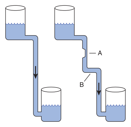
Two structural facts about metabolism follow. First, metabolite flow tends to be unidirectional. Harper's uses a hydrostatic analogy: flow through a pathway with a large overall negative ΔG is like water through a pipe whose far end is lower — it only runs one way. Glycolysis, for instance, has an overall ΔG of about −96 kJ/mol, far too large to simply run in reverse, which is why gluconeogenesis needs separate enzymes for the three most unfavourable steps (Unit 13).
Second, a few reactions — isomerisations, where the free-energy difference between substrate and product is close to zero — can act as bidirectional catalysts in vivo. But Harper's is explicit that these represent the exception rather than the rule.
- Define homeostasis in biochemical terms → Maintaining a constant internal environment despite external fluctuation, by changing the rates of biochemical reactions
- Why is metabolite flow unidirectional? → Pathways proceed with a large overall negative ΔG — like water down a sloping pipe
- Which reactions can run both ways in vivo? → Those with ΔG close to zero, such as isomerisations — the exception, not the rule
Passive control — why [S] sits near Km ★★
Before any active regulation, there is a passive mechanism built into the kinetics of Unit 6 — and it is an elegant piece of design worth understanding rather than memorising.
An enzyme working on the plateau is deaf to how much substrate is present. An enzyme working near its Km reports substrate concentration faithfully in its output.
That is why, for most enzymes, the average intracellular concentration of substrate tends to be close to the Km: it places the enzyme at the point of maximum sensitivity, so that changes in substrate concentration automatically generate corresponding changes in metabolite flux — with no regulatory machinery required at all.
Passive control is a consequence of kinetics: substrate levels rise, the enzyme speeds up, and nothing had to decide anything. Active control is the cell deliberately changing an enzyme's quantity or catalytic efficiency in response to a signal. The rest of this unit is about the active mechanisms — but the passive one is doing quiet work underneath all of them.
- Where on the curve is an enzyme most responsive to [S]? → Near Km, where the curve is steepest
- Why? → Far above Km the curve has flattened, so extra substrate barely changes the rate
- What is active control? → Changing the concentration, catalytic activity, or both, of an enzyme catalysing a committed, rate-limiting reaction
![The same increment in substrate concentration produces a large change in rate at [S] near K<sub>m</sub> (ΔV<sub>A</sub>) but almost none far above it (ΔV<sub>B</sub>) — which is why intracellular substrate levels sit close to K<sub>m</sub>](images/bc/bc07_km_sensitivity.png)
Compartmentation
In eukaryotes, anabolic and catabolic pathways that interconvert common products may take place in separate subcellular compartments. This ensures metabolic efficiency and simplifies regulation — you cannot have futile cycling between two pathways that never meet.
| Pathway | Compartment | Unit |
|---|---|---|
| Fatty acid biosynthesis | Cytosol | 16 |
| Fatty acid oxidation | Mitochondria | 17 |
| Protein degradation | Lysosomes (and the proteasome) | — |
Physical barriers are not the only way. Antagonistic pathways can coexist in the same space provided each proceeds via one or more unique intermediates. And there is a third, subtler form of separation: enzymes discriminate between the structurally similar coenzymes NAD⁺ and NADP⁺, even though their reduction potentials are similar.
Enzymes generating electrons destined for the electron transport chain reduce NAD⁺. Enzymes catalysing reductive steps in biosynthetic pathways generally use NADPH as the electron donor.
In one line: NADH is for making ATP; NADPH is for building things. That single distinction runs through Units 9, 14, 16 and 19, and it is a form of compartmentation achieved chemically rather than physically.
- Where do fatty acid synthesis and oxidation occur? → Cytosol and mitochondria respectively
- How can antagonistic pathways coexist without a barrier? → If each proceeds via one or more unique intermediates
- NAD⁺ versus NADPH? → NAD⁺ is reduced by enzymes feeding the electron transport chain; NADPH donates electrons in biosynthesis
The rate-limiting step ★★★
A pathway may involve twenty enzymes, but active control is exerted over only a select subset of them. The ideal target is the enzyme whose quantity or catalytic efficiency makes its reaction slow relative to all the others — the bottleneck.
The slowest reaction in a pathway. Decreasing the catalytic efficiency or quantity of the enzyme responsible immediately reduces metabolite flux through the entire pathway; increasing either enhances flux through the pathway as a whole. Such enzymes are the natural “governors” of metabolic flux.
| Enzyme | Pathway | Why it is the target | Unit |
|---|---|---|---|
| Acetyl-CoA carboxylase | Fatty acid biosynthesis | Catalyses synthesis of malonyl-CoA, the first committed reaction. Inhibit it and the subsequent reactions cease for lack of substrate | 16 |
| HMG-CoA reductase | Cholesterol biosynthesis | The rate-limiting reaction of cholesterogenesis — and therefore the target of the statin drugs | 19 |
Rate-limiting enzymes are efficient targets for regulatory intervention by drugs. That is not a passing remark — it is the reason a whole class of medicines exists. Statins work because HMG-CoA reductase is the governor of cholesterol synthesis, so inhibiting one enzyme shuts down an entire pathway. Whenever you meet a new pathway in Modules B–E, find its rate-limiting enzyme first: that is where the regulation is, and that is where the drugs are.
- Why regulate the rate-limiting enzyme? → Changing it immediately alters flux through the entire pathway
- Give two examples → Acetyl-CoA carboxylase (fatty acid synthesis) and HMG-CoA reductase (cholesterol synthesis)
- Why are they drug targets? → They are the natural governors of flux — inhibiting one enzyme controls a whole pathway
Regulating the QUANTITY of enzyme ★★
There are two ways to change how much enzyme is present: make more, or destroy it faster. Both are used.
Control of synthesis
Constitutive enzymes are those whose concentrations remain essentially constant over time.
By contrast, the concentrations of many enzymes depend on the presence of inducers — typically substrates or structurally related compounds — that initiate their synthesis.
Notice what an inducer usually is: the enzyme's own substrate. A cell that suddenly encounters a compound makes the enzyme needed to deal with it, and stops making it when the compound is gone. This is the same principle you will meet formally as the lac operon in Unit 26 — induction is gene regulation seen from the enzyme's side.
Control of degradation
Mammalian proteins are degraded both by ATP- and ubiquitin-dependent pathways and by ATP-independent pathways. The major route is the ubiquitin-proteasome pathway, whose discovery earned Ciechanover, Hershko and Rose a Nobel Prize.
| Component | Detail |
|---|---|
| Ubiquitin | A small, approximately 8.5 kDa protein, highly conserved among eukaryotes |
| Ubiquitination | Covalent attachment of one or more ubiquitin molecules, catalysed by a large family of E3 ligases, which attach ubiquitin to the side-chain amino group of lysyl residues |
| The 26S proteasome | A large complex of more than 30 subunits arranged as a hollow cylinder, with the active sites of its proteolytic subunits facing the interior — which prevents indiscriminate degradation of cellular proteins |
The system's selectivity comes from the variety of E3 ligases and their ability to discriminate between the physical and conformational states of target proteins. It therefore removes not only regulated proteins such as the cyclins, but also proteins damaged by loss of a prosthetic group, oxidation of cysteine or histidine residues, or deamidation of asparagine or glutamine.
Harper's notes that dysfunction of the ubiquitin-proteasome pathway contributes to the accumulation of misfolded proteins characteristic of several neurodegenerative diseases. Unit 3 described what happens when folding fails; this is what happens when the cell's clean-up system fails too.
- What is a constitutive enzyme? → One whose concentration remains essentially constant over time
- What is an inducer? → Typically a substrate or structurally related compound that initiates synthesis of the enzyme
- How are proteins targeted for degradation? → By ubiquitination — E3 ligases attach ubiquitin to lysyl side-chain amino groups
- Why do the proteasome's active sites face inward? → To prevent indiscriminate degradation of cellular proteins
Long-term versus short-term regulation ★★★
This is the organising idea of the whole unit, and the answer to the TMU study question “how is homeostasis achieved?”. Two timescales, two mechanisms, two purposes.
| Long-term regulation | Short-term regulation | |
|---|---|---|
| Mechanism | Induction of protein synthesis — changing the amount of enzyme | Changing intrinsic catalytic efficiency, by binding of dissociable ligands (allosteric regulation) or by covalent modification |
| Timescale | A complex multistep process typically requiring hours | Within seconds |
| Best suited to | Long-term adaptive requirements | Rapid and transient alterations in metabolite flux |
A cell that has just started sprinting cannot wait an hour for transcription and translation — it needs glycogen breakdown now, which is what a phosphorylation cascade delivers in seconds. But a cell adapting to a permanently high-protein diet does not need a fast response; it needs more enzyme, and building more is worth the wait.
Speed versus capacity. Whenever you meet a regulated enzyme in the rest of the course, ask which of the two it is under — and usually the answer is both.
- How long does induction of protein synthesis take? → Hours — long-term regulation
- How fast is allosteric or covalent regulation? → Within seconds — short-term regulation
- Which suits long-term adaptation, and which rapid change? → Changes in protein level; changes in catalytic efficiency
Allosteric regulation ★★★
The first TMU study question. Learn the definition, the origin of the word, and the two kinetic classes.
Regulation in which a small molecule (an effector) binds at an allosteric site, spatially distinct from the catalytic site of the target enzyme, producing a change in the enzyme's intrinsic catalytic efficiency.
Allosteric enzymes are therefore those whose catalysis at the active site may be modulated by the presence of effectors at an allosteric site.
So these effectors are not isosteric with the substrate but allosteric — “occupying another space”. The hypothesis has since been confirmed by X-ray crystallography and site-directed mutagenesis, which demonstrated spatially distinct active and allosteric sites in many enzymes.
Note how neatly this contrasts with Unit 6: a competitive inhibitor resembles the substrate because it binds the same site; an allosteric effector does not, because it does not.
K-series — the inhibitor raises Km without affecting Vmax. The conformational change may weaken the bonds between substrate and the substrate-binding residues.
V-series — the inhibitor lowers Vmax without affecting Km. The primary effect may be to alter the orientation or charge of the catalytic residues.
In both cases the change is induced by binding of the effector at its own site and propagates to the active site. Intermediate effects on both constants may also be seen.
Aspartate transcarbamoylase — the model allosteric enzyme
ATCase catalyses the first reaction unique to pyrimidine biosynthesis and is the standard worked example. It is feedback-inhibited by CTP, an end product of that pathway.
| Evidence / feature | Detail |
|---|---|
| The mercurial experiment | After treatment with mercurials, ATCase loses its sensitivity to CTP but retains full activity for the synthesis of carbamoyl aspartate. Since the catalytic and regulatory functions can be separated, CTP must bind at a different site from the substrate — the original evidence for allosterism |
| Subunit structure | Multiple catalytic and regulatory subunits. Your slide states that each catalytic subunit has four aspartate (substrate) sites and each regulatory subunit at least two CTP sites; the Harper's edition in your folder describes the E. coli enzyme as six catalytic and six regulatory subunits |
| Reciprocal control | Additional in Harper's: CTP inhibits, while the purine nucleotide ATP activates — and high ATP can overcome CTP inhibition, so pyrimidine synthesis can proceed when purine levels are high. This keeps purine and pyrimidine production balanced |
Two further points. Many hormones act through allosteric second messengers — a bridge to §10 and to Unit 22. And allosteric regulation is the mechanism by which most feedback inhibition operates, which is the next section.
- Define allosteric regulation → A small effector binds at a site spatially distinct from the catalytic site, changing intrinsic catalytic efficiency
- Where does the word come from? → Monod — effectors are not isosteric with substrate but allosteric, 'occupying another space'
- What are K-series and V-series enzymes? → K-series: Km raised, Vmax unaffected. V-series: Vmax lowered, Km unaffected
- What does the mercurial experiment on ATCase show? → Sensitivity to CTP is lost while catalytic activity is retained — so CTP binds a separate site
- What inhibits and what activates ATCase? → CTP inhibits; ATP activates and can overcome CTP inhibition
Feedback inhibition ★★★
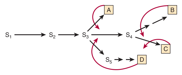
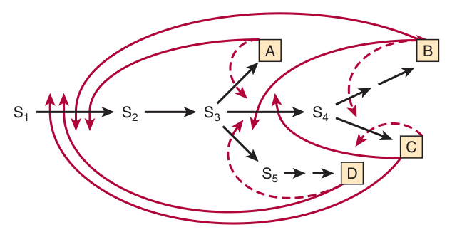
The process by which the end product of a multistep biosynthetic pathway binds to and inhibits an enzyme catalysing one of the early steps in that pathway.
In most cases it operates by allosteric regulation.
Because inhibiting the final enzyme would leave every intermediate accumulating uselessly behind it. Shutting off the first committed step stops the whole assembly line at the point where raw material would otherwise be committed to a product the cell already has enough of. It is the difference between switching off the factory gate and switching off the packing department.
Branched pathways — where it becomes clever
A single pathway with one end product is straightforward. Real biosynthetic pathways branch, so several end products share an early common segment — and shutting that segment down because one end product is plentiful would starve the cell of the other three.
| Solution | How it works |
|---|---|
| Multiple feedback loops | Superimposed on the simple loops are additional loops that regulate the enzymes common to the biosynthesis of several end products, providing fine control |
| Cooperative feedback inhibition | The effect of an excess of two or more end products may be strictly additive, or greater than their individual effects |
| Multiple isoforms | Several isoforms of one enzyme evolve, each sensitive to a different end product. A high level of any one product inhibits only its own isoform, thereby reducing but not eliminating flux through the shared segment. The classic case is the bacterial pathway making phenylalanine, tyrosine and tryptophan |
Feedback inhibition names a specific mechanism for the regulation of enzyme activity: the end product binds and inhibits an early enzyme.
So all feedback inhibition is feedback regulation, but feedback regulation may also be achieved by, for example, repressing the synthesis of the enzyme rather than inhibiting the enzyme itself. Getting this distinction into an answer shows you have read the chapter rather than the summary.
- Define feedback inhibition → The end product of a multistep biosynthetic pathway binds and inhibits an enzyme catalysing an early step
- By what mechanism does it usually work? → Allosteric regulation
- How do branched pathways avoid shutting down shared segments? → Multiple feedback loops, cooperative inhibition, or multiple isoforms each sensitive to a different end product
- Feedback regulation vs feedback inhibition? → Regulation is a phenomenological term; inhibition names a specific mechanism
Proenzymes and zymogens ★★★
The third TMU study question, and one of the most clinically useful definitions in Module A.
Certain proteins are synthesised as inactive precursor proteins called proproteins. Selective, or “partial”, proteolysis — one or more successive proteolytic “clips” — converts a proprotein to the form showing the characteristic activity of the mature protein.
The proprotein forms of enzymes are termed proenzymes or zymogens.
| Mature protein | Its proprotein |
|---|---|
| Insulin | Proinsulin |
| Pepsin | Pepsinogen |
| Trypsin | Trypsinogen |
| Chymotrypsin | Chymotrypsinogen |
| Collagen | Procollagen (Unit 3) |
| Blood clotting and complement factors | Their respective proproteins |
Why cells bother — three reasons, all examinable
| Reason | Explanation |
|---|---|
| 1 · Protection of the tissue of origin | Synthesis and secretion of proteases as catalytically inactive proenzymes protects the tissue of origin (e.g. the pancreas) from autodigestion — as occurs in pancreatitis |
| 2 · Rapid mobilisation on demand | Some processes — digestion — are intermittent but predictable; others — blood clot formation and dissolution, tissue repair — are brought “on line” only in response to pressing physiological or pathophysiological need. Induction of synthesis would be far too slow to respond to something like the loss of blood |
| 3 · Economy | Zymogen activation is a simple and economical, albeit one-way, mechanism for restraining the latent activity of a protein until the appropriate circumstances arise |
Contrast with phosphorylation, which is a reversible modification — which is precisely why the two mechanisms are used for different jobs. Irreversible activation suits a one-off emergency such as clotting; reversible modification suits a process that must be switched on and off repeatedly.
Chymotrypsin — how proteolysis builds an active site
This is the detail that makes the mechanism intelligible rather than magical. Selective proteolysis does not merely remove a blocking peptide — it causes conformational changes that properly configure the active site.
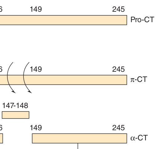
In α-chymotrypsin the catalytic triad is Asp 102 – His 57 – Ser 195. The catalytically essential His 57 and Asp 102 reside on the B peptide, while Ser 195 resides on the C peptide. The conformational changes accompanying selective proteolysis of prochymotrypsin align the three residues of the charge-relay network, thereby forming the catalytic site. Successive proteolysis gives pro-CT → π-CT → α-CT, whose three peptides remain associated by interchain disulfide bonds.
The active site is three-dimensional and its residues are far apart in the primary structure — Unit 5 said exactly that. Here they are not merely far apart in sequence but on different peptide chains, held within bond-forming distance by interchain disulfide bonds — Unit 3's covalent cross-links. And the activation step is a conformational change, Unit 3 again.
If you can explain chymotrypsin activation, you have demonstrated command of half of Module A.
- Define a zymogen → The inactive proprotein form of an enzyme, converted to the active form by selective (partial) proteolysis
- Name four zymogens → Pepsinogen, trypsinogen, chymotrypsinogen, proinsulin (also procollagen and clotting factors)
- Give three reasons cells use zymogens → Protection from autodigestion, rapid mobilisation on demand, economy
- Why is the activation irreversible? → Cells cannot rejoin the two portions of a hydrolysed peptide bond; reunification is entropically disfavoured
- Name the chymotrypsin catalytic triad → Asp 102 – His 57 – Ser 195, aligned by the conformational change of activation
Reversible covalent modification — phosphorylation ★★★
The second great mechanism of short-term control, and by far the commonest covalent modification regulating protein function. A typical mammalian cell contains thousands of phosphorylated proteins and several hundred protein kinases and protein phosphatases that interconvert them.
| Direction | Enzyme | Chemistry |
|---|---|---|
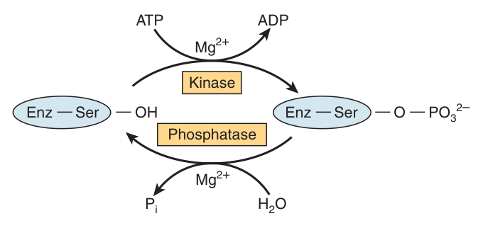
| Phosphorylation | Protein kinases | Transfer of the terminal (γ) phosphoryl group of ATP to the hydroxyl groups of seryl, threonyl or tyrosyl residues, forming O-phosphoseryl, O-phosphothreonyl or O-phosphotyrosyl residues. Thermodynamically spontaneous |
| Dephosphorylation | Protein phosphatases | Hydrolytic removal of the phosphoryl group — not recombination with ADP to re-form ATP. Equally spontaneous |
Thermodynamics forbids the obvious route: if the reaction introducing the modification is favourable, simply running it backwards would be rendered impractical by the correspondingly unfavourable free-energy change. Phosphorylation is favourable because it uses the high-energy γ-phosphoryl group of ATP — so the reverse would have to re-make ATP, which will not happen spontaneously.
The solution is to use a different reaction: phosphate is removed by hydrolysis, catalysed by protein phosphatases, which is itself favourable. Two spontaneous reactions in opposite directions, not one reaction run both ways. The same logic applies to acetylation, where acetyltransferases use the high-energy donor NAD⁺ and deacetylases catalyse a direct hydrolysis generating free acetate.
Why is phosphorylation used so heavily? Two reasons Harper's gives. First, the ease of interconversion: the functional properties of an enzyme can be altered only for as long as it serves a specific need, and then converted back, poised to respond to the next stimulus. Second, the chemical properties of the phosphoryl group itself — it is bulky and carries a substantial negative charge, so adding one substantially changes the local structure.
| Category | Modifications |
|---|---|
| Reversible | Phosphorylation, acetylation, methylation, ADP-ribosylation |
| Irreversible | Partial (selective) proteolysis — §9 |
| Essentially permanent (structural, not regulatory) | Prenylation, glycosylation, hydroxylation, fatty acid acylation — these introduce features that persist for the lifetime of the protein |
Histones and other DNA-binding proteins in chromatin are extensively modified by acetylation, methylation, ADP-ribosylation and phosphorylation. These alter how chromatin proteins interact with each other and with DNA, either rendering genes more accessible to the transcription machinery or, conversely, silencing gene expression.
This is the “histone code” — a classic example of epigenetics, the hereditary transmission of information by a means other than the nucleotide sequence. The pattern of gene expression in a daughter cell is determined in part by the set of histone modifications inherited from the parent cell.
The protein kinases and phosphatases participating in regulatory cascades that respond to hormonal or second-messenger signals constitute, in Harper's phrase, a “bio-organic computer” — a network able to process and integrate complex environmental information to produce an appropriate and comprehensive cellular response.
That is the point of this entire unit. Regulation is not a collection of switches; it is an information-processing system. Second messengers include cAMP, cGMP, Ca²⁺, nitric oxide and the polyphosphoinositols.
- Which residues are phosphorylated? → Seryl, threonyl and tyrosyl (some kinases also target histidyl, lysyl, arginyl, aspartyl)
- Which enzymes add and remove the phosphate? → Protein kinases and protein phosphatases
- What is the phosphate donor? → The terminal (γ) phosphoryl group of ATP
- How is the phosphate removed? → By HYDROLYSIS, not by re-forming ATP
- Name four reversible covalent modifications → Phosphorylation, acetylation, methylation, ADP-ribosylation
- Which covalent modification is irreversible? → Partial proteolysis
- Name four second messengers → cAMP, cGMP, Ca²⁺, nitric oxide (also polyphosphoinositols)
Revision layer
Three study questions on this deck — what is allosteric regulation? how is homeostasis achieved? what are proenzymes? — and four printed MCQs, two of which revisit Units 1 and 5. Module A ends here, so the last table below is a bridge into Module B.
How homeostasis is achieved — the master answer
| Level | Mechanism | Timescale |
|---|---|---|
| Passive | Substrate concentrations sit close to Km, where the curve is steepest, so changes in [S] automatically generate corresponding changes in flux | Instant |
| Compartmentation | Antagonistic pathways separated into different organelles, or by unique intermediates, or by using NAD⁺ versus NADPH | Structural |
| Enzyme quantity | Control of synthesis (constitutive vs inducible) and of degradation (ubiquitin-proteasome pathway) | Hours — long-term |
| Allosteric regulation | Effector binds a site distinct from the active site, altering Km (K-series) or Vmax (V-series); mostly feedback inhibition | Seconds — short-term |
| Reversible covalent modification | Phosphorylation by protein kinases, reversed hydrolytically by protein phosphatases | Seconds — short-term |
| Irreversible covalent modification | Selective proteolysis of zymogens | Seconds, but one-way |
And in all of it, the target is the same: the enzyme catalysing the committed, rate-limiting step of the pathway.
Definitions from this unit — Section I material
| Term | Definition |
|---|---|
| Allosteric regulation | Regulation in which a small molecule binds at an allosteric site, spatially distinct from the catalytic site, to change the enzyme's intrinsic catalytic efficiency; allosteric enzymes are those whose catalysis at the active site is modulated by effectors at an allosteric site |
| Feedback inhibition | The process by which the end product of a multistep biosynthetic pathway binds to and inhibits an enzyme catalysing one of the early steps of that pathway; in most cases it operates allosterically |
| Proenzyme (zymogen) | The inactive precursor form of an enzyme, converted to the active enzyme by selective (partial) proteolysis, which produces conformational changes that properly configure the active site; the modification is physiologically irreversible |
| Constitutive enzyme | An enzyme whose concentration remains essentially constant over time, as distinct from an inducible enzyme whose synthesis is initiated by an inducer — typically its substrate or a structurally related compound |
| Rate-limiting step | The slowest reaction of a pathway, whose enzyme acts as the natural governor of metabolic flux; altering its quantity or catalytic efficiency immediately alters flux through the whole pathway |
| K-series and V-series allosteric enzymes | In a K-series enzyme the allosteric inhibitor raises Km without affecting Vmax; in a V-series enzyme it lowers Vmax without affecting Km |
| Ubiquitination | The covalent attachment of one or more molecules of ubiquitin — a small, highly conserved ~8.5 kDa protein — to the side-chain amino groups of lysyl residues by E3 ligases, targeting the protein for degradation in the 26S proteasome |
| Second messenger | An intracellular signalling molecule generated in response to an extracellular signal, which regulates enzymes allosterically; examples include cAMP, cGMP, Ca²⁺, nitric oxide and the polyphosphoinositols |
Reversible versus irreversible modification
| Reversible | Irreversible | |
|---|---|---|
| Examples | Phosphorylation, acetylation, methylation, ADP-ribosylation | Partial (selective) proteolysis |
| Why | The modification can be undone by a separate favourable reaction — e.g. hydrolysis by a phosphatase | Cells cannot rejoin a hydrolysed peptide bond; reunification is entropically disfavoured |
| Suited to | Processes switched on and off repeatedly | One-way emergencies — digestion, clotting, tissue repair |
Rate-limiting enzymes to carry into Modules B–E
| Pathway | Rate-limiting enzyme | Unit |
|---|---|---|
| Fatty acid biosynthesis | Acetyl-CoA carboxylase (makes malonyl-CoA, the first committed step) | 16 |
| Cholesterol biosynthesis | HMG-CoA reductase — target of the statins | 19 |
| Pyrimidine biosynthesis | Aspartate transcarbamoylase — inhibited by CTP, activated by ATP | 21 |
- Define allosteric regulation and explain Monod's isosteric/allosteric reasoning
- Distinguish K-series from V-series allosteric enzymes
- Explain why substrate concentrations sit near Km, and what that achieves
- Give the full answer to “how is homeostasis achieved?” — all six levels
- Define feedback inhibition and distinguish it from feedback regulation
- Define a zymogen, name four, and give three reasons cells use them
- Explain how selective proteolysis creates the chymotrypsin catalytic triad
- Explain why phosphorylation is reversible although both directions are spontaneous
- Name three rate-limiting enzymes and the pathways they govern