Cranial Nerves I – XII
Skull Base — Cranial Nerve Foramina
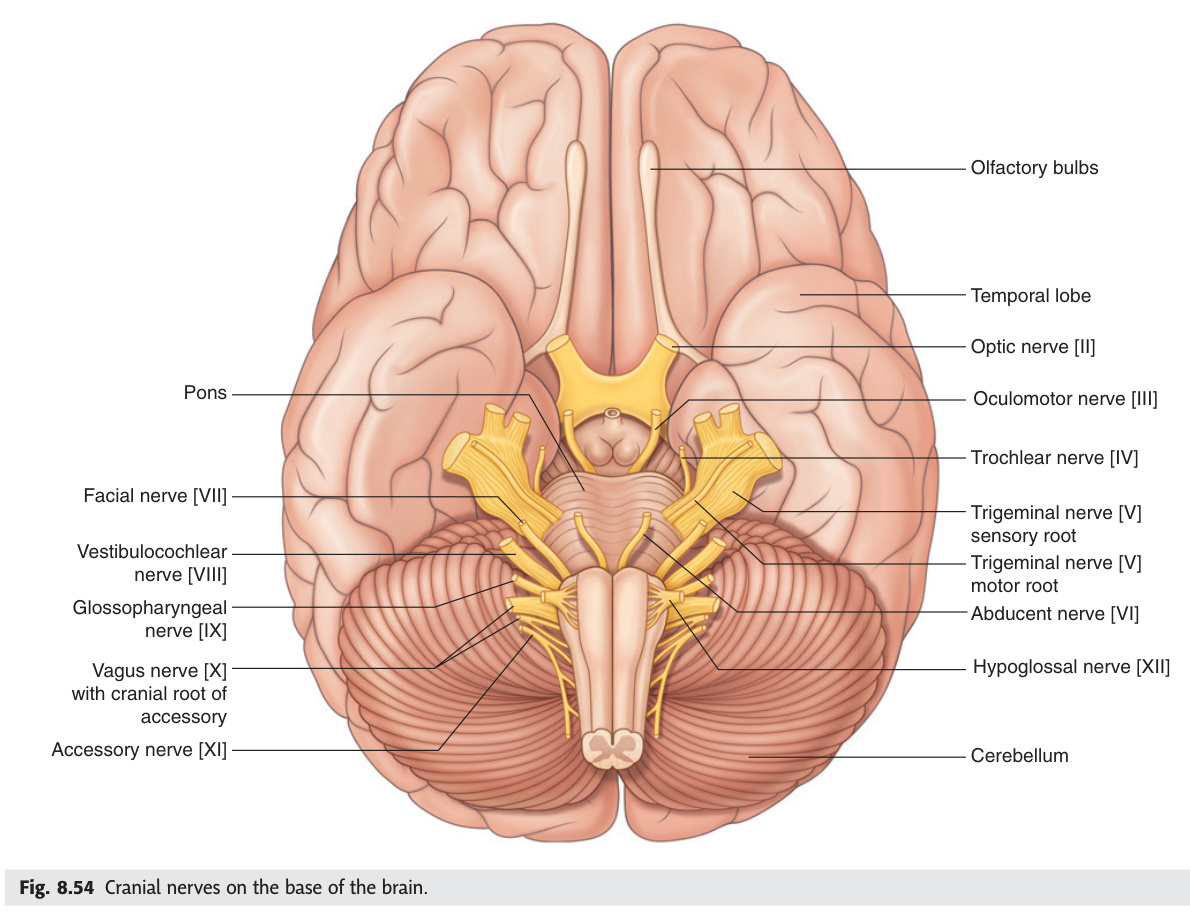
Mnemonic — foramen ovale vs rotundum: OVALE = V3 (Oval = bottom of triangle of V), ROTUNDUM = V2 (Round = middle)
Master Table & Mnemonics
Twelve cranial nerves emerge directly from the brain and brainstem, each with a fixed number, name, and exit foramen that must be memorised before clinical neurology can make sense. The mnemonic "Oh Oh Oh To Touch And Feel Very Good Velvet And Hair" sequences all twelve names, while "Some Say Marry Money But My Brother Says Big Business Matters More" codes each nerve's fibre type — purely sensory (I, II, VIII), purely motor (III, IV, VI, XI, XII), or mixed (V, VII, IX, X). Knowing which foramina share multiple nerves — the superior orbital fissure (III, IV, V1, VI) and the jugular foramen (IX, X, XI) — immediately reveals which nerve clusters fall together in skull-base pathology.
CN I Olfactory · II Optic · III Oculomotor · IV Trochlear · V Trigeminal · VI Abducens · VII Facial · VIII Vestibulocochlear · IX Glossopharyngeal · X Vagus · XI Accessory · XII Hypoglossal
I=S · II=S · III=M · IV=M · V=B · VI=M · VII=B · VIII=S · IX=B · X=B · XI=M · XII=M
| CN | Name | Foramen | Fibre types | Key test |
|---|---|---|---|---|
| I | Olfactory | Cribriform plate (ethmoid) | SVA (smell) | Smell each nostril separately |
| II | Optic | Optic canal | SSA (vision) | Snellen chart + visual fields |
| III | Oculomotor | Superior orbital fissure | GSE (eye muscles) + GVE (para) | Eye movements + pupil light reflex |
| IV | Trochlear | Superior orbital fissure | GSE | Failure of downward gaze when eye adducted |
| V | Trigeminal | V1: SOF; V2: foramen rotundum; V3: foramen ovale | GSA (3 divisions) + SVE (mastication) | Touch/pain face + corneal reflex + jaw clench |
| VI | Abducens | Superior orbital fissure | GSE | Lateral gaze (abduction of eye) |
| VII | Facial | Internal acoustic meatus → stylomastoid foramen | SVE (facial muscles) + GVE (para: lacrimal/salivary) + SVA (taste ant. 2/3 tongue) + GSA (ear) | Face movement (raise eyebrow, close eye, smile) |
| VIII | Vestibulocochlear | Internal acoustic meatus | SSA (hearing + balance) | Rinne + Weber; Hallpike for vestibular |
| IX | Glossopharyngeal | Jugular foramen | SVE (stylopharyngeus) + GVE (para: parotid) + GVA (carotid body/sinus) + SVA (taste post. 1/3) + GSA (middle ear) | Gag reflex (sensory limb) |
| X | Vagus | Jugular foramen | SVE (pharynx/larynx) + GVE (para: thorax + abdomen) + GVA + SVA (epiglottis taste) + GSA (dura + ear) | Gag reflex (motor); say "Ah" (palate rises) |
| XI | Accessory | Jugular foramen (cranial root via vagus) + foramen magnum (spinal root) | SVE (sternocleidomastoid + trapezius) | Head turning against resistance; shoulder shrug |
| XII | Hypoglossal | Hypoglossal canal | GSE (tongue muscles) | Protrude tongue (deviates to side of lesion) |
- Which foramen transmits CN III, IV, V1, and VI? The superior orbital fissure.
- Which three CNs exit via the jugular foramen? CN IX, X, and XI.
- Name the five purely motor cranial nerves. CN III, IV, VI, XI, and XII.
- Which foramen does CN V3 exit through? Foramen ovale (NOT the superior orbital fissure — that is V1 and V2 is foramen rotundum).
- Which CN exits via both the foramen magnum and the jugular foramen? CN XI — spinal root ascends through foramen magnum; both roots exit via jugular foramen.
Olfactory Nerve
The olfactory nerve is unique among all twelve cranial nerves because its fibres bypass the thalamus entirely — smell reaches the uncus and entorhinal cortex directly, explaining why a familiar scent can trigger an instant, vivid memory without conscious filtering. The fila olfactoria (20–30 delicate filaments) cross the cribriform plate of the ethmoid, making them the most vulnerable nerve fibres to shearing in frontal head trauma. Foster Kennedy syndrome — ipsilateral optic atrophy from direct compression plus contralateral papilloedema from raised ICP — signals an olfactory groove meningioma and is a classic exam presentation linking CN I and CN II pathology in one patient.
Special sensory: smell. Bipolar neurons in olfactory epithelium (roof of nasal cavity) → fila olfactoria (20–30 filaments) → cribriform plate of ethmoid → olfactory bulb (on orbital surface of frontal lobe) → olfactory tract → primary olfactory cortex (uncus + entorhinal cortex — pyriform cortex — not relayed through thalamus: the only sensory modality that bypasses thalamus).
Anosmia: Head injury (cribriform plate shearing — the fila olfactoria are delicate), viral infection, zinc deficiency, ageing, early Alzheimer's / Parkinson's. Always test each nostril separately. Cacosmia/olfactory hallucinations: uncal herniation (uncinate fits) → temporal lobe seizures often begin with smell aura. Olfactory groove meningioma: unilateral anosmia → bilateral anosmia → Foster Kennedy syndrome (ipsilateral optic atrophy from direct compression + contralateral papilloedema from raised ICP).
- Why does smell bypass the thalamus? Olfactory tract projects directly to pyriform cortex (uncus/entorhinal cortex) — the only sensory modality without a thalamic relay.
- What causes anosmia after frontal head injury? Shearing of the fila olfactoria at the cribriform plate of the ethmoid.
- Describe Foster Kennedy syndrome. Ipsilateral optic atrophy (direct nerve compression) + contralateral papilloedema (raised ICP) — classic of olfactory groove meningioma.
- What is an uncinate fit? Temporal lobe seizure originating in the uncus/medial temporal lobe, often beginning with an olfactory hallucination (cacosmia).
- Name three non-traumatic causes of anosmia. Viral URTI, ageing, and early Alzheimer's/Parkinson's disease.
Optic Nerve
The optic nerve carries visual information from retinal ganglion cells, but its clinical importance at exam level lies at the optic chiasm: temporal fibres stay ipsilateral while nasal fibres cross, creating the hallmark pattern where lesions at different pathway points produce predictably different field defects. Optic neuritis — demyelination of the optic nerve, most commonly in multiple sclerosis — presents with the classic triad of painful vision loss (pain on eye movement is the key distinguishing feature), central scotoma, and a relative afferent pupillary defect (RAPD). Because approximately 50% of isolated optic neuritis cases will go on to develop MS, every first episode demands urgent MRI brain and orbits.
Retinal ganglion cell axons → optic nerve → optic chiasm (nasal fibres decussate, temporal fibres stay ipsilateral) → optic tracts → lateral geniculate nucleus (thalamus) → optic radiations → primary visual cortex (calcarine fissure, occipital lobe). Visual fields covered in detail in Unit 13 (Eye).
Demyelination of optic nerve (most common cause: MS). Presentation: painful loss of vision in one eye (pain on eye movement), reduced colour vision, central scotoma, RAPD. Marcus Gunn pupil (RAPD) = optic nerve or severe retinal lesion. Key distinguishing feature of optic neuritis from other causes: pain with eye movement. 50% will go on to develop MS. Urgent MRI brain + orbits. Treat with IV methylprednisolone.
- Which fibres decussate at the optic chiasm? Nasal fibres decussate (cross); temporal fibres remain ipsilateral.
- What is a RAPD and what does it indicate? Relative afferent pupillary defect (Marcus Gunn pupil) — indicates an optic nerve lesion or severe retinal disease on the affected side.
- What distinguishes optic neuritis from other causes of acute visual loss? Pain on eye movement is the hallmark feature of optic neuritis.
- What percentage of isolated optic neuritis cases progress to MS? Approximately 50%.
- Which thalamic nucleus relays visual information? The lateral geniculate nucleus (LGN) of the thalamus.
Oculomotor Nerve
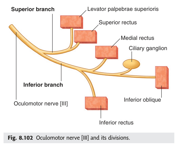
The oculomotor nerve controls four of the six extraocular muscles plus the levator palpebrae and the pupillary sphincter, making a complete CN III palsy instantly recognisable: eye "down and out" (unopposed CN IV and VI), complete ptosis, and a dilated fixed pupil. The single most critical distinction is surgical vs medical CN III palsy — in surgical (compressive) palsy the parasympathetic fibres on the outside of the nerve are damaged first, producing a dilated unreactive pupil, while medical (ischaemic/diabetic) palsy damages the core motor fibres while sparing the superficial parasympathetics. A dilated pupil in CN III palsy is a neurological emergency until a posterior communicating artery aneurysm is excluded by CT angiography.
Nucleus in midbrain tegmentum (at superior colliculus level). Nerve exits between cerebral peduncles → through cavernous sinus (superior + lateral wall) → superior orbital fissure → orbit. Supplies: superior rectus, inferior rectus, medial rectus, inferior oblique, levator palpebrae superioris. Parasympathetic (Edinger-Westphal nucleus) → ciliary ganglion → sphincter pupillae + ciliary muscle.
Surgical CN III palsy (compression): pupil INVOLVED (dilated, unreactive). Causes: posterior communicating artery aneurysm (PCoA), herniation (uncal). EMERGENCY: immediate CT angiography to exclude PCoA aneurysm.
Medical CN III palsy (ischaemic/diabetic): pupil SPARING (intact light reflex). Cause: microvascular disease (DM, HTN) damages core fibres (motor) but spares peripheral parasympathetic fibres. Mnemonic: "Surgical = Scary = Surgical emergency = pupil involved".
- Why is the eye "down and out" in complete CN III palsy? Unopposed action of superior oblique (CN IV) and lateral rectus (CN VI).
- What distinguishes surgical from medical CN III palsy? Surgical (compressive) = pupil involved (dilated, unreactive); medical (ischaemic/diabetic) = pupil sparing (intact light reflex).
- Which artery most commonly causes surgical CN III palsy by compression? Posterior communicating artery (PCoA) aneurysm — a neurosurgical emergency.
- Through which structure does CN III enter the orbit? Superior orbital fissure.
- Which nucleus provides parasympathetic fibres via CN III? Edinger-Westphal nucleus → ciliary ganglion → sphincter pupillae and ciliary muscle.
Trochlear Nerve
The trochlear nerve holds three anatomical records: only cranial nerve to exit the brainstem posteriorly (dorsal midbrain surface), the only one to completely decussate within the brainstem, and the one with the longest intracranial course — all three features making it uniquely vulnerable to head trauma. It supplies only the superior oblique, whose primary function is intorsion and depression of the adducted eye; the practical clinical test is asking the patient to look "down and in" (as in reading or descending stairs), which is the exact movement that fails and causes vertical diplopia. The Bielschowsky head-tilt test localises the lesion: tilting toward the affected side worsens diplopia because the eye is forced into further extorsion.
Only CN to exit POSTERIORLY from brainstem (dorsal surface of midbrain, below inferior colliculus). Only CN that decussates COMPLETELY in the brainstem (contralateral nucleus controls ipsilateral superior oblique). Longest intracranial course → most vulnerable to trauma. Passes through cavernous sinus → superior orbital fissure → superior oblique muscle.
Superior oblique action: intorsion (primary), depression in adduction (tested: ask patient to look down-in e.g. "look at your nose"). CN IV palsy: vertical diplopia worse when looking down + head tilt away from lesion (to compensate for extorsion). Bielschowsky test: head tilt ipsilateral worsens diplopia.
Most common cause of vertical diplopia. Commonest congenital cause (uncompensated for years). Acquired: head trauma (blow to forehead shearing dorsal midbrain), DM, microvascular. Symptom: difficulty reading/descending stairs (both require downgaze in adduction). Often compensated by contralateral head tilt (Parks-Bielschowsky 3-step test to differentiate).
- What makes CN IV unique in its brainstem exit? Only CN to exit posteriorly (dorsal midbrain surface), and the only one to fully decussate within the brainstem.
- What is the primary action of superior oblique? Intorsion (primary), plus depression of the adducted eye (tested by asking patient to look down-and-in).
- Describe the diplopia in CN IV palsy. Vertical diplopia, worse on downward gaze in adduction (reading, descending stairs).
- What does the Bielschowsky head-tilt test demonstrate? Tilting the head toward the affected side worsens vertical diplopia, confirming superior oblique palsy on that side.
- Why is CN IV uniquely vulnerable to trauma? Longest intracranial course; exits at the dorsal midbrain — sheared by sudden acceleration/deceleration (coup-contrecoup).
Trigeminal Nerve
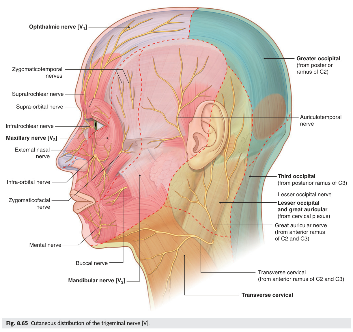
The trigeminal nerve is the largest cranial nerve and provides all sensation to the face, scalp (anterior vertex), cornea, and anterior two-thirds of the tongue (general sensation via V3), divided into three branches that exit through three distinct foramina — V1 through the superior orbital fissure, V2 through foramen rotundum, and V3 through foramen ovale. The Gasserian ganglion sitting in Meckel's cave at the petrous apex houses the first-order sensory cell bodies for all three divisions, making it the target for percutaneous rhizotomy procedures. Trigeminal neuralgia — paroxysmal electric-shock pain in V2/V3 territory triggered by eating or light face touch — is managed first with carbamazepine and, when medication fails, by microvascular decompression (Jannetta procedure) separating the offending vessel from the nerve root.
Largest CN. Sensory nucleus extends entire brainstem. Motor nucleus (pons) → V3 → muscles of mastication + tensor tympani + tensor veli palatini. Sensory: V1 (ophthalmic), V2 (maxillary), V3 (mandibular). Trigeminal ganglion (Gasserian, semilunar) in Meckel's cave (lateral to cavernous sinus, petrous apex). NOT sensation: scalp behind vertex (C2,3), angle of jaw (C2,3), parotid skin (great auricular, C2,3).
| Division | Foramen | Distribution |
|---|---|---|
| V1 (ophthalmic) | Superior orbital fissure | Forehead + scalp (vertex) + cornea + bridge of nose + upper eyelid + dura. Branches: frontal (supraorbital + supratrochlear) + lacrimal + nasociliary |
| V2 (maxillary) | Foramen rotundum → pterygopalatine fossa | Cheek + upper lip + upper teeth + lower eyelid + lateral nose. Branches: infraorbital + posterior superior alveolar + zygomatic |
| V3 (mandibular) | Foramen ovale | Lower lip + lower teeth + chin + anterior 2/3 of tongue (general sensation — taste from chorda tympani/CN VII) + ear. Motor: masseter + temporalis + pterygoids + tensor tympani + tensor veli palatini. Branches: inferior alveolar + lingual + auriculotemporal + buccal |
Severe paroxysmal electric-shock/stabbing unilateral facial pain in V2 + V3 distribution (rarely V1). Triggers: talking, chewing, touching face. Duration: seconds. Pathophysiology: vascular compression of trigeminal root entry zone by superior cerebellar artery (usually). >50 years, more common in women. Secondary: MS (young, bilateral), tumour. Treatment: carbamazepine (1st line) → oxcarbazepine → MVD (microvascular decompression — separating vessel from nerve root, Jannetta procedure).
- Through which foramina do V1, V2, and V3 exit? V1: superior orbital fissure; V2: foramen rotundum; V3: foramen ovale.
- Where does the Gasserian (trigeminal) ganglion lie? Meckel's cave — a dural recess at the petrous apex of the temporal bone.
- Which muscles does V3 supply motor fibres to? Muscles of mastication (masseter, temporalis, pterygoids) + tensor tympani + tensor veli palatini.
- What is first-line treatment for trigeminal neuralgia? Carbamazepine; surgical option when drugs fail = microvascular decompression (Jannetta procedure).
- Which facial areas are NOT supplied by CN V? Angle of jaw (great auricular nerve, C2/C3) and scalp posterior to vertex (C2/C3).
Abducens Nerve
The abducens nerve has the longest subarachnoid course of any cranial nerve, running from the dorsal pons all the way over the petrous apex through Dorello's canal to the cavernous sinus — this distance is why raised intracranial pressure can stretch it against the petrous apex, producing a CN VI palsy that looks like a localising sign but is actually a "false localising sign." Its nucleus shares the floor of the fourth ventricle with looping facial nerve fibres forming the facial colliculus, so a single pontine lesion can simultaneously knock out horizontal gaze and ipsilateral facial movement. Gradenigo syndrome (petrous apicitis from mastoiditis) produces the memorable triad of CN VI palsy, retro-orbital pain (V1), and purulent ear discharge.
Nucleus in dorsal pons (floor of 4th ventricle) — facial nerve fibres loop around it (facial colliculus). Long subarachnoid course → vulnerable to raised ICP (false localising sign). Passes over petrous apex (Dorello's canal) → cavernous sinus → superior orbital fissure → lateral rectus (abducts eye). CN VI palsy: convergent squint (eye turned medially by unopposed medial rectus), failure of abduction, horizontal diplopia worse on ipsilateral gaze.
CN VI palsy as false localising sign: raised ICP stretches CN VI over petrous apex (Dorello's canal) → unilateral or bilateral CN VI palsy without a lesion at the nerve itself. Always exclude raised ICP. Gradenigo syndrome: petrous apicitis (mastoiditis complication) → triad of ipsilateral CN VI palsy + retro-orbital pain (CN V) + purulent otorrhoea.
- What is Dorello's canal and why is it clinically important? The fibro-osseous canal over the petrous apex where CN VI runs — raised ICP stretches the nerve here, producing a "false localising" CN VI palsy.
- Why does a pontine lesion cause both ipsilateral CN VI and facial palsy? Facial nerve fibres loop around the CN VI nucleus (the facial colliculus) in the floor of the 4th ventricle.
- Describe Gradenigo syndrome. Petrous apicitis triad: ipsilateral CN VI palsy + retro-orbital pain (V1) + purulent otorrhoea.
- What does CN VI palsy look like clinically? Convergent squint, failure of abduction, horizontal diplopia worse on ipsilateral gaze.
- Which structure within the cavernous sinus is CN VI most vulnerable — and why? CN VI runs within the sinus itself (not just the lateral wall), giving it no bony protection.
Facial Nerve
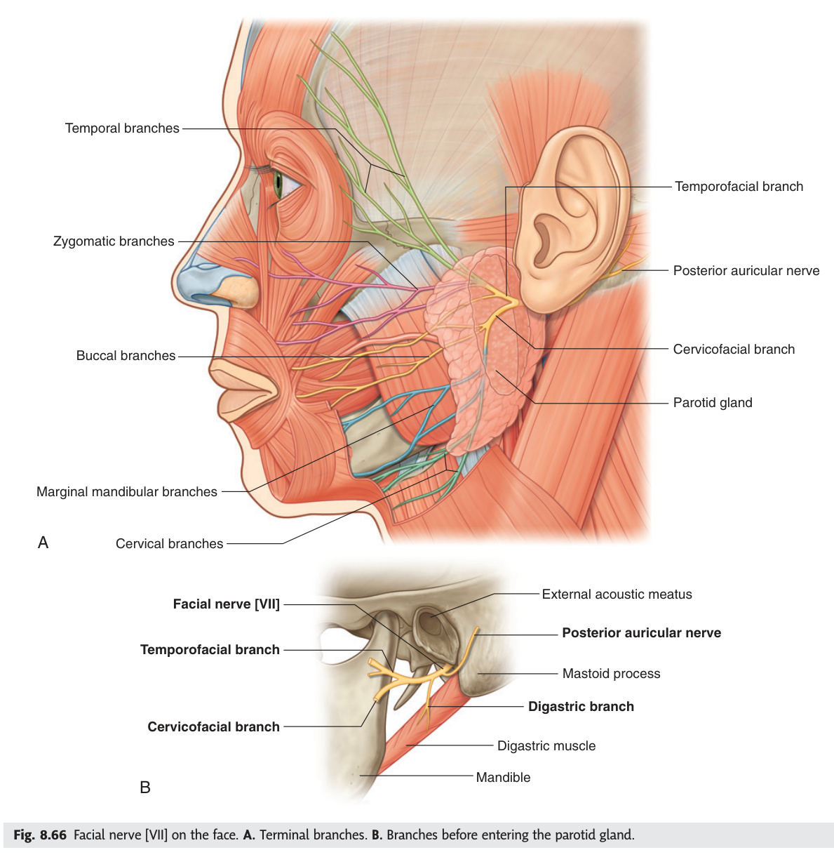
The facial nerve's long course through the petrous bone — from the internal acoustic meatus, through the facial canal, to the stylomastoid foramen — means that a lesion at different points along this route produces a different deficit: damage proximal to the geniculate ganglion loses lacrimation, damage proximal to the stapedius nerve causes hyperacusis, and damage proximal to the chorda tympani removes taste from the anterior two-thirds of the tongue and reduces salivation. The most critical clinical question is forehead sparing: an upper motor neurone lesion (cortical stroke) spares the forehead because frontalis receives bilateral cortical input, while a lower motor neurone lesion (Bell's palsy) paralyses the entire ipsilateral face including the forehead, preventing eye closure. Bell's palsy requires oral prednisolone within 72 hours and meticulous eye care to prevent corneal ulceration from exposure.
Motor nucleus: lower pons (caudal). Nerve enters IAM → facial canal in petrous bone → stylomastoid foramen → parotid gland (NOT parotid secretion — just traverses) → 5 terminal branches: Temporal, Zygomatic, Buccal, Marginal mandibular, Cervical ("Ten Zebras Bit My Cat").
LMN facial palsy (nerve nucleus or peripheral nerve): Forehead INVOLVED. Entire ipsilateral face paralysed (can't raise eyebrow, can't close eye — Bell's phenomenon: eye rolls up on attempted closure). Bell's palsy: idiopathic LMN, probably HSV-1 reactivation. Features: acute unilateral facial weakness, pain behind ear (chorda tympani), taste loss (ant. 2/3 tongue via chorda tympani), hyperacusis (stapedius). Treat: oral prednisolone within 72h; aciclovir if HSV confirmed; eye care (lubricants + tape at night).
| CN VII branch in petrous | Gives off |
|---|---|
| At geniculate ganglion | Greater petrosal nerve (to pterygopalatine ganglion → lacrimal + nasal secretion) + communicating branch to CN IX |
| In facial canal | Nerve to stapedius (stapedius reflex, hyperacusis if lost) |
| In facial canal (just above stylomastoid) | Chorda tympani (joins lingual nerve → taste anterior 2/3 tongue + submandibular/sublingual secretion) |
- Why is the forehead spared in UMN but not LMN facial palsy? Frontalis receives bilateral cortical input — one UMN lesion is compensated by the other hemisphere; LMN cuts all input to the entire ipsilateral face.
- Name the three intrapetrous branches of CN VII in order. Greater petrosal nerve (at geniculate ganglion) → nerve to stapedius → chorda tympani (just above stylomastoid foramen).
- Name the five terminal branches using the mnemonic. Temporal, Zygomatic, Buccal, Marginal mandibular, Cervical — "Ten Zebras Bit My Cat."
- What does chorda tympani carry? Taste from anterior 2/3 tongue + parasympathetic to submandibular and sublingual glands (via lingual nerve).
- What is Bell's palsy and how is it treated? Idiopathic LMN facial palsy (likely HSV-1 reactivation); treat with oral prednisolone within 72h + eye lubricants to prevent corneal ulceration.
Vestibulocochlear Nerve
The vestibulocochlear nerve carries two distinct modalities in one trunk through the internal acoustic meatus: the cochlear division relays sound from the spiral ganglion to bilateral auditory cortices (explaining why unilateral lesions rarely cause total deafness), while the vestibular division connects via the medial longitudinal fasciculus to the extraocular muscle nuclei, which is why vestibular disease disrupts both balance and gaze simultaneously. An acoustic neuroma (vestibular schwannoma) growing in the internal acoustic meatus classically presents with unilateral sensorineural hearing loss and tinnitus as its earliest symptoms — precisely because the cochlear fibres share the confined canal with the tumour. Detailed clinical content including Rinne/Weber tests, BPPV, and Ménière's disease is covered in Unit 14 (Ear).
Purely sensory. Enters skull via IAM alongside CN VII. Cochlear division: spiral ganglion (organ of Corti) → cochlear nuclei (pons) → bilateral (both inferior colliculi → both medial geniculate → both auditory cortices) → hearing. Vestibular division: Scarpa's ganglion → vestibular nuclei (pons + medulla) → cerebellum + spinal cord + CN III/IV/VI (via MLF) → balance + eye stabilisation. See Unit 14 (Ear) for detailed clinical content (acoustic neuroma, BPPV, Ménière's, Rinne/Weber).
- Why doesn't a unilateral CN VIII lesion usually cause complete deafness? The auditory pathway relays to both inferior colliculi and both auditory cortices above the cochlear nuclei (bilateral representation).
- Via which tract do vestibular nuclei connect to extraocular muscles? Medial longitudinal fasciculus (MLF) → CN III, IV, VI nuclei.
- Where do CN VII and CN VIII both enter the skull? Internal acoustic meatus (IAM).
- What are the earliest symptoms of acoustic neuroma (vestibular schwannoma)? Unilateral sensorineural hearing loss + tinnitus on the affected side.
- Name the two ganglia of CN VIII. Cochlear division: spiral ganglion; vestibular division: Scarpa's ganglion.
Glossopharyngeal Nerve
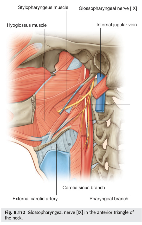
The glossopharyngeal nerve carries five different fibre types simultaneously — monitoring oxygen and CO₂ at the carotid body, detecting blood pressure at the carotid sinus via Hering's nerve, mediating the afferent limb of the gag reflex, relaying taste from the posterior tongue, and supplying the sole motor target stylopharyngeus. Its most clinically dangerous reflex arc is through the carotid sinus: hypersensitive baroreceptors send impulses via CN IX to the nucleus tractus solitarius, which activates the vagus to brake the heart — explaining why neck pressure (tight collar, head turning) can trigger a syncopal episode. Glossopharyngeal neuralgia mirrors trigeminal neuralgia in its electric-shock character but is triggered by swallowing or yawning rather than touching the face, and can produce dangerous vagally-mediated bradycardia at the same time.
Exits medulla as multiple rootlets (lateral sulcus) alongside CN X + XI → jugular foramen. Key functions: Motor — stylopharyngeus (elevates pharynx during swallowing — only muscle). Parasympathetic — inferior salivatory nucleus → tympanic nerve (Jacobson's nerve) → middle ear plexus → lesser petrosal nerve → otic ganglion → parotid. Sensory — posterior 1/3 tongue (taste + general sensation), tonsil, pharynx (gag reflex afferent), middle ear, carotid body (chemoreception: O₂, CO₂) + carotid sinus (baroreception: BP — via Hering's nerve).
Glossopharyngeal neuralgia: electric shock pain in posterior tongue/throat/ear, triggered by swallowing, yawning, coughing. Rare; may cause bradycardia + syncope (vagal). Treatment: carbamazepine (as trigeminal neuralgia). Carotid sinus syncope: hypersensitive baroreceptors in carotid sinus (CN IX → nucleus tractus solitarius → vagus → heart) → bradycardia + hypotension on neck pressure (turning head, tight collar) → syncope. Demonstrate by carotid sinus massage (with resuscitation available); treat with dual-chamber pacemaker.
- Which muscle is the only motor target of CN IX? Stylopharyngeus — elevates the pharynx during swallowing.
- What is the parasympathetic pathway of CN IX to the parotid? Inferior salivatory nucleus → tympanic nerve (Jacobson's) → middle ear plexus → lesser petrosal nerve → otic ganglion → parotid gland.
- CN IX is the afferent limb of which two important reflexes? Gag reflex (afferent) + carotid sinus reflex (baroreception via Hering's nerve).
- What triggers carotid sinus syncope and how is it treated? Pressure on the carotid sinus → vagally-mediated bradycardia + hypotension; treated with dual-chamber pacemaker in recurrent cases.
- How does glossopharyngeal neuralgia differ from trigeminal neuralgia in its trigger? Triggered by swallowing, yawning, or coughing — not by touching the face; may also trigger vagal bradycardia.
Vagus Nerve
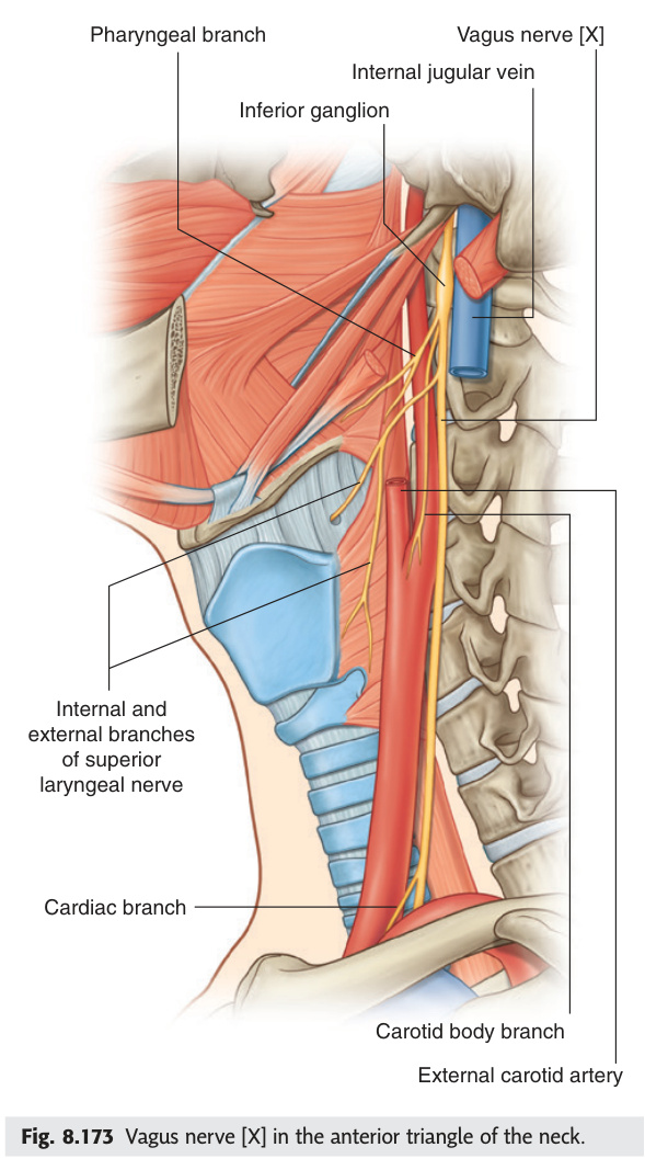
The vagus nerve is the body's longest cranial nerve, running from the medulla to the left colic flexure — its parasympathetic preganglionic fibres from the dorsal motor nucleus innervate the SA and AV nodes, the lungs, and essentially the entire abdominal viscera down to the midgut/hindgut boundary. The recurrent laryngeal nerve is its most surgically significant branch: the left RLN loops under the aortic arch (making it vulnerable to lung cancer, aortic aneurysm, and oesophageal pathology), while the right RLN loops under the right subclavian — both must be identified and preserved in thyroid surgery. The external branch of the superior laryngeal nerve is the "singer's nerve," supplying cricothyroid for pitch control, and its silent damage during thyroid surgery leaves the patient unable to produce high-pitched phonation.
Widest distribution of all CNs. Exits medulla → jugular foramen (superior [jugular] ganglion + inferior [nodose] ganglion) → descends in carotid sheath (between ICA/CCA + IJV) → thorax + abdomen. Supplies: pharynx + larynx + trachea + oesophagus + thoracic + abdominal viscera (to left colic flexure). Parasympathetic preganglionic: dorsal motor nucleus → heart (SA + AV nodes) + lungs + GI.
| Branch | Level | Function |
|---|---|---|
| Pharyngeal branches | Neck | With CN IX + sympathetics → pharyngeal plexus → pharyngeal constrictors + levator veli palatini (palate elevation) |
| Superior laryngeal nerve (SLN) | Neck | Internal branch: sensory mucosa above vocal cords (cough reflex). External branch (EBSLN): cricothyroid (pitch control). EBSLN at risk in thyroid surgery → monotone voice, no high-pitched phonation |
| Recurrent laryngeal nerve (RLN) | Thorax (loops under arch of aorta left; under subclavian artery right) | Motor all intrinsic laryngeal muscles except cricothyroid. Sensory below vocal cords. RLN palsy: hoarse voice + bovine cough. Bilateral RLN palsy: stridor + respiratory distress (emergency). Causes: thyroid cancer, thyroid surgery, aortic aneurysm, lung cancer (left RLN — crosses aortic arch) |
| Cardiac branches | Neck + thorax | Parasympathetic to SA + AV nodes (bradycardia, slows AV conduction) |
- Which three nuclei contribute to CN X? Dorsal motor nucleus (parasympathetic to thorax/abdomen) + nucleus ambiguus (motor to pharynx/larynx) + nucleus tractus solitarius (visceral sensation + taste).
- Why is the left RLN more clinically vulnerable than the right? Left RLN loops under the aortic arch — exposed to lung cancer (left hilum), aortic aneurysm, and mediastinal pathology.
- Which laryngeal muscle does the EBSLN supply, and what is lost when damaged? Cricothyroid (pitch control); loss = inability to increase vocal pitch, monotone voice.
- Signs of unilateral vs bilateral RLN palsy? Unilateral: hoarse voice + bovine cough; bilateral: stridor + respiratory distress (emergency — may need tracheostomy).
- What is the extent of vagal parasympathetic supply in the abdomen? Foregut and midgut to the left (splenic) colic flexure; hindgut supplied by pelvic splanchnics (S2–S4).
Accessory Nerve
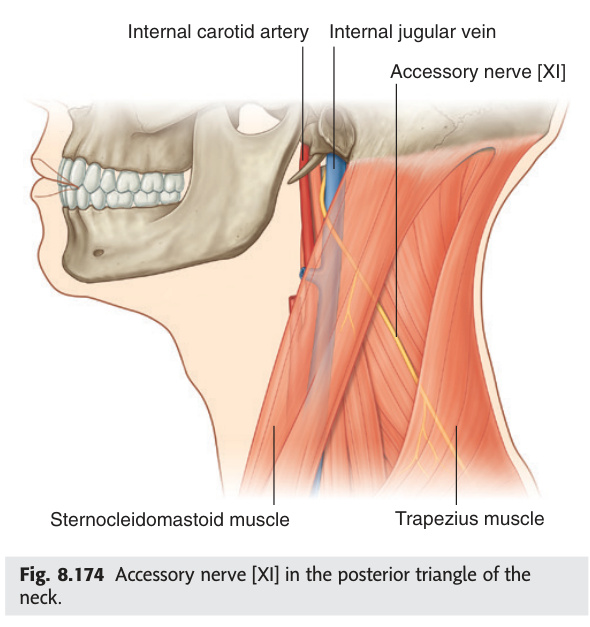
The accessory nerve is the only cranial nerve with a major contribution from the spinal cord: its spinal root arises from anterior horn cells at C1–C5, ascends through the foramen magnum, briefly joins the cranial root at the jugular foramen, and then swings through the posterior triangle of the neck to reach sternocleidomastoid and trapezius. This posterior triangle course is surgically treacherous — the nerve runs superficially just beneath the investing fascia, and even a routine lymph node biopsy can divide it, leaving the patient with a dropped shoulder, inability to shrug, and difficulty abducting the arm above 90°. The cranial root of CN XI (from nucleus ambiguus) is now considered functionally part of the vagus, contributing to pharyngeal and laryngeal muscles via the pharyngeal plexus.
Two parts: Cranial root (C. accessory): arises from nucleus ambiguus → exits medulla → joins spinal root briefly → immediately joins vagus trunk → innervates pharyngeal + laryngeal muscles via pharyngeal plexus (some texts consider this part purely vagal). Spinal root (spinal accessory): C1–C5 anterior horn → ascends through foramen magnum → exits via jugular foramen → posterior triangle of neck → sternocleidomastoid (SCM) + trapezius.
Most commonly injured during posterior triangle surgery (lymph node biopsy, neck dissection). The nerve runs superficially in the posterior triangle — vulnerable to careless dissection. Result: SCM weakness (weakened head turning to contralateral side) + trapezius weakness (dropped shoulder, inability to shrug, difficulty abducting arm above 90° due to inability to rotate scapula). Winging of scapula (but lateral winging, different from long thoracic nerve palsy which gives medial winging). Treatment: careful identification during surgery; if damaged, rerouting or nerve grafting.
- Where does the spinal root of CN XI originate? Anterior horn cells of C1–C5; ascends through foramen magnum to join cranial root at the jugular foramen.
- Which two muscles does the spinal accessory nerve supply? Sternocleidomastoid (SCM) and trapezius.
- Why is CN XI commonly injured in neck surgery? It runs superficially in the posterior triangle of the neck — at risk during lymph node biopsy or neck dissection.
- What are the clinical features of spinal accessory nerve damage? Dropped shoulder + inability to shrug + weakened contralateral head turning + difficulty abducting arm above 90° (lateral scapular winging).
- What happens to the cranial root of CN XI at the jugular foramen? It immediately joins the vagus trunk to supply pharyngeal and laryngeal muscles via the pharyngeal plexus.
Hypoglossal Nerve
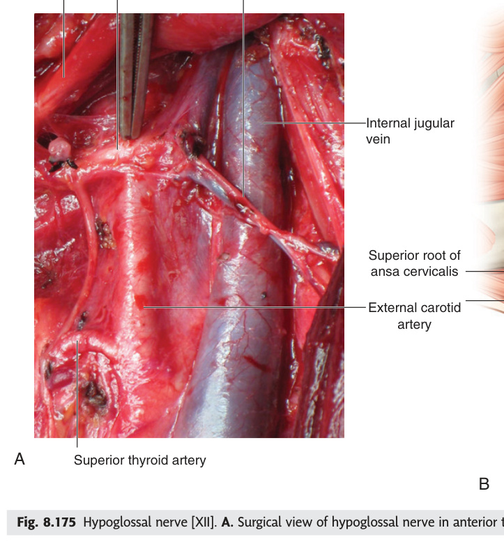
The hypoglossal nerve supplies every intrinsic tongue muscle and all extrinsic tongue muscles except palatoglossus (supplied by CN X), making it the motor workhorse of speech, chewing, and swallowing. The key clinical rule is that the tongue deviates toward a lower motor neurone lesion — the paralysed genioglossus on that side cannot protrude, so the healthy contralateral genioglossus pushes the tongue across — but deviates away from an upper motor neurone lesion (toward the hemiplegic side of the body). In motor neuron disease, bilateral CN XII degeneration produces a wasted, fasciculating tongue that is the bedside hallmark of progressive bulbar palsy.
Nucleus in medulla (hypoglossal triangle in floor of 4th ventricle). Exits anterior medulla between olive + pyramid → hypoglossal canal → loops around occipital artery → passes lateral to hyoglossus → enters tongue. Supplies ALL intrinsic tongue muscles + all extrinsic except palatoglossus (CN X). Genioglossus (protrudes tongue — largest extrinsic) is the key muscle tested.
Test Cranial Nerves
All 12 cranial nerves — clinical palsies, pathways, and exam scenarios.
- Which tongue muscle does CN XII NOT supply, and what supplies it? Palatoglossus — supplied by CN X (vagus via pharyngeal plexus).
- Which way does the tongue deviate in LMN vs UMN CN XII lesion? LMN: toward the lesion (paralysed genioglossus); UMN: away from the lesion (toward the hemiplegic side).
- Through which foramen does CN XII exit the skull? Hypoglossal canal (anterior condylar canal).
- What causes bilateral CN XII involvement in MND? Progressive bulbar palsy (lower motor neuron form of ALS) — wasted, fasciculating tongue + dysarthria + dysphagia.
- Name the largest extrinsic tongue muscle and its function. Genioglossus — protrudes the tongue; the muscle tested by asking the patient to "stick out your tongue."
Cavernous Sinus Syndrome ★★
The cavernous sinus is the most anatomically crowded dural venous sinus — CN III, IV, V1, V2, and VI plus the internal carotid artery and its perivascular sympathetic plexus all share a trabeculated venous lake at the skull base. This crowding means one lesion produces multiple simultaneous cranial nerve palsies, and the key clinical exercise is separating cavernous sinus syndrome (CN III/IV/V1/V2/VI) from orbital apex syndrome (adds CN II — optic nerve involvement giving visual loss) from superior orbital fissure syndrome (no CN II — optic canal separate). A carotid-cavernous fistula — direct (post-traumatic, high-flow) or indirect (spontaneous dural AVF) — classically presents with the unmistakable triad of pulsating exophthalmos, chemosis, and a bruit heard over the eye.
A paired venous sinus on either side of the sella turcica (body of sphenoid). Contents (medial to lateral in lateral wall + within sinus): CN III (superior lateral wall), CN IV (lateral wall, below III), CN V1 (lateral wall, below IV), CN V2 (lateral wall, lowest), CN VI (runs within the sinus, not just the wall — most vulnerable), Internal carotid artery (with its sympathetic plexus). CN V3 does NOT pass through the cavernous sinus (exits via foramen ovale directly). Drainage: receives superior + inferior ophthalmic veins, sphenoparietal sinus; drains into superior + inferior petrosal sinuses → sigmoid sinus / IJV.
| Syndrome | CNs involved | Distinguishing feature | Causes |
|---|---|---|---|
| Cavernous sinus syndrome | CN III, IV, V1, V2, VI ± Horner's (sympathetic plexus on ICA) | Multiple ipsilateral CN III/IV/VI palsies + facial numbness (V1/V2) + chemosis + proptosis. Bilateral involvement = cavernous sinus thrombosis. CN VI palsy often earliest/most prominent (lies within the sinus, no bony protection). | Pituitary apoplexy (haemorrhage into pituitary tumour — sudden, with headache + visual loss + ophthalmoplegia), ICA aneurysm (intracavernous), carotid-cavernous fistula (pulsating exophthalmos + bruit + chemosis), cavernous sinus thrombosis (from facial/sinus infection — Staphylococcus aureus most common; signs: fever + bilateral involvement + proptosis), meningioma, metastasis |
| Orbital apex syndrome | CN II + CN III + IV + V1 + VI | As cavernous sinus syndrome plus CN II involvement (visual loss + RAPD) — optic canal is adjacent to the superior orbital fissure at the orbital apex. Vision loss distinguishes orbital apex from cavernous sinus syndrome (optic nerve does not pass through cavernous sinus). | Trauma (orbital apex fracture), fungal sinusitis (Mucormycosis — especially in diabetics; angioinvasive, rapidly fatal), metastasis, granuloma (sarcoidosis, Wegener's / GPA) |
| Superior orbital fissure syndrome (Rochon-Duvigneaud) | CN III, IV, V1, VI (no CN II — optic canal not involved) | Same as orbital apex but no visual loss (optic nerve in separate optic canal, not through the SOF). Proptosis + ophthalmoplegia + V1 sensory loss. | Trauma, metastasis, sphenoid wing meningioma, nasopharyngeal carcinoma |
Abnormal communication between the internal carotid artery (high pressure) and the cavernous sinus (low pressure venous). Direct CCF: usually post-traumatic (rupture of ICA within sinus); high-flow. Indirect CCF: dural arteriovenous fistula via meningeal branches; lower flow, may be spontaneous (in elderly women). Classic presentation of direct CCF: pulsating exophthalmos (proptosis that pulsates) + chemosis (conjunctival oedema) + bruit over the eye (auscultate with stethoscope) + ipsilateral CN VI palsy (first/most common) ± CN III/IV palsies + raised intraocular pressure. Mechanism: arterial pressure transmitted to superior ophthalmic vein → retrograde venous hypertension → proptosis + venous engorgement. Treatment: endovascular embolisation (coiling or Onyx injection).
Q: A diabetic patient develops severe headache, right ptosis, right eye "down and out" with a dilated pupil, and right facial numbness over forehead and cheek. There is also a right CN VI palsy. Where is the lesion? What is the immediate concern?
A: Right cavernous sinus syndrome — involvement of CN III (ptosis + "down and out" + dilated pupil), CN VI (abduction palsy), and CN V1 + V2 (facial numbness over forehead = V1, cheek = V2). The immediate concern with a dilated unreactive pupil is cavernous sinus ICA aneurysm or pituitary apoplexy — both require urgent MRI/MRA. In a diabetic, also consider cavernous sinus thrombosis (fever would suggest this) and mucormycosis (check nasal mucosa for black eschar). Note: CN VI palsy within cavernous sinus does NOT indicate "false localising" — it is truly localised here; CN VI runs within the sinus and is most vulnerable. Unlike CN VI palsy from raised ICP (which would be bilateral and without other CN signs).
- List the nerves and structures passing through the cavernous sinus. CN III, IV, V1, V2, VI + internal carotid artery + perivascular sympathetic plexus (CN V3 does NOT pass through).
- How does cavernous sinus syndrome differ from orbital apex syndrome? Orbital apex adds CN II involvement (visual loss + RAPD) — optic nerve does not pass through the cavernous sinus.
- Classic triad of carotid-cavernous fistula? Pulsating exophthalmos + chemosis (conjunctival oedema) + bruit heard over the eye.
- Which CN within the cavernous sinus is first and most affected, and why? CN VI — runs within the sinus itself (not the lateral wall), with no bony protection.
- Name three causes of cavernous sinus syndrome. Pituitary apoplexy, cavernous sinus thrombosis (septic, Staphylococcus), ICA aneurysm/CCF — plus meningioma/nasopharyngeal carcinoma metastasis.
Jugular Foramen Syndromes ★★
The jugular foramen transmits CN IX, X, and XI together — these three nerves exiting as a cluster explain why a single lesion here produces Vernet's syndrome: dysphagia, dysphonia, unilateral palate drop, absent gag afferent, and SCM/trapezius weakness all at once. When the lesion extends anteromedially to the nearby hypoglossal canal, CN XII is added to give Collet-Sicard syndrome (tongue deviates toward the lesion), and further extension into the retroparotid sympathetic chain adds Horner's syndrome to complete Villaret's syndrome. The most important single cause to know is glomus jugulare tumour (paraganglioma): its "salt and pepper" T1 MRI signal, pulsatile tinnitus, and conductive hearing loss make it the classic teaching case for jugular foramen pathology.
The jugular foramen lies between the petrous part of the temporal bone (anterolaterally) and the occipital bone (posteromedially). It transmits: CN IX (glossopharyngeal — anterior part), CN X (vagus — middle), CN XI (spinal accessory — posterior), Internal jugular vein (IJV — posterior/largest compartment), and the inferior petrosal sinus. Closely related structures: hypoglossal canal (CN XII) is just anteromedial to the jugular foramen (but separate). The superior and inferior ganglia of CN IX and X lie within the foramen.
| Syndrome | CNs involved | Site / Key | Causes |
|---|---|---|---|
| Vernet's syndrome | CN IX, X, XI | Jugular foramen proper. The three CNs exiting together. Features: dysphagia + dysphonia (CN IX + X — pharyngeal/laryngeal), unilateral palate drooping + absent gag reflex afferent (CN IX) + efferent (CN X), ipsilateral SCM + trapezius weakness (CN XI). | Glomus jugulare tumour (paraganglioma — most common; pulsatile tinnitus + conductive hearing loss + CN palsies), jugular foramen meningioma, metastasis (nasopharyngeal carcinoma), skull base fracture through occipital condyle, thrombosis of IJV |
| Collet-Sicard syndrome | CN IX, X, XI, XII | Jugular foramen + nearby hypoglossal canal. Adds CN XII (tongue deviation towards lesion) to Vernet's triad. | Retroparotid space tumour, metastasis at skull base, trauma, nasopharyngeal carcinoma. CN XII involvement suggests lesion extends to/beyond jugular foramen into the retrocondylar space |
| Villaret's syndrome | CN IX, X, XI, XII + Horner's syndrome | Collet-Sicard + ipsilateral Horner's syndrome (sympathetic chain involvement in retroparotid space). The sympathetic chain descends adjacent to the internal carotid artery through the retroparotid/retropharyngeal space — lesion here can involve both the jugular foramen contents and the sympathetic trunk. | Retroparotid/retropharyngeal mass (parapharyngeal space tumour), deep cervical lymph node metastasis (level II/III), glomus jugulare extension, ICA dissection with retroparotid haematoma |
| Schmidt's syndrome | CN X, XI (only) | Partial jugular foramen lesion or involvement of just nucleus ambiguus + CN XI. Features: ipsilateral vocal cord palsy (dysphonia) + palatal palsy (CN X) + SCM/trapezius weakness (CN XI). CN IX spared. | Partial jugular foramen lesion; nucleus ambiguus lesion (lateral medullary infarct can produce a similar picture) |
| Tapia's syndrome | CN X, XII (only) | Below the jugular foramen — in the retropharyngeal/retrostyloid space where CN X (RLN + pharyngeal branch) and CN XII run in proximity. Features: hoarseness (CN X recurrent laryngeal) + tongue deviation towards lesion (CN XII). CN IX + XI spared. | Difficult orotracheal intubation (compression of nerves in lateral pharyngeal space), carotid artery surgery, penetrating neck trauma |
Vernet = IX + X + XI (jugular foramen = "VX9" — V for Vernet, IX X XI)
Collet-Sicard = IX + X + XI + XII (add the tongue — "the collector adds XII")
Villaret = IX + X + XI + XII + Horner's (the "villain" takes everything — even the sympathetics)
Glomus jugulare is the #1 cause of jugular foramen syndrome — know its features: pulsatile tinnitus + conductive hearing loss + multiple lower CN palsies. MRI: "salt and pepper" appearance on T1. Very vascular tumour (paraganglioma) — pre-op embolisation before surgical resection.
Q: A 55-year-old woman presents with pulsatile tinnitus, right-sided hearing loss, hoarseness, difficulty swallowing, and deviation of the tongue to the right on protrusion. On examination, the right shoulder is dropped and she cannot shrug. Name the syndrome and most likely cause.
A: Collet-Sicard syndrome (CN IX, X, XI, XII involvement). Features: hoarseness + dysphagia = CN X (nucleus ambiguus/vagus); tongue deviation to right = right CN XII palsy (genioglossus paralysed on right); shoulder drop + inability to shrug = right CN XI palsy (trapezius + SCM). Pulsatile tinnitus + conductive hearing loss + multiple lower CN palsies = glomus jugulare tumour (paraganglioma) until proven otherwise. Investigations: MRI head/neck (salt-and-pepper T1 signal), CT temporal bone (bony destruction), DSA/MRA (highly vascular). Treatment: pre-operative embolisation + surgical excision ± radiotherapy. Secretory paragangliomas (catecholamine-secreting) require pre-op alpha-blockade.
- Name the three CNs exiting the jugular foramen proper (Vernet's triad). CN IX, X, and XI — dysphagia + dysphonia + SCM/trapezius weakness + absent gag afferent.
- Which syndrome adds CN XII to Vernet's, and what does this add clinically? Collet-Sicard syndrome — adds tongue deviation toward the lesion (CN XII palsy of genioglossus).
- What distinguishes Villaret's from Collet-Sicard syndrome? Villaret's adds ipsilateral Horner's syndrome — sympathetic chain involvement in the retroparotid space.
- Most common cause of jugular foramen syndrome, and its imaging hallmark? Glomus jugulare tumour (paraganglioma) — "salt and pepper" T1 signal on MRI from vascular flow voids.
- In Tapia's syndrome, which two nerves are involved and at what level? CN X + CN XII, below the jugular foramen in the retropharyngeal/retrostyloid space (e.g. post-intubation injury).