Cerebellum, Brainstem & Cerebral Cortex
Basal Ganglia — Direct & Indirect Pathways
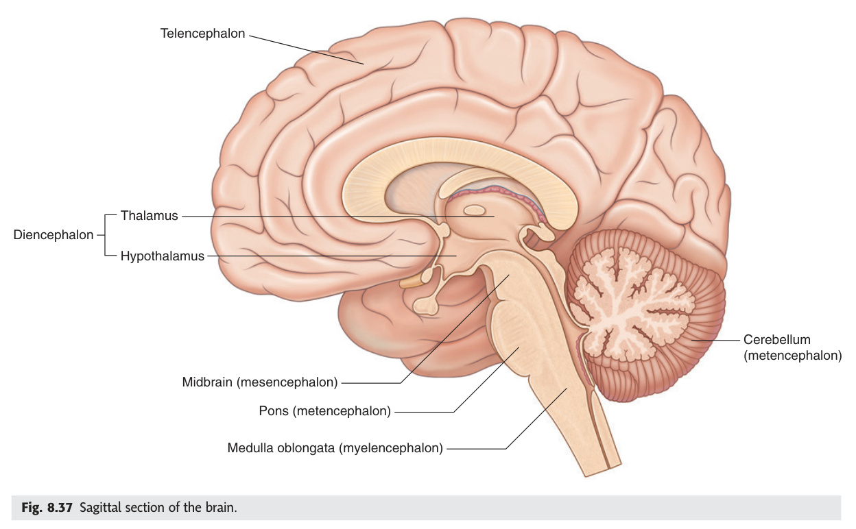
Cerebellum — Structure
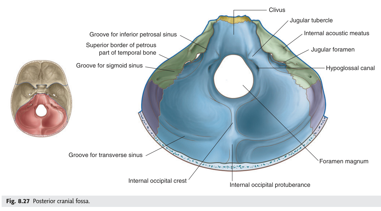
The cerebellum never initiates movement — it compares what the cortex intended with what the body is actually doing, and silently corrects the error before you even notice it. Think of it as the brain's autocorrect: without it, every reaching movement overshoots (dysmetria), every repetitive tap becomes irregular (dysdiadochokinesia), and every step is uncertain (ataxia). The cerebellum is divided into three functional zones that correspond to evolutionary age: the archicerebellum (flocculonodular lobe, oldest) receives balance and vestibular input and controls posture and eye movements — damage here causes truncal ataxia and nystagmus, the hallmark of midline tumours like medulloblastoma in children. The paleocerebellum (spinocerebellum, vermis + paravermis) processes proprioceptive feedback from the limbs via spinocerebellar tracts and fine-tunes axial and proximal movement. The neocerebellum (cerebrocerebellum, lateral hemispheres) receives cortical plans via the pontine nuclei and is responsible for timing and sequencing skilled voluntary movements — lesions here produce the classic ipsilateral limb signs. The critical anatomical rule: cerebellar output decussates in the superior cerebellar peduncle (SCP), then the corticospinal tract decussates again at the medulla, so a right cerebellar lesion causes right-sided limb signs (double-cross = same side).
| Part | Lobules / Regions | Input from | Function |
|---|---|---|---|
| Archicerebellum (vestibulocerebellum) | Flocculonodular lobe | Vestibular nuclei + labyrinths | Balance + eye movements. Lesion: truncal ataxia + nystagmus (wide-based gait, can't tandem walk, fall to either side — midline lesion). Classic: medulloblastoma in children |
| Paleocerebellum (spinocerebellum) | Vermis + paravermal cortex (anterior lobe + pyramis + uvula) | Spinocerebellar tracts (proprioception from muscles/joints) | Coordination of axial + proximal limb movements. Vermis → body midline. Paravermis → limbs. Lesion: gait ataxia dominant, limb ataxia mild |
| Neocerebellum (pontocerebellum / cerebrocerebellum) | Lateral hemispheres (posterior lobe) | Pontine nuclei (relay from cerebral cortex via corticopontine fibres) | Planning + timing of skilled voluntary limb movements. Lesion: ipsilateral limb ataxia, intention tremor, dysmetria, dysdiadochokinesia |
Dentate (largest, lateral hemispheres) · Emboliform · Globose (together = interposed) · Fastigial (vermis, oldest). Output path: deep nuclei → superior cerebellar peduncle → decussate in midbrain tegmentum → contralateral red nucleus + thalamus (VL) → motor cortex. Because peduncle decussates: right cerebellar lesion → right-sided limb ataxia (ipsilateral to lesion, crossed at peduncle then crossed again by corticospinal tract).
| Peduncle | Fibres (direction) |
|---|---|
| Superior cerebellar peduncle (SCP) | Mainly OUTPUT: dentate/emboliform/globose → thalamus + red nucleus. Decussates in midbrain. Also: anterior spinocerebellar tract (input) |
| Middle cerebellar peduncle (MCP) | Largest. INPUT only: pontine nuclei → cerebellar cortex (corticopontocerebellar fibres — from contralateral cortex) |
| Inferior cerebellar peduncle (ICP) | INPUT dominant: posterior spinocerebellar tract + cuneocerebellar + vestibular nuclei + olivocerebellar. OUTPUT: cerebellovestibular + cerebelloreticular |
- Name the three functional divisions of the cerebellum, their input, and their lesion pattern. Archicerebellum (flocculonodular) ← vestibular: truncal ataxia + nystagmus. Paleocerebellum (vermis + paravermis) ← spinocerebellar: gait ataxia. Neocerebellum (lateral hemispheres) ← cortex via pons: ipsilateral limb ataxia + intention tremor + dysdiadochokinesia.
- Why do right-sided cerebellar lesions cause right-sided limb signs? Output leaves via SCP → decussates in midbrain → reaches contralateral thalamus → motor cortex → corticospinal tract decussates again at medulla. Double-cross = ipsilateral (same side as lesion).
- Recall the deep cerebellar nuclei medial to lateral with mnemonic. "Don't Eat Greasy Food": Dentate (largest, lateral hemispheres) → Emboliform → Globose (emboliform + globose = interposed) → Fastigial (vermis, oldest). All project via SCP.
- Which peduncle is purely INPUT, and from where? Middle cerebellar peduncle (MCP) — largest peduncle, carries only INPUT from ipsilateral pontine nuclei (which relay from contralateral cerebral cortex via corticopontine fibres).
- A child presents with truncal ataxia and hydrocephalus. Which cerebellar division and tumour? Archicerebellum (vermis/flocculonodular). Tumour: medulloblastoma (posterior fossa, WHO grade 4) — compresses 4th ventricle → hydrocephalus.
Cerebellar Cortex Circuit
The cerebellar cortex is a remarkably uniform three-layer processor — the same microcircuit repeated across every folium — yet its logic is counterintuitive: the sole output neuron (the Purkinje cell) is inhibitory. Every signal the cerebellum sends to the deep nuclei is a GABA-ergic brake; the deep nuclei are tonically active and the cerebellar cortex modulates them by releasing that brake at precisely the right moment. Information reaches the cortex via two fibre types with fundamentally different roles: mossy fibres carry the bulk of sensorimotor input (from spinal cord, pons, vestibular nuclei) and synapse on granule cells, which fan out as parallel fibres to activate thousands of Purkinje cells — a broad, diffuse signal for ongoing movement. Climbing fibres from the inferior olivary nucleus are the error signal: each climbing fibre contacts exactly one Purkinje cell with an extraordinarily powerful synapse, delivering a precise "you just got it wrong" signal that modifies future Purkinje cell responses — the cellular basis of motor learning. This is why repeated practice improves coordination: each error is registered by climbing fibre discharge, driving long-term depression of the active parallel fibre synapses (cerebellar LTD).
Cerebellar cortex has 3 layers: Molecular (outer: stellate + basket cells) → Purkinje (middle: single Purkinje cell layer — sole OUTPUT of cortex; inhibitory, GABA-ergic, project to deep nuclei) → Granule (inner/deepest: granule cells — receive mossy fibre input; send parallel fibres up to molecular layer).
| Input fibre type | Source | Synapses on |
|---|---|---|
| Mossy fibres | Spinal cord (spinocerebellar) + pons (corticopontine) + vestibular nuclei | Granule cells in granular layer |
| Climbing fibres | Inferior olivary nucleus (medulla) only | Directly on Purkinje cell dendrites (powerful 1:1 synapse — one climbing fibre per Purkinje cell). Carry error signals for motor learning |
- Name the three layers of cerebellar cortex from outer to inner. Molecular (outer: stellate + basket cells) → Purkinje (single layer — sole cortical output, inhibitory/GABAergic) → Granular (inner: granule cells receive mossy fibre input, send parallel fibres upward).
- What is the functional difference between mossy fibres and climbing fibres? Mossy fibres: bulk sensorimotor input (spinal cord, pons, vestibular) → granule cells → parallel fibres → broad Purkinje activation — ongoing movement signal. Climbing fibres: inferior olive only → one-to-one Purkinje contact — error signal for motor learning.
- Why is the Purkinje cell described as paradoxical output? It is the sole output of the cerebellar cortex yet is inhibitory (GABAergic). It inhibits deep cerebellar nuclei; timed release of this inhibition at the right moment drives the deep nuclei output to the thalamus.
- What is cerebellar LTD and what drives it? Long-term depression of parallel fibre → Purkinje cell synapses, driven by coincident climbing fibre (error) + parallel fibre (motor command) activation. This is the cellular mechanism by which the cerebellum learns from repeated errors — each climbing fibre discharge weakens the active parallel fibre synapses.
- Which nucleus is the sole source of climbing fibres, and where is it? Inferior olivary nucleus, located in the anterior (ventral) medulla, lateral to the pyramids (forms the visible olive externally).
Cerebellar Lesions
DANISH is medicine's neatest mnemonic because it captures the entire cerebellar examination in six letters — and every sign on the list reflects a failure of the cerebellum's core job: the smooth, timed, well-calibrated movement. Dysdiadochokinesia is the failure of timing in repetitive acts; ataxia is uncoordinated gait; nystagmus reflects failure of the vestibulo-ocular reflex gain; intention tremor is an error-correction overshoot that worsens as the limb approaches target (opposite to Parkinson's resting tremor, which stops on movement); scanning dysarthria is cerebellar timing failure in speech muscles; hypotonia reflects loss of deep nuclear tonic output. The Romberg test is the critical differentiator — cerebellar ataxia is equally bad with eyes open or closed (Romberg negative), while posterior column sensory ataxia is compensated by vision so closing the eyes makes it much worse (Romberg positive). Knowing the age and lesion site narrows the diagnosis sharply: a child with truncal ataxia and hydrocephalus has a midline vermis medulloblastoma until proven otherwise; an adult alcoholic with gait ataxia but preserved finger-nose test has selective anterior vermis degeneration; Friedreich's ataxia combines spinocerebellar + dorsal column loss with pes cavus and cardiomyopathy — a multi-system signature.
Dysdiadochokinesia (inability to perform rapid alternating movements — tap hand rapidly) · Ataxia (incoordination of gait/limbs) · Nystagmus (horizontal, towards side of lesion) · Intention tremor (tremor worsens on approaching target, tested with finger-nose test; absent at rest — unlike Parkinson's rest tremor) · Slurred speech (dysarthria — scanning/staccato speech) · Hypotonia (decreased muscle tone — pendular knee jerk)
Children: medulloblastoma (vermis → truncal ataxia + hydrocephalus; posterior fossa, WHO grade 4), cerebellar pilocytic astrocytoma (lateral hemisphere → limb ataxia, cystic with mural nodule, WHO grade 1). Adults: metastases (posterior fossa common — lung, breast, melanoma), haemangioblastoma (von Hippel-Lindau), alcoholic cerebellar degeneration (vermis predominantly → gait ataxia), MS plaques, stroke (PICA or SCA territory). Hereditary: Friedreich's ataxia (GAA repeat in frataxin gene, AR; onset adolescence; spinocerebellar + dorsal column degeneration; pes cavus + cardiomyopathy).
- Expand DANISH and state what each sign reflects anatomically. Dysdiadochokinesia (timing failure — lateral hemisphere); Ataxia (coordination — spinocerebellum/hemispheres); Nystagmus (VOR gain — flocculonodular); Intention tremor (error-correction overshoot — neocerebellum); Slurred speech / scanning dysarthria (cerebellar timing of speech muscles); Hypotonia (loss of deep nuclear tonic output — pendular knee jerk).
- Distinguish cerebellar ataxia from sensory (posterior column) ataxia using the Romberg test. Cerebellar: broad-based reeling gait, Romberg NEGATIVE (equally unsteady eyes open or closed — vision cannot compensate for cerebellar loss). Sensory: stamping high-stepping gait, patient watches feet, Romberg POSITIVE (stable eyes open, falls eyes closed — using vision to compensate for lost proprioception).
- What is intention tremor and how does it differ from Parkinson's resting tremor? Intention tremor worsens as the limb approaches the target (finger-nose test), absent at rest — reflects cerebellar error-correction overshoot. Parkinson's resting tremor is present at rest (4–6 Hz, pill-rolling) and diminishes with voluntary movement — reflects basal ganglia dopamine deficiency, not cerebellar pathology.
- Name two cerebellar tumours in children, their location, and their WHO grade. Medulloblastoma: posterior fossa/vermis, WHO grade 4, highly malignant, CSF spread — truncal ataxia + hydrocephalus. Pilocytic astrocytoma: lateral hemisphere, WHO grade 1, cystic with mural nodule — limb ataxia, best prognosis.
- What are the key features of Friedreich's ataxia beyond cerebellar signs? Autosomal recessive (GAA trinucleotide repeat in frataxin gene); onset adolescence; combined spinocerebellar + dorsal column degeneration (ataxia + proprioception loss + absent ankle jerks); pes cavus; scoliosis; hypertrophic cardiomyopathy (leading cause of death); diabetes mellitus in ~10%.
Medulla Oblongata
The medulla oblongata is where the spinal cord becomes the brain — a dense 3 cm cylinder packed with life-sustaining centres and the crossing points for almost every major tract. Two anatomical rules govern medullary stroke interpretation: the motor (pyramidal) decussation is at the lower medulla and the sensory (medial lemniscal) decussation is at the upper medulla, meaning that by the time a signal leaves the medulla heading rostrally, it is already on the opposite side. This creates the two classic alternating stroke syndromes. Wallenberg (lateral medullary) syndrome from PICA occlusion is one of the most tested strokes in medicine: the lateral medulla contains the spinothalamic tract (contralateral body pain/temperature loss), the CN V spinal nucleus (ipsilateral face pain/temperature loss), the nucleus ambiguus (ipsilateral dysphagia/dysphonia), descending sympathetic fibres (ipsilateral Horner's), and the inferior cerebellar peduncle (ipsilateral ataxia) — but the pyramids are medial and intact, so there is NO motor weakness. Déjérine's medial medullary syndrome from anterior spinal artery occlusion is the mirror image: ipsilateral CN XII palsy + contralateral hemiplegia (pyramids) + contralateral fine touch/proprioception loss (medial lemniscus) — but no Horner's, no dysphagia, no face sensory loss (lateral structures intact).
| Structure | Location | Function / Clinical |
|---|---|---|
| Pyramids | Anterior, flanking anterior median fissure | Corticospinal tract fibres. Pyramidal (motor) decussation at lower medulla (85–90% cross). Pyramid = UMN tract |
| Olives (inferior olivary nuclei) | Lateral, posterior to pyramids | Relay for cerebellar motor learning. Hypertrophic olivary degeneration: rubral tremor, palatal tremor |
| Nucleus gracilis + cuneatus | Posterior (dorsal), form gracile + cuneate tubercles | Relay for dorsal columns (fine touch/vibration/proprioception). Sensory (medial lemniscus) decussation here |
| Reticular formation | Central core of entire brainstem | Arousal (ascending reticular activating system — ARAS) + autonomic centres (cardiac, vasomotor, respiratory in medulla). Lesion → coma |
| Nucleus ambiguus | Lateral reticular formation | Motor nucleus for CN IX, X, XI (swallowing + phonation). Lesion → dysphagia + dysphonia (bulbar palsy) |
| Dorsal motor nucleus of vagus | Floor of 4th ventricle | Parasympathetic preganglionic for CN X (thoracic + abdominal viscera) |
Occlusion of PICA (posterior inferior cerebellar artery) → infarction of lateral medulla. The classic posterior fossa stroke. Features: ipsilateral: facial pain/temperature loss (CN V spinal nucleus), Horner's syndrome (descending sympathetic fibres), cerebellar signs (ataxia, nystagmus from inferior cerebellar peduncle), dysphagia + dysphonia (nucleus ambiguus — CN IX, X), vertigo/vomiting (vestibular nuclei). Contralateral: limb + trunk pain/temperature loss (spinothalamic tract). Fine touch/proprioception INTACT (medial medulla — pyramids + medial lemniscus — spared). Mnemonic: "PICA is LATERAL" = crossed sensory loss (face vs body). NOT associated with pyramidal (motor) weakness because pyramids are in the medial medulla.
Occlusion of anterior spinal artery (ASA) or medullary branches of vertebral artery → infarction of medial medulla (pyramids + medial lemniscus + CN XII fascicles). Contrast with Wallenberg (lateral medulla = different artery, different deficits).
Features (the medial triad):
(1) Ipsilateral CN XII palsy (tongue deviates TOWARDS lesion on protrusion — LMN; fascicles or nucleus in medial medulla).
(2) Contralateral hemiplegia (arm + leg, face spared — pyramids, UMN; the decussation is at the lower medulla so fibres above the decussation are ipsilateral to their future territory — lesion above decussation = contralateral weakness).
(3) Contralateral fine touch + proprioception loss (medial lemniscus already decussated at this level — carries contralateral body signals upward).
Pain/temperature (spinothalamic) = INTACT (spinothalamic in lateral medulla — not infarcted).
Facial sensation INTACT (CN V spinal nucleus in lateral medulla — not infarcted).
Exam trick: Wallenberg = lateral = pain/temperature deficits + Horner's + dysphagia, NO motor weakness; Déjérine's = medial = motor weakness + fine touch loss + CN XII, NO Horner's, NO dysphagia, NO face sensory loss.
- Which artery causes Wallenberg syndrome, and name five ipsilateral features. PICA (posterior inferior cerebellar artery). Ipsilateral: (1) facial pain/temperature loss (CN V spinal nucleus), (2) Horner's syndrome (descending sympathetics), (3) ataxia/nystagmus (ICP/vestibular), (4) dysphagia + dysphonia (nucleus ambiguus — CN IX/X), (5) vertigo/vomiting (vestibular nuclei). Contralateral: body pain/temperature loss (spinothalamic). NO motor weakness.
- What is Déjérine's medial medullary syndrome — artery and triad? Anterior spinal artery occlusion → medial medulla. Triad: (1) ipsilateral CN XII LMN palsy (tongue deviates to lesion side), (2) contralateral hemiplegia (pyramids, above decussation), (3) contralateral fine touch + proprioception loss (medial lemniscus, already decussated). Pain/temperature and face sensation INTACT (lateral structures spared).
- Where do the two major sensory/motor decussations occur in the medulla? Motor (pyramidal) decussation: lower medulla (85–90% of corticospinal fibres cross). Sensory (medial lemniscal) decussation: upper medulla at nucleus gracilis/cuneatus level. Both cross before fibres ascend, so all signals leaving the medulla rostrally are already contralateral.
- What does the nucleus ambiguus contain and what is the clinical deficit when damaged? LMN cell bodies for CN IX (glossopharyngeal), X (vagus), and XI (accessory) — controls swallowing and phonation. Damage (e.g., Wallenberg) → ipsilateral dysphagia (palatal droop, food/liquid aspiration) + dysphonia (hoarse voice). Bilateral damage = bulbar palsy.
- Where is the ascending reticular activating system (ARAS) and what happens if it is damaged? Reticular formation runs through the central core of the entire brainstem; ARAS projects from midbrain reticular formation to the thalamus and cortex. Bilateral damage (e.g., large basilar artery stroke, herniation) → coma (loss of consciousness by interrupting arousal pathways).
Pons
The pons is a bridge in name and function — it connects the cerebrum above to the cerebellum laterally and the medulla below, while housing some of the most clinically important cranial nerve nuclei in the brainstem. Its anatomy divides cleanly into ventral (basis pontis: highway for descending motor fibres + pontine nuclei relaying to cerebellum) and dorsal (tegmentum: CN V, VI, VII, VIII nuclei + MLF + locus coeruleus). The facial colliculus is the classic anatomy viva landmark: facial nerve fibres loop around the abducens nucleus to form a bump on the floor of the 4th ventricle — damage here hits both CN VI (lateral rectus, abduction) and CN VII (entire ipsilateral face) simultaneously, explained by their intimate anatomical relationship. The pontine stroke syndromes turn on a single anatomical principle — "eyes look away from pontine lesions, towards cortical lesions": a cortical lesion destroys the ipsilateral frontal eye field, leaving the contralateral one to push gaze towards the lesion; a pontine PPRF/CN VI nucleus lesion destroys the ipsilateral horizontal gaze centre, so eyes are pushed to the opposite side. Central pontine myelinolysis from over-rapid sodium correction is the classic cause of locked-in syndrome: basis pontis demyelination paralyses corticospinal + corticobulbar fibres bilaterally while the midbrain and consciousness are preserved.
| Feature | Detail |
|---|---|
| Basis pontis (ventral/basal pons) | Corticospinal + corticobulbar fibres pass through (scattered among pontine nuclei). Pontine nuclei relay cortical signals via MCP to cerebellum |
| Pontine tegmentum (dorsal pons) | CN V (trigeminal — mid-pons), CN VI (abducens — caudal pons), CN VII (facial — caudal pons at pontomedullary junction), CN VIII (vestibulocochlear — pontomedullary junction). Medial longitudinal fasciculus (MLF), locus coeruleus (noradrenaline — arousal) |
| 4th ventricle | Floor shared by pons + medulla. Facial colliculus = facial nerve fibres looping over abducens nucleus (CN VI). Damage to facial colliculus → ipsilateral CN VI + CN VII palsy |
| Respiratory centres | Pneumotaxic (rostral pons — inhibits inspiration) + Apneustic (caudal pons — prolongs inspiration) |
Rapid correction of hyponatraemia → osmotic stress → demyelination of pontine fibres. Features: locked-in syndrome (quadriplegia + lower cranial nerve palsies, but consciousness intact because tegmentum/cortex spared — corticospinal + corticobulbar in basis pontis demyelinated). Patients can communicate only by vertical eye movements + blinking (CN III and IV nuclei in midbrain — above the lesion). Prevention: correct Na⁺ no faster than 8–10 mmol/L per 24 hours.
Millard-Gubler syndrome: Infarction of ventral caudal pons (basilar artery perforators). Involves basis pontis + CN VI & VII fascicles as they pass through.
Ipsilateral: CN VI palsy (lateral rectus — eye cannot abduct, diplopia on lateral gaze), CN VII palsy (LMN facial — entire ipsilateral face, including forehead; cannot close eye or smile).
Contralateral: hemiplegia (corticospinal in basis pontis, above its decussation).
Key: "alternating hemiplegia" pattern — ipsilateral CN, contralateral body.
Foville's syndrome: Similar but more dorsal lesion — involves CN VI nucleus (not just fascicles) + ipsilateral PPRF (paramedian pontine reticular formation = horizontal gaze centre).
Result: conjugate gaze palsy towards the side of the lesion (PPRF damage — neither eye can look towards lesion side; eyes deviate away from lesion = towards the hemiplegia) + ipsilateral CN VII + contralateral hemiplegia. A CN VI nucleus lesion always causes gaze palsy (not just abduction palsy) because the nucleus contains interneurons projecting via the MLF to the contralateral CN III nucleus.
Mnemonic — "Eyes look away from pontine lesions, towards cortical lesions": Cortical hemisphere lesion → eyes deviate towards the lesion (ipsilateral gaze preference). Pontine PPRF/CN VI nucleus lesion → eyes deviate away from lesion.
- What is the facial colliculus and what is the deficit when it is damaged? A bump on the floor of the 4th ventricle formed by CN VII fascicles looping over the CN VI (abducens) nucleus. Damage → combined ipsilateral CN VI palsy (cannot abduct eye) + ipsilateral LMN CN VII palsy (entire ipsilateral face including forehead).
- Describe Millard-Gubler syndrome — location, structures, deficits. Ventral caudal pons infarct (basilar perforators). Ipsilateral: CN VI palsy + CN VII LMN palsy. Contralateral: hemiplegia (corticospinal in basis pontis). Classic "alternating hemiplegia" pattern — ipsilateral cranial nerve, contralateral body.
- How does a CN VI nucleus lesion differ clinically from a CN VI fascicle lesion? Fascicle lesion → isolated abduction palsy (one eye can't abduct). Nucleus lesion → conjugate horizontal gaze palsy (neither eye looks towards the lesion side) because the nucleus contains interneurons that project via the MLF to the contralateral CN III nucleus to coordinate both eyes.
- Why does rapid correction of hyponatraemia cause locked-in syndrome? Osmotic demyelination of basis pontis (central pontine myelinolysis) → bilateral corticospinal + corticobulbar fibre disruption → quadriplegia + lower CN palsies. Tegmentum and consciousness (ARAS via midbrain) spared → patient is awake but can only communicate via vertical eye movements (CN III/IV above the lesion).
- What do the pneumotaxic and apneustic centres do, and where are they? Pneumotaxic (rostral pons): inhibits inspiration, shortens breath duration — damage causes slow deep breathing. Apneustic (caudal pons): prolongs inspiration — damage causes apneustic breathing (prolonged inspiratory hold). Both modulate the medullary respiratory rhythm generator.
Midbrain
The midbrain is the most rostral brainstem segment and the level where the cerebellum's output (via the superior cerebellar peduncle) enters the motor system — it is also where CN III exits, making it the key level for the two classic midbrain stroke syndromes. Weber's syndrome (ventral midbrain, peduncle + CN III fascicle) gives the most recognisable pattern in neurology: an ipsilateral dilated fixed pupil and ptosis (CN III — exiting between the peduncles) combined with contralateral hemiplegia (cerebral peduncle carrying corticospinal tract above its decussation). Parinaud's syndrome (dorsal midbrain compression, e.g., from a pinealoma pressing on the superior colliculi) blocks upward gaze and produces light-near dissociation — a pattern also seen in neurosyphilis (Argyll Robertson pupil). The substantia nigra pars compacta sits at the junction of peduncle and tegmentum; its dopaminergic projection to the striatum is what Parkinson's disease destroys, and its loss of neuromelanin pigment makes the midbrain look pale at autopsy. The periaqueductal grey (PAG) surrounding the cerebral aqueduct is rich in opioid receptors and is the anatomical target for endogenous pain suppression — stimulation here activates descending inhibitory pathways that gate pain signals at the dorsal horn.
| Structure | Location | Clinical significance |
|---|---|---|
| Tectum (roof): superior + inferior colliculi | Dorsal midbrain | Superior colliculi: visual reflex (eye movements to visual stimuli). Parinaud's syndrome (dorsal midbrain compression, e.g., pinealoma): failure of upward gaze + pupil light-near dissociation. Inferior colliculi: auditory reflex (turning head to sound) |
| Cerebral peduncles (crus cerebri) | Ventral midbrain | Corticospinal + corticopontine + corticobulbar fibres. CN III exits between peduncles (interpeduncular fossa). Weber's syndrome: CN III palsy + contralateral hemiplegia (ipsilateral peduncle compression) |
| Substantia nigra | Tegmentum, between peduncle and tegmentum | Dopaminergic neurons → striatum (dopamine inhibits inhibitory striatal neurons → allows movement). Degeneration → Parkinson's disease. Lewy bodies (alpha-synuclein inclusions). Loss of pigmentation grossly (neuromelanin) |
| Red nucleus | Tegmentum | Receives input from cerebellum (dentate via SCP). Contributes to rubrospinal tract. Role in motor coordination |
| Periaqueductal grey (PAG) | Around cerebral aqueduct | Endogenous pain modulation (opioid receptors, descending inhibition). Stimulation → analgesia |
- Describe Weber's syndrome — location, structures, and deficits. Ventral midbrain infarct (posterior cerebral or basilar artery perforator). Ipsilateral: CN III palsy (dilated fixed pupil, ptosis, eye down-and-out — fascicles in interpeduncular fossa). Contralateral: hemiplegia (cerebral peduncle/crus cerebri). The alternating pattern — ipsilateral CN, contralateral body — is the midbrain equivalent of Millard-Gubler.
- What is Parinaud's syndrome and what causes it? Dorsal midbrain compression (classically pinealoma/pineal gland tumour, also MS, hydrocephalus). Features: failure of upward gaze (superior colliculi/pretectal area compressed), convergence-retraction nystagmus on attempted upgaze, and light-near dissociation (pupil responds to accommodation but not light).
- Why does the substantia nigra appear pale in Parkinson's disease at autopsy? SNc neurons contain neuromelanin (a dark pigment derived from dopamine oxidation); neurodegeneration of these dopaminergic cells causes loss of this pigmentation → pale SNc macroscopically. Lewy bodies (alpha-synuclein inclusions) are the histological hallmark in surviving neurons.
- What is the periaqueductal grey, and what is its clinical relevance? Grey matter surrounding the cerebral aqueduct; rich in mu-opioid receptors. Part of the descending pain modulation pathway: PAG → nucleus raphe magnus (medulla) → dorsal horn (inhibits pain via serotonin + enkephalin). Deep brain stimulation of PAG produces analgesia; this is also how exogenous opioids partially work.
- CN III exits between which two structures, and why does this make it vulnerable in uncal herniation? CN III exits between the posterior cerebral artery (PCA) and the superior cerebellar artery (SCA), then runs along the tentorium. In uncal herniation, the medial temporal lobe herniates through the tentorial notch → compresses CN III from above → early sign is a unilaterally dilated unreactive pupil (parasympathetic fibres on the outside of the nerve are compressed first).
Cerebral Cortex — Functional Areas
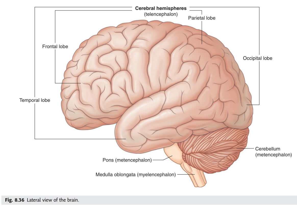
The cerebral cortex is functionally organised around a central sulcus axis — precentral gyrus (BA 4) executes movement, postcentral gyrus (BA 1,2,3) receives sensation — with language, vision, and auditory processing layered around it in the dominant hemisphere. The language axis is one of the highest-yield examination topics in neurology: Broca's area (BA 44,45, dominant inferior frontal gyrus) handles speech production, so damage produces non-fluent, effortful, telegraphic speech with intact comprehension — the patient understands everything but cannot get the words out ("frustrated patient"). Wernicke's area (BA 22, dominant posterior superior temporal gyrus) handles comprehension, so damage produces fluent but semantically empty jargon speech with severely impaired comprehension — the patient talks freely but makes no sense ("happy patient who talks rubbish"). The primary visual cortex (BA 17, calcarine fissure) has a clinically important anatomical feature: the macula projects to the posterior occipital pole where it receives dual blood supply from both the PCA and MCA, explaining macular sparing in PCA territory infarcts. The prefrontal cortex is where Phineas Gage became famous — a tamping iron through his frontal lobe left his intellect intact but destroyed his personality and judgment, the first evidence that the frontal lobes house what we now call executive function.
| Area (Brodmann) | Location | Function & Clinical |
|---|---|---|
| Primary motor cortex (M1, BA 4) | Precentral gyrus (anterior to central sulcus) | Somatotopic map — homunculus (medial: leg, lateral: face). Voluntary movement commands via corticospinal tract. Lesion → contralateral spastic hemiplegia (UMN pattern) |
| Primary somatosensory cortex (S1, BA 1,2,3) | Postcentral gyrus | Receives fine touch/proprioception from thalamus (VPL). Somatotopic homunculus mirrors motor. Lesion → contralateral sensory loss + astereognosis |
| Broca's area (BA 44,45) | Inferior frontal gyrus, dominant (usually left) hemisphere | Speech production (motor aspect). Broca's aphasia: non-fluent, effortful speech; comprehension relatively intact ("frustrated patient"). Lesion: dominant inferior frontal gyrus ← MCA branch |
| Wernicke's area (BA 22) | Posterior superior temporal gyrus, dominant hemisphere | Speech comprehension. Wernicke's aphasia: fluent but nonsensical (jargon aphasia); poor comprehension ("happy patient who talks rubbish"). Lesion: dominant posterior temporal ← MCA |
| Primary visual cortex (V1, BA 17) | Calcarine fissure (medial occipital), around calcarine sulcus | Receives input from LGN (thalamus). Macular representation at posterior pole (spared in PCA infarcts because dual blood supply from MCA + PCA). Cortical blindness: bilateral V1 damage |
| Primary auditory cortex (BA 41,42) | Superior temporal gyrus (Heschl's gyri) | Receives bilateral auditory input from medial geniculate body (thalamus). Unilateral lesion → minimal hearing deficit (bilateral representation) |
| Prefrontal cortex | Anterior frontal lobe | Executive function, personality, decision-making. Lesion → disinhibition, perseveration, personality change (Phineas Gage) |
- Distinguish Broca's from Wernicke's aphasia in one sentence each. Broca's (BA 44/45, dominant inferior frontal): non-fluent, effortful, telegraphic speech; comprehension relatively intact — patient is frustrated. Wernicke's (BA 22, dominant posterior superior temporal): fluent jargon speech with neologisms; comprehension severely impaired — patient is unaware of errors.
- Why is macular vision spared in posterior cerebral artery infarcts? The macula projects to the posterior occipital pole, which receives dual blood supply from both the PCA and the MCA. When the PCA is occluded, MCA collaterals preserve macular representation even as peripheral vision (anterior calcarine) is lost → homonymous hemianopia with macular sparing.
- Where is the primary motor cortex and what is its somatotopic organisation? Precentral gyrus (BA 4), anterior to the central sulcus. Homunculus: medial surface = lower limb (leg/foot — supplied by ACA); lateral surface = upper limb; most lateral = face. Areas with fine motor control (hand, face) have disproportionately large representation.
- What artery supplies Broca's and Wernicke's areas, and what is the clinical implication? Both are supplied by branches of the middle cerebral artery (MCA). Large MCA territory stroke can cause global aphasia (both production and comprehension lost). Broca's = anterior MCA branch (inferior frontal); Wernicke's = posterior MCA branch (posterior temporal).
- What is astereognosis and which cortex lesion causes it? Inability to identify objects by touch alone (with eyes closed) despite intact primary sensation. Caused by lesion of the primary somatosensory cortex (S1, BA 1,2,3, postcentral gyrus) or adjacent parietal association cortex — the patient feels the object but cannot interpret the sensory pattern into recognition.
Basal Ganglia
The basal ganglia circuit is best understood as a gating system: it runs a loop from cortex → striatum → GPi/SNr → thalamus → back to cortex, and its job is to open the gate for wanted movements (direct pathway) while keeping the gate shut for competing, unwanted ones (indirect pathway). Dopamine from the substantia nigra pars compacta simultaneously facilitates the direct pathway (D1 receptors) and suppresses the indirect pathway (D2 receptors) — both actions increase net movement. Lose dopamine (Parkinson's) and both the gate-opener fails and the gate-closer overactivates: the result is poverty of movement, rigidity, and resting tremor. Gain too much activity in the direct pathway (Huntington's, from indirect pathway neuron loss) and the gate is permanently open: chorea — excessive, involuntary, irregular movement. Lose the subthalamic nucleus (STN, the accelerator pedal of the indirect pathway) unilaterally and the GPi is no longer driven, the thalamus is disinhibited on one side, and the contralateral limb flings wildly in hemiballismus. This single circuit — understood in terms of what gets activated and what gets inhibited — explains Parkinson's, Huntington's, hemiballismus, and the rationale for deep brain stimulation of the STN or GPi in one unified framework.
| Structure | Components / Notes |
|---|---|
| Striatum | Caudate + putamen (+ nucleus accumbens). Main input structure. Receives from cortex + substantia nigra (dopamine) |
| Globus pallidus | GPe (external) + GPi (internal). GPi = main output of basal ganglia → thalamus |
| Subthalamic nucleus (STN) | Thalamic region. Excitatory output to GPi/GPe (glutamate). Hemiballismus from unilateral STN lesion |
| Substantia nigra | Pars reticulata (SNr) = output, similar to GPi; Pars compacta (SNc) = dopaminergic input to striatum |
Indirect pathway (suppresses unwanted movements): Cortex → Striatum → GPe (inhibited) → STN disinhibited → GPi/SNr excited → Thalamus inhibited → Motor cortex suppressed. Dopamine (D2 receptor) INHIBITS indirect pathway.
Net dopamine effect: Excites direct (more movement) + Inhibits indirect (less suppression) → net facilitation of movement.
In Parkinson's (dopamine loss): Direct pathway underactive → Thalamus not facilitated. Indirect pathway overactive → Thalamus over-inhibited. Result: poverty of movement (hypokinesia, bradykinesia, akinesia) + rigidity (lead-pipe / cogwheel) + resting tremor (4–6 Hz, "pill-rolling").
Treatment: L-DOPA (crosses BBB, converted to dopamine) + dopamine agonists + deep brain stimulation (STN or GPi).
Indirect pathway loss → GPe disinhibited → STN inhibited → GPi/SNr underactive → Thalamus disinhibited → Cortex over-excited → Chorea (excessive, involuntary, irregular, dance-like movements). Later: direct pathway also fails → hypokinesia in advanced disease.
Clinical: Chorea + psychiatric (personality change, depression, psychosis precede motor) + dementia (subcortical). Onset 30–50 years. MRI: caudate atrophy (loss of caudate head → "bat wing" lateral ventricles). No treatment modifies disease; tetrabenazine (VMAT2 inhibitor, depletes dopamine) for chorea.
Hemiballismus: Sudden violent flinging movements of one limb (proximal). Cause: acute ischaemic infarction or haemorrhage of contralateral subthalamic nucleus (STN). Mechanism: STN loss → GPi not excited → GPi/SNr underactive → Thalamus disinhibited → excessive motor output (ipsilateral limb = contralateral STN, because corticospinal decussates). Self-resolves in weeks (reorganisation). Treat symptomatically: haloperidol, tetrabenazine. Acute STN stroke can present as dramatic monolimb ballismus.
Test Unit 18 knowledge
Cerebellum, brainstem syndromes, cortical areas, and basal ganglia pathways.
- Trace the direct pathway step by step and state how dopamine modulates it. Cortex → Striatum (D1 receptors) → GPi/SNr inhibited → Thalamus disinhibited → Motor cortex activated → movement facilitated. Dopamine (via D1) excites this pathway. Loss of dopamine (Parkinson's) → direct pathway underactive → thalamus not facilitated → poverty of movement.
- Trace the indirect pathway and explain how dopamine suppresses it. Cortex → Striatum (D2 receptors) → GPe inhibited → STN disinhibited → GPi/SNr excited → Thalamus inhibited → motor cortex suppressed → unwanted movement suppressed. Dopamine (via D2) inhibits striatum in this pathway → GPe less inhibited → STN inhibited → net movement facilitation. Loss = overactive indirect → over-suppression of movement.
- Huntington's disease: gene, mechanism, and first clinical features. Autosomal dominant CAG repeat expansion in HTT gene (chromosome 4); anticipation. Selective loss of indirect pathway striatal neurons (D2/enkephalin-expressing) → GPe disinhibited → STN inhibited → GPi underactive → thalamus disinhibited → chorea. Psychiatric symptoms (personality change, depression) typically precede chorea by years.
- What causes hemiballismus and what is the mechanism? Acute infarct/haemorrhage of the contralateral STN. STN loss → GPi not driven → GPi/SNr underactive → thalamus disinhibited on that side → excessive ipsilateral cortical motor output → violent proximal flinging of the contralateral limb. Typically self-resolves in weeks; treat with haloperidol/tetrabenazine.
- Name the main output nucleus of the basal ganglia and its target. GPi (globus pallidus internus) and SNr (substantia nigra pars reticulata) — both GABAergic. Project to the VL/VA thalamus → motor cortex. DBS of STN or GPi in Parkinson's modulates this output to restore thalamic facilitation of movement.
Thalamus — Relay Nuclei ★★
Every sensory signal reaching the cortex — touch, pain, temperature, vision, hearing, taste — passes through the thalamus first, making it the brain's grand central station. The key to learning thalamic nuclei is matching the modality to the relay nucleus: VPL for body sensation (medial lemniscus + spinothalamic), VPM for face sensation and taste (trigeminal lemniscus), LGN for vision (optic tract → V1), MGN for hearing (inferior colliculus → auditory cortex), VL/VA for motor (cerebellum + basal ganglia → motor cortex). The two most clinically tested features are thalamic pain syndrome (Déjérine-Roussy) — a VPL infarct initially causes contralateral hemisensory loss then evolves into agonising spontaneous burning pain and allodynia as the thalamus reorganises — and the internal capsule, which passes just lateral to the thalamus and carries all thalamocortical sensory fibres and all corticospinal motor fibres in a millimetre-thin compact band. A small lacunar infarct in the posterior limb of the internal capsule (from hypertensive lenticulostriate artery disease) can produce pure motor hemiplegia with no other deficits — one of medicine's most anatomically instructive strokes. The anterior nucleus is part of the Papez memory circuit (hippocampus → mammillary bodies → mammillothalamic tract → anterior thalamus → cingulate gyrus); destruction in Korsakoff's syndrome from thiamine deficiency explains the anterograde amnesia.
The thalamus is the main relay station for almost all sensory and motor information passing to the cerebral cortex. Located at the centre of the diencephalon, flanking the 3rd ventricle. The internal medullary lamina (Y-shaped sheet of white matter) divides it into major groups.
| Nucleus | Input | Output | Function / Clinical |
|---|---|---|---|
| VPL (Ventral posterolateral) | Medial lemniscus (fine touch/vibration/proprioception) + spinothalamic tract (pain/temperature) — from body/limbs | Primary somatosensory cortex (S1, postcentral gyrus) | Contralateral body sensation relay. PCA/lacunar infarct → Déjérine-Roussy syndrome (thalamic pain): initially contralateral hemisensory loss → later spontaneous burning hyperpathia + allodynia (even light touch = agonising). Treat: tricyclics or lamotrigine |
| VPM (Ventral posteromedial) | Trigeminal lemniscus (CN V — face) + taste (CN VII/IX via nucleus tractus solitarius) | Primary somatosensory cortex (face area, lateral) + taste cortex (insula) | Face sensation + taste relay. Infarction → contralateral face numbness ± loss of taste |
| VL/VA (Ventral lateral/anterior) | Cerebellum (dentate via SCP) + basal ganglia (GPi/SNr) | Primary motor cortex (M1) + premotor/SMA | Motor relay. DBS (deep brain stimulation) targets VIM (ventral intermediate nucleus, part of VL) for essential tremor + Parkinson's tremor. STN-DBS → modulates GPi → thalamus for PD |
| LGN (Lateral geniculate body) | Optic tract (retinal ganglion cells via optic nerve + chiasm) | Primary visual cortex (V1, calcarine sulcus) via optic radiation | Visual relay. Meyer's loop (temporal lobe, inferior fibres) carries superior visual field. LGN lesion = contralateral homonymous hemianopia (no macular sparing) |
| MGN (Medial geniculate body) | Inferior colliculus (bilateral auditory pathways) | Primary auditory cortex (Heschl's gyri, superior temporal gyrus) | Auditory relay. Bilateral representation → unilateral MGN/cortex lesion gives minimal hearing loss |
| Pulvinar | Superior colliculus + association cortices | Parieto-occipito-temporal association cortex | Visual attention + multimodal integration. Largest thalamic nucleus |
| Anterior nucleus | Mammillary bodies (via mammillothalamic tract) + hippocampus | Cingulate gyrus | Part of Papez circuit (limbic memory). Lesion (Korsakoff's, Wernicke's) → anterograde amnesia |
The internal capsule is a compact white matter structure through which all corticospinal, corticobulbar, thalamocortical, and corticothalamic fibres pass. Supplied by lenticulostriate arteries (perforating branches of MCA). Lacunar infarct here (from HTN small vessel disease) → pure motor hemiplegia (posterior limb: corticospinal) or pure sensory stroke (if thalamocortical fibres affected).
Anatomy: Anterior limb (between caudate + lentiform nucleus): frontopontine fibres + anterior thalamic radiation. Genu (bend): corticobulbar fibres (face/tongue/throat — "you can kiss with your genu"). Posterior limb (between thalamus + lentiform nucleus): corticospinal (arm anterior, leg posterior) + thalamocortical sensory fibres. Retrolenticular: optic radiation. Sublenticular: Meyer's loop + auditory radiation.
- Match each thalamic nucleus to its sensory modality and cortical destination. VPL → body touch/pain/proprioception (medial lemniscus + STT) → S1 postcentral gyrus. VPM → face sensation + taste (CN V + NTS) → S1 face area + insula. LGN → vision (optic tract) → V1 calcarine. MGN → hearing (inferior colliculus) → Heschl's gyri. VL/VA → motor (cerebellum + basal ganglia) → M1/premotor.
- What is Déjérine-Roussy (thalamic pain) syndrome — cause and features? VPL thalamic infarct (PCA or lenticulostriate territory). Initial: contralateral hemisensory loss (all modalities). Later (weeks–months): spontaneous burning hyperpathia + allodynia (even light touch causes agonising pain) as thalamic reorganisation generates aberrant signals. Treat with tricyclics (amitriptyline) or lamotrigine.
- What runs in each limb of the internal capsule? Anterior limb: frontopontine + anterior thalamic radiation. Genu: corticobulbar (face/throat control). Posterior limb: corticospinal (arm anterior, leg posterior) + thalamocortical sensory. Retrolenticular: optic radiation. Sublenticular: Meyer's loop + auditory radiation. Blood supply: lenticulostriate arteries (MCA perforators).
- Why does a small posterior limb internal capsule infarct cause pure motor hemiplegia without sensory loss? The corticospinal tract is packed into the posterior limb (motor fibres only); a small lacune can interrupt these fibres selectively without reaching the adjacent thalamocortical sensory fibres. The compactness of the capsule means even a tiny infarct produces dense hemiplegia — unlike cortical strokes where the deficit is partial (homunculus is spread out).
- Which thalamic nucleus is part of the Papez memory circuit and what is damaged in Korsakoff's? Anterior thalamic nucleus — receives input from mammillary bodies (via mammillothalamic tract) and projects to cingulate gyrus. In Korsakoff's syndrome (thiamine deficiency, chronic alcoholism), haemorrhagic necrosis of the mammillary bodies + anterior thalamus disrupts this circuit → anterograde amnesia (cannot form new declarative memories).
Hypothalamus ★★★
The hypothalamus is less than 4 cm³ of tissue yet it controls virtually everything the body does automatically: temperature, hunger, thirst, circadian rhythm, fluid balance, sexual drive, and the entire pituitary endocrine axis. Its relationship with the pituitary is dual-tracked — releasing hormones flow through the portal bloodstream to the anterior pituitary, while ADH and oxytocin travel down axons to be stored and released directly from the posterior pituitary. The two nuclei most tested clinically are the supraoptic nucleus (primary ADH synthesis) and the arcuate nucleus (tuberoinfundibular dopamine pathway — dopamine is prolactin's physiological brake). Damage or stalk compression removes dopamine inhibition and prolactin rises — the mechanism behind antipsychotic-induced galactorrhoea and amenorrhoea. The feeding centres are a classic viva topic: the ventromedial nucleus is the satiety centre (VMN lesion → uncontrolled hyperphagia + obesity), while the lateral hypothalamic area is the hunger centre with orexin neurons (LHA destruction → anorexia; orexin neuron loss → narcolepsy with cataplexy). Temperature regulation is split by location: the anterior hypothalamus dissipates heat (sweating, vasodilation), while the posterior hypothalamus conserves heat (shivering, vasoconstriction) — a posterior hypothalamic lesion causes poikilothermia, where body temperature simply tracks the environment.
The hypothalamus forms the floor and inferior lateral walls of the 3rd ventricle. It is the master regulator of the autonomic nervous system, endocrine system, homeostasis, and circadian rhythms. Supplies the pituitary gland via two routes: hypothalamic–hypophyseal portal system (anterior pituitary) and direct axonal transport (posterior pituitary — ADH and oxytocin synthesised in hypothalamus, stored/released from neurohypophysis).
| Nucleus | Region | Function | Clinical Significance |
|---|---|---|---|
| Suprachiasmatic nucleus (SCN) | Anterior | Circadian rhythm master clock; receives direct retinal input via retinohypothalamic tract | Disrupted in jet lag, shift work, blind patients; target for melatonin therapy |
| Supraoptic nucleus (SON) | Anterior | Synthesises ADH (vasopressin); major site. Also oxytocin | SON lesion (surgery, trauma, infiltration) → cranial diabetes insipidus: polyuria + polydipsia + low urine osmolality + high serum osmolality |
| Paraventricular nucleus (PVN) | Anterior | Synthesises oxytocin (primary site) + ADH. Also CRH → ACTH release | Shared role in DI; CRH secretion → Cushing's if PVN pathologically activated (e.g. CRH-secreting tumour) |
| Anterior hypothalamus / preoptic area | Anterior | Heat dissipation (sweating, vasodilation); GnRH pulse generator (preoptic area) | Lesion → hyperthermia (loss of heat dissipation). Kallmann syndrome: GnRH neurons fail to migrate from olfactory placode → anosmia + hypogonadotropic hypogonadism |
| Ventromedial nucleus (VMN) | Medial | Satiety centre ("do not eat"); destruction → hyperphagia and obesity | VMN lesion = obesity + hyperphagia. VMN stimulation = anorexia |
| Lateral hypothalamic area (LHA) | Lateral | Hunger centre; contains orexin/hypocretin neurons; wakefulness | LHA lesion = anorexia + weight loss. Orexin neuron loss → narcolepsy type 1 (sudden sleep attacks + cataplexy). LHA stimulation = feeding |
| Dorsomedial nucleus | Medial | Feeding + aggression regulation; autonomic integration | Rage behaviour in animals with lesions above this |
| Posterior hypothalamus | Posterior | Heat conservation (shivering, vasoconstriction) | Lesion → poikilothermia (inability to regulate temperature, fluctuates with environment) |
| Mammillary bodies | Posterior | Memory (Papez circuit): hippocampus → fornix → mammillary bodies → mammillothalamic tract → anterior thalamus → cingulate gyrus | Destroyed in Wernicke–Korsakoff syndrome (thiamine deficiency): Wernicke's = acute encephalopathy (confusion + ophthalmoplegia + ataxia); Korsakoff's = chronic anterograde amnesia + confabulation. Treat: IV thiamine before glucose |
| Arcuate nucleus | Medial | Contains dopaminergic (tuberoinfundibular) neurons → inhibit prolactin; also GnRH; NPY (appetite) | Dopamine = prolactin inhibiting factor. Stalk compression or dopamine antagonists (antipsychotics, metoclopramide) → hyperprolactinaemia → galactorrhoea + amenorrhoea + infertility |
Craniopharyngioma: benign (WHO Grade 1) epithelial tumour arising from Rathke's pouch remnants (adamantinomatous type in children; papillary type in adults). Located at the pituitary stalk / suprasellar region. Classic triad: (1) bitemporal hemianopia (compression of optic chiasm — see visual fields section), (2) hypopituitarism (GH deficiency most common in children → growth failure; then FSH/LH, TSH, ACTH, ADH), (3) raised intracranial pressure (hydrocephalus from 3rd ventricle obstruction → headache + vomiting). Calcification visible on CT (key discriminator — "Rathke's cleft cyst" does not calcify). MRI: cystic + solid with ring enhancement. Treatment: surgical resection (high recurrence) ± radiotherapy. Hypothalamic damage from surgery → severe obesity + metabolic syndrome in children.
| Feature | Central DI (ADH deficiency) | SIADH (ADH excess) |
|---|---|---|
| Urine output | ↑↑ (polyuria, >3 L/day) | ↓ (concentrated) |
| Serum sodium | ↑ (hypernatraemia) | ↓ (hyponatraemia) |
| Serum osmolality | ↑ (>295 mOsm/kg) | ↓ (<275 mOsm/kg) |
| Urine osmolality | ↓ (<300 mOsm/kg — inappropriately dilute) | ↑ (>100 mOsm/kg — inappropriately concentrated) |
| KEY bedside test | Water deprivation test → no urine concentration; desmopressin (ADH analogue) → urine concentrates (central, not nephrogenic) | Fluid restriction → hyponatraemia corrects |
| Causes | Pituitary surgery, head trauma, craniopharyngioma, infiltration (sarcoid, Langerhans cell histiocytosis), autoimmune | CNS disease (meningitis, SAH, TBI), lung (small cell carcinoma — ectopic ADH), drugs (SSRIs, carbamazepine, vincristine, cyclophosphamide, NSAIDs) |
| Treatment | Desmopressin (synthetic ADH) | Fluid restriction ± hypertonic saline (if severe <120 mEq/L + symptomatic) ± tolvaptan (V2 receptor antagonist). MAX correction 8–10 mEq/L/day to avoid osmotic demyelination (central pontine myelinolysis) |
- Which hypothalamic nucleus synthesises ADH and what happens when it is damaged? Supraoptic nucleus (primary, anterior hypothalamus) — also paraventricular nucleus. Damage (surgery, trauma, infiltration) → cranial diabetes insipidus: polyuria (>3 L/day) + polydipsia + low urine osmolality + high serum Na⁺. Diagnose with water deprivation test; treat with desmopressin.
- Contrast the ventromedial nucleus (VMN) and lateral hypothalamic area (LHA) in feeding regulation. VMN = satiety centre: stimulation stops eating; lesion → hyperphagia + obesity. LHA = hunger centre (contains orexin neurons): stimulation drives eating; lesion → anorexia + weight loss; orexin neuron loss (autoimmune) → narcolepsy type 1 (sleep attacks + cataplexy).
- Why do antipsychotics cause galactorrhoea and amenorrhoea? Arcuate nucleus dopaminergic neurons (tuberoinfundibular pathway) normally travel via portal blood to inhibit prolactin release from anterior pituitary. D2 receptor blockers (haloperidol, metoclopramide) block this → prolactin rises → galactorrhoea + menstrual disruption + infertility. Pituitary stalk compression (craniopharyngioma, macroadenoma) has the same effect.
- What is the classic triad of craniopharyngioma and its key radiological sign? (1) Bitemporal hemianopia (chiasm compression), (2) hypopituitarism (GH deficiency most common in children → growth failure), (3) raised ICP (hydrocephalus from 3rd ventricle obstruction). Key CT/MRI sign: calcification (adamantinomatous type) — distinguishes it from Rathke's cleft cyst.
- What is Wernicke–Korsakoff syndrome and which specific structures are destroyed? Thiamine (B1) deficiency (classically alcoholism). Wernicke's (acute): confusion + ophthalmoplegia + ataxia. Korsakoff's (chronic): anterograde amnesia + confabulation. Structures destroyed: mammillary bodies (+ anterior thalamic nuclei, periaqueductal grey, mammillothalamic tract). Treatment: IV thiamine BEFORE glucose (giving glucose first depletes remaining thiamine → precipitates Wernicke's).
Visual Pathway & Field Defects ★★★
The visual pathway is anatomy's best lesion-localisation exercise: each site from retina to cortex produces a unique field defect, and knowing why is more useful than memorising the pattern. The critical rule at the chiasm is that nasal retinal fibres (which see the temporal fields) cross, while temporal fibres (nasal fields) do not — so chiasmal compression destroys the crossing fibres and knocks out both temporal fields simultaneously (bitemporal hemianopia). Beyond the chiasm, every lesion produces a homonymous defect (same side of visual space in both eyes) because fibres from corresponding retinal points are now travelling together. The macular sparing rule distinguishes cortical from pre-cortical lesions: the macula projects to the posterior occipital pole which has dual PCA+MCA supply, so a PCA stroke knocks out the peripheral homonymous field but spares central vision — the patient can still read but walks into furniture. At the tract or LGN, there is no dual supply and no macular sparing. Meyer's loop (inferior optic radiation looping into the temporal lobe) carries the superior visual field — damage here from temporal lobe surgery or tumour causes a contralateral superior quadrantanopia ("pie in the sky"). The parietal lobe carries the inferior field — damage causes inferior quadrantanopia ("pie on the floor"). Bilateral occipital destruction causes cortical blindness, and if the patient denies it (Anton's syndrome), it means awareness areas are also disconnected.
The visual pathway runs from retinal ganglion cells → optic nerve → optic chiasm → optic tract → lateral geniculate nucleus (LGN, thalamus) → optic radiation → primary visual cortex (V1, calcarine sulcus, occipital lobe). Each lesion along this pathway produces a characteristic visual field defect — essential for clinical localisation.
| Lesion Site | Field Defect | Laterality | Key Cause / Notes |
|---|---|---|---|
| 1. Optic nerve | Total monocular blindness (one eye only) | Ipsilateral eye | MS (optic neuritis: painful visual loss, RAPD — relative afferent pupillary defect, Marcus Gunn pupil, MRI: enhancement of optic nerve), AION (arteritic = GCA, or non-arteritic), retinal artery occlusion, trauma. RAPD test: swing light → affected eye shows paradoxical dilatation (consensual response preserved, direct response lost) |
| 2. Optic chiasm — central lesion | Bitemporal hemianopia (both temporal fields lost = "tunnel vision") | Bilateral (both eyes) | Pituitary adenoma (most common — compresses chiasm from below), craniopharyngioma, meningioma (compresses from above), aneurysm (anterior communicating artery). REMEMBER: nasal fibres (seeing temporal fields) decussate at chiasm; temporal fibres (nasal fields) do NOT cross. Chiasm compression destroys crossing nasal fibres → temporal fields lost bilaterally. May present as car accidents, difficulty reading edge of page |
| 3. Optic chiasm — lateral lesion (rare) | Binasal hemianopia (both nasal fields lost) | Bilateral | Bilateral ICA aneurysms or atherosclerosis compressing uncrossed temporal fibres. Very rare. Opposite pattern to bitemporal |
| 4. Optic tract | Contralateral homonymous hemianopia (same half of visual field lost in both eyes, NO macular sparing) | Contralateral to lesion | Rare; MCA stroke affecting temporal lobe, trauma. No macular sparing (macula has dual blood supply only at cortex, not tract). RAPD may be present in contralateral eye (more fibres from contralateral retina cross at chiasm) |
| 5. Optic radiation — temporal lobe (Meyer's loop) | Contralateral superior quadrantanopia ("pie in the sky") | Contralateral superior quadrant | Temporal lobe lesion (MCA branch, glioma, abscess). Meyer's loop carries inferior retinal fibres (seeing the superior visual field) as they loop anteriorly into temporal lobe. Mnemonic: "Temporal people look down at their feet → inferior fibres → superior field lost when temporal lobe damaged". Common after temporal lobe surgery or lobectomy |
| 6. Optic radiation — parietal lobe | Contralateral inferior quadrantanopia ("pie in the floor") | Contralateral inferior quadrant | Parietal lobe lesion (MCA superior division stroke). Superior retinal fibres (inferior visual field) run directly through parietal lobe. Parietal lobe also: visuospatial processing — damage → contralateral neglect, Gerstmann's syndrome (dominant hemisphere: finger agnosia + acalculia + agraphia + left–right disorientation) |
| 7. Primary visual cortex (V1) — unilateral | Contralateral homonymous hemianopia with MACULAR SPARING | Contralateral; central vision intact | PCA (posterior cerebral artery) stroke = most important cause. Macular sparing occurs because the macular cortical representation is supplied by BOTH PCA AND MCA (dual blood supply at calcarine sulcus). Key exam point: macular sparing = cortical (PCA) lesion; no macular sparing = pre-cortical (tract/LGN) lesion. Patient can still read (central vision preserved) but bumps into things on one side |
| 8. V1 — bilateral occipital lesions | Cortical blindness (no light perception, normal pupil reflexes, normal fundoscopy) | Complete bilateral visual loss | Bilateral PCA strokes (vertebrobasilar occlusion), cardiac arrest/hypoxia, eclampsia. Anton's syndrome: patient is cortically blind but DENIES blindness (confabulates visual experiences) — due to disconnection of V1 from language/awareness areas. Normal pupillary light reflex (afferent = optic nerve/LGN intact → pretectal → Edinger-Westphal = pupil reflex preserved even with cortical blindness) |
O-C-T-L-P-V (One, Chiasm, Tract, Loop, Parietal, Visual cortex):
One eye = optic Nerve (monocular blindness)
Chiasm = Bitemporal hemianopia (B for both + Bi)
Tract + LGN = homonymous hemianopia, No macular sparing
Meyer's loop (temporal) = superior quad ("sky pie") — M up top
Parietal = inferior quad ("floor pie") — P at the bottom
V1 cortex = homonymous hemianopia + Macular Sparing (MS = PCA Stroke)
Pituitary adenoma is the most common cause of bitemporal hemianopia. As the adenoma enlarges superiorly out of the sella, it compresses the optic chiasm from below, destroying the crossing nasal fibres first. Characteristically: upper temporal quadrant lost first (inferior chiasm fibres most vulnerable), then full bitemporal hemianopia. If the lesion extends laterally into the cavernous sinus: CN III, IV, V1, V2, VI palsies (cavernous sinus syndrome). Investigations: MRI pituitary (T1 + gadolinium), formal visual field testing (Humphrey automated perimetry — essential for monitoring), hormonal profile (prolactin, GH/IGF-1, cortisol, TSH, LH/FSH). Prolactinomas (>200 μg/L strongly suggests): treat medically with cabergoline (D2 agonist) — visual fields improve as tumour shrinks. Non-functioning macroadenomas: trans-sphenoidal surgery.
- Why does a pituitary macroadenoma cause bitemporal hemianopia specifically? Nasal retinal fibres (which detect the temporal visual field) decussate at the optic chiasm. A pituitary adenoma compressing the chiasm from below destroys these crossing fibres bilaterally → both temporal fields lost. Temporal retinal fibres (nasal field) do not cross and are spared.
- Homonymous hemianopia WITH macular sparing vs WITHOUT — what does each localise? With macular sparing = cortical lesion (V1, calcarine sulcus), most often PCA stroke — the macular occipital pole receives dual PCA+MCA supply so central vision survives. Without macular sparing = pre-cortical lesion (optic tract, LGN) — no dual supply at that level, macula representation is also lost.
- What field defect does a left temporal lobe lesion produce and why ("pie in the sky")? Right superior quadrantanopia. Meyer's loop (inferior optic radiation) loops anteriorly into the temporal lobe carrying inferior retinal fibres (which see the superior visual field). Temporal lobe damage destroys these fibres → contralateral superior quadrant lost.
- A patient has cortical blindness but insists they can see. Name the syndrome and explain the mechanism. Anton's syndrome. Bilateral V1 destruction (bilateral PCA strokes/cardiac arrest) eliminates visual perception, but also disrupts connections to self-monitoring (frontal-parietal awareness) areas → patient confabulates. Pupillary light reflex is intact (pretectal pathway via CN II is preserved independent of V1).
- What is a RAPD (Marcus Gunn pupil) and what does it indicate? Relative afferent pupillary defect: on swinging torch test, the affected eye shows paradoxical dilatation (consensual response from the good eye preserved, but direct response from damaged optic nerve is reduced). Indicates unilateral optic nerve or severe retinal disease — classic in optic neuritis (MS) and AION.
CN V — Trigeminal Nerve ★★★
The trigeminal nerve is the face's entire sensory supply and the jaw's motor supply rolled into one — the largest cranial nerve, and arguably the most clinically important for day-to-day examination. Its three divisions (V1 ophthalmic, V2 maxillary, V3 mandibular) all converge on the trigeminal ganglion in Meckel's cave before entering the pons, but each has its own foramen (superior orbital fissure, foramen rotundum, foramen ovale) and its own clinical territory. The four brainstem nuclei span an extraordinary distance from the midbrain to C2: the mesencephalic nucleus (proprioception from jaw muscles — unique because cell bodies are actually inside the CNS, not in a ganglion), the principal sensory nucleus (fine touch), and the spinal nucleus descending all the way to C2 (pain and temperature — continuously with the dorsal horn, which is why a high cervical cord lesion can produce facial pain/temperature loss). The spinal nucleus explains the onion-skin pattern of facial sensory loss in syringobulbia and Wallenberg syndrome — lesions at different levels knock out different concentric facial zones. Trigeminal neuralgia is the highest-yield clinical application: electric-shock lancinating pain in V2/V3 territory triggered by touch, no objective sensory deficit, caused by superior cerebellar artery pulsating on the root entry zone — carbamazepine is first-line.
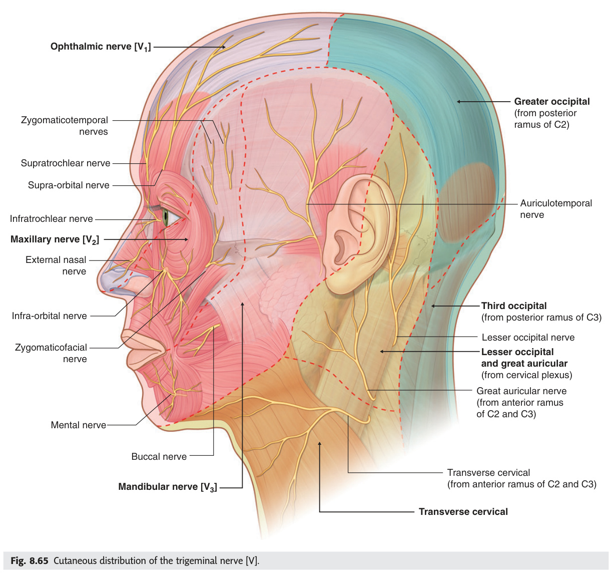
Largest cranial nerve. Emerges from pons. Three divisions: V1 (ophthalmic), V2 (maxillary), V3 (mandibular). V1 + V2 = purely sensory; V3 = sensory + motor. Trigeminal ganglion (semilunar/Gasserian ganglion) = cell bodies for V1–V3; located in Meckel's cave (dural pocket on petrous temporal bone — petrous apex).
| Division | Foramen | Sensory territory | Important branches |
|---|---|---|---|
| V1 Ophthalmic | Superior orbital fissure | Forehead + upper eyelid + nose (dorsum + tip) + cornea + anterior scalp | Lacrimal (lateral orbit) + Frontal (supratrochlear + supraorbital) + Nasociliary (corneal reflex afferent + ethmoidal nerves). V1 shingles (herpes zoster ophthalmicus): vesicles on forehead + nose tip (Hutchinson's sign = nasociliary branch → at risk for ocular herpes) |
| V2 Maxillary | Foramen rotundum → pterygopalatine fossa → infraorbital foramen | Cheek + lower eyelid + nose (lateral) + upper lip + upper teeth + hard palate | Infraorbital nerve (main terminal branch, exits infraorbital foramen → cheek/lip); zygomatic nerve (anastomoses with lacrimal for lacrimation); nasopalatine; greater palatine |
| V3 Mandibular | Foramen ovale | Lower lip + chin + lower teeth + tongue (anterior 2/3 general sensation) + auricle + TMJ + external auditory canal | Inferior alveolar (enters mandibular foramen → mental foramen → chin/lower lip); lingual nerve (general sensation anterior 2/3 tongue; joined by chorda tympani for taste + submandibular parasympathetics); auriculotemporal; buccal nerve; motor: masticatory muscles (masseter, temporalis, medial + lateral pterygoids) |
| Nucleus | Location | Modality |
|---|---|---|
| Chief (principal) sensory nucleus | Pons | Fine touch + pressure from face (projects to contralateral VPM via trigeminal lemniscus) |
| Spinal nucleus | Medulla → C2 (continuous with dorsal horn) | Pain + temperature from face. Lesion here (lateral medullary infarct, MS, syrinx, Wallenberg's) → ipsilateral facial pain/temperature loss. The nuclear sub-division has onion-skin pattern: peri-oral region (most caudal nucleus subnucleus caudalis) → ear area most rostral. Crossed fibres = trigeminal lemniscus → VPM contralateral |
| Mesencephalic nucleus | Midbrain | Proprioception from jaw muscles (unique: cell bodies in CNS, not ganglion). Jaw jerk reflex (masseter stretch → mesencephalic → motor nucleus) |
| Motor nucleus | Pons (medial to main sensory) | Motor: masticatory muscles (masseter, temporalis, pterygoids, mylohyoid, ant. digastric). Exiting via foramen ovale in V3 |
Trigeminal neuralgia: severe, lancinating, electric-shock-like facial pain, lasting seconds, in the distribution of V2 or V3 (most common) or V2 alone. Triggered by light touch (eating, talking, cold wind — trigger zones). No sensory deficit (pure pain, not numbness). Age >50, female predominance. Most common cause: neurovascular compression (tortuous superior cerebellar artery pulsating on trigeminal root entry zone at pons — demyelination at root entry zone). MRI may show vessel loop. Secondary TN: MS (in young patients → bilateral), posterior fossa tumour, skull base metastases.
Treatment: Carbamazepine 1st line (sodium channel blocker — 80% respond). Alternatives: oxcarbazepine, gabapentin, lamotrigine, baclofen. Surgical: microvascular decompression (MVD — Jannetta procedure, open surgery, definitive for vascular compression), stereotactic radiosurgery (Gamma Knife), balloon compression / glycerol rhizotomy / radiofrequency thermocoagulation (for unfit/elderly).
- Name the three divisions of CN V, their foramina, and whether each is sensory or mixed. V1 (ophthalmic): superior orbital fissure — purely sensory (forehead, scalp, nose, cornea). V2 (maxillary): foramen rotundum → inferior orbital fissure — purely sensory (cheek, upper lip, palate). V3 (mandibular): foramen ovale — mixed sensory + motor (lower face + masseter/temporalis/pterygoids).
- Name the four trigeminal nuclei from rostral to caudal and what each processes. Mesencephalic nucleus (midbrain): proprioception from jaw muscles — unique, cell bodies inside CNS. Principal/main sensory nucleus (pons): fine touch + pressure. Spinal nucleus (pons → medulla → C2): pain + temperature — continuous with dorsal horn. Motor nucleus (pons): masticatory muscle innervation via V3.
- Why does Wallenberg syndrome cause ipsilateral facial pain/temperature loss? The lateral medullary infarct destroys the spinal trigeminal nucleus and tract (in the lateral medulla) → ipsilateral facial pain/temperature loss. Simultaneously, the spinothalamic tract (contralateral body) is damaged → crossed sensory loss pattern (ipsilateral face + contralateral body).
- Describe trigeminal neuralgia — features, cause, first-line treatment. Severe lancinating electric-shock pain in V2/V3 territory, lasts seconds, triggered by light touch (trigger zones: upper lip, nasal ala, cheek). No objective sensory deficit. Most common cause: neurovascular compression of root entry zone at pons (superior cerebellar artery). First-line: carbamazepine. Surgical: microvascular decompression (Jannetta) if refractory.
- What is the jaw jerk reflex and which nuclei mediate it? Tap chin downward → jaw closes. Afferent: mesencephalic trigeminal nucleus (stretch receptors in masseters). Efferent: trigeminal motor nucleus → V3 → masseter contraction. Brisk jaw jerk = UMN lesion above pons (bilateral corticobulbar tract disease, e.g., pseudobulbar palsy); absent = LMN/peripheral CN V lesion or normal variant.
Cranial Nerve Nuclei — Complete Reference ★★★
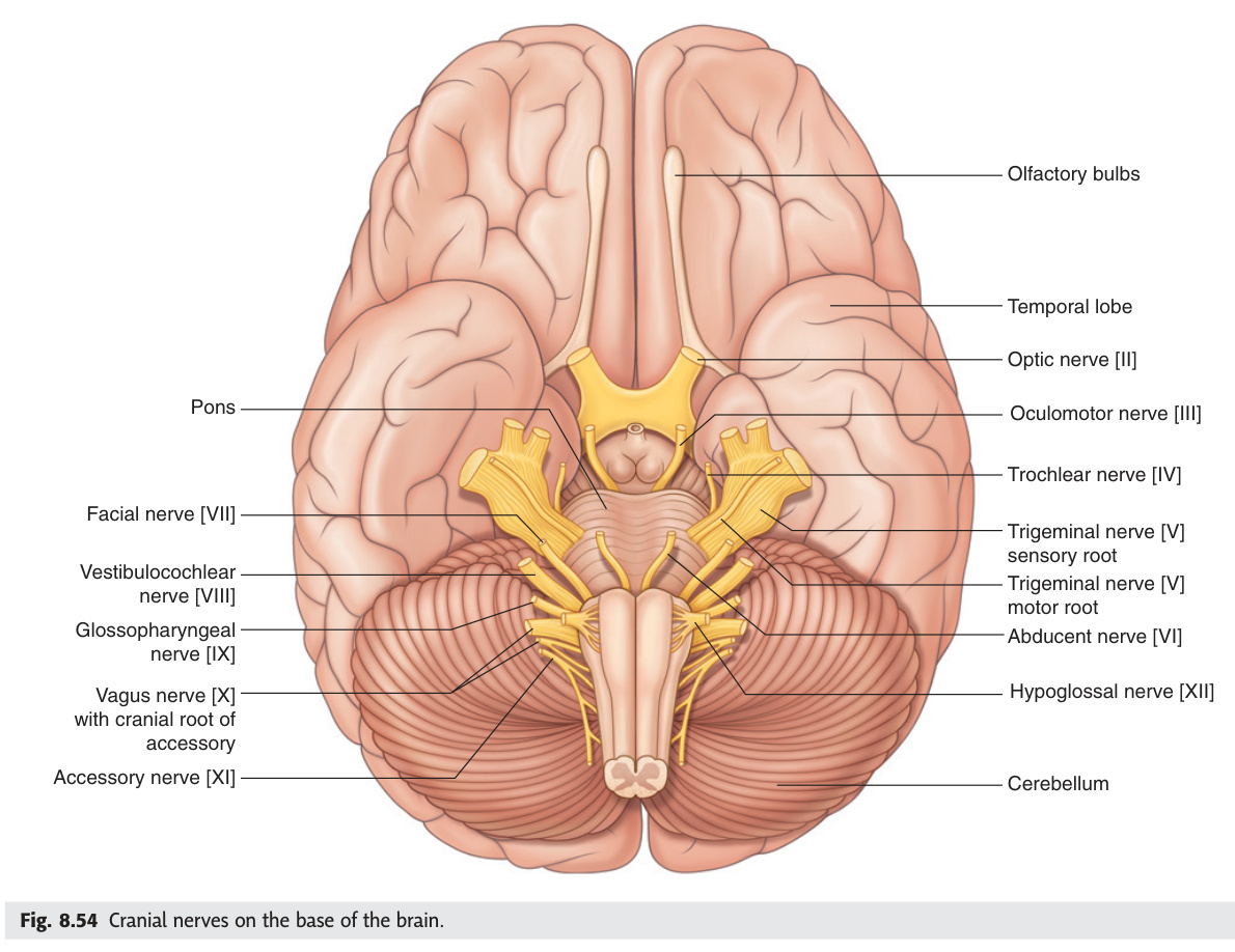
The twelve cranial nerves are best learned by their brainstem level first — CN III–IV in the midbrain, CN V–VIII in the pons, CN IX–XII in the medulla — and then by their functional column. The functional column concept explains why nearby nuclei serve unrelated functions: the brainstem is organised in longitudinal columns (GSA/touch, SSA/special senses, GVA, SVA/taste, GVE/parasympathetic, SVE/branchial muscles, GSE/eye and tongue muscles), and a lesion at a given level hits multiple columns simultaneously, producing the characteristic multi-cranial-nerve syndromes. The most clinically tested paired lesions are the pontomedullary junction syndromes: CN VI + VII palsy together (facial colliculus lesion, as CN VII loops over CN VI nucleus), and CN IX + X together (nucleus ambiguus in lateral medulla — dysphagia + dysphonia). The pupil light reflex is a high-yield circuit: CN II afferent → pretectal nucleus (midbrain, just anterior to superior colliculus) → bilateral Edinger-Westphal nuclei → CN III efferent → pupillary sphincter. A lesion of CN II causes a RAPD (afferent defect, both pupils fail to constrict properly on direct stimulation of the affected eye); a lesion of CN III causes a fixed dilated pupil (efferent defect, that one pupil won't constrict). The pretectal lesion in Parinaud's syndrome disrupts the light reflex while sparing the near-response pathway (which bypasses the posterior commissure) — light-near dissociation.
CN III–IV = Midbrain · CN V–VIII = Pons · CN IX–XII = Medulla. (CN I = olfactory bulb; CN II = diencephalon/LGN — not brainstem.)
Functional columns in brainstem (dorsal → ventral in cross-section): GSA (general somatic afferent — touch/pain/temp) · SSA (special somatic afferent — hearing/balance/vision) · GVA (general visceral afferent) · SVA (special visceral afferent — taste/smell) · GVE (general visceral efferent — parasympathetic) · SVE (special visceral efferent — branchial arch muscles) · GSE (general somatic efferent — eye/tongue muscles).
| CN | Name | Nucleus / location | Functional component | Key clinical correlate |
|---|---|---|---|---|
| I | Olfactory | Olfactory bulb (telencephalon, NOT brainstem) | SVA (smell) | Anosmia: cribriform plate fracture, meningioma of olfactory groove, Parkinson's (early), COVID-19. Foster Kennedy syndrome: ipsilateral optic atrophy + contralateral papilloedema (olfactory groove meningioma) |
| II | Optic | Lateral geniculate nucleus (thalamus); optic tract → V1 (calcarine cortex) | SSA (vision) | NOT a true CN (no Schwann cells — oligodendrocytes; vulnerable to MS). Optic neuritis: MS, NMO. Pupil light reflex: CN II afferent → pretectal nucleus (midbrain) → bilateral Edinger-Westphal → CN III efferent → pupil constriction |
| III | Oculomotor | Somatic motor: oculomotor nucleus (midbrain, periaqueductal grey, level of superior colliculus). Parasympathetic: Edinger-Westphal nucleus (dorsal to oculomotor nucleus) | GSE (eye movement) + GVE (pupil constriction/accommodation) | CN III palsy: ptosis (levator palpebrae), eye "down and out" (SR/IR/MR/IO paralysed → unopposed SO + LR), dilated fixed pupil (parasympathetic fibres on outer surface of nerve — first compressed by PComm aneurysm or uncal herniation). "Surgical CN III" = pupil-involving = aneurysm/herniation. "Medical CN III" = pupil-sparing = DM (vasa nervorum ischaemia spares outer fibres) |
| IV | Trochlear | Trochlear nucleus (midbrain, periaqueductal grey, level of inferior colliculus — caudal to CN III nucleus). ONLY CN to exit from dorsal brainstem; crosses in anterior medullary velum | GSE (superior oblique) | CN IV palsy: head tilt to opposite side (compensates for intorsion deficit). Test: ask patient to look down and in — diplopia worsens. Longest intracranial course → vulnerable in head trauma (most common CN damaged in closed head injury) |
| V | Trigeminal | Motor: motor V nucleus (mid-pons). Fine touch: chief (principal) sensory nucleus (mid-pons). Pain/temp: spinal nucleus (pons → medulla → C2). Proprioception: mesencephalic nucleus (midbrain) — unique: 1st-order neuron cell bodies in CNS | SVE (mastication) + GSA (face sensation) | See section 18.10. Jaw deviation towards lesion (LMN motor V — weak pterygoid). Jaw jerk ↑ in bilateral UMN lesions (pseudobulbar palsy). Corneal reflex: V1 afferent → chief sensory nucleus → bilateral facial motor nucleus → CN VII efferent → orbicularis oculi blink |
| VI | Abducens | Abducens nucleus (caudal pons, floor of 4th ventricle = facial colliculus; CN VII fibres loop over it) | GSE (lateral rectus) | CN VI palsy: medial deviation of eye, cannot abduct, horizontal diplopia worse on looking to affected side. Longest intracranial course after CN IV → false localising sign in raised ICP. Lesion at facial colliculus: CN VI + CN VII palsy + ipsilateral gaze palsy (PPRF fibres also damaged) = Foville's syndrome |
| VII | Facial | Motor: facial nucleus (caudal pons). Parasympathetic: superior salivatory nucleus (caudal pons) → submandibular + sublingual + lacrimal glands. Taste (ant. 2/3 tongue): nucleus tractus solitarius (NTS), medulla — via chorda tympani | SVE (facial expression) + GVE (parasympathetic) + SVA (taste) | UMN (central) vs LMN (peripheral) facial palsy: UMN spares forehead (bilateral cortical representation of frontalis); LMN = entire face (Bell's palsy). Bell's palsy: acute LMN CN VII (HSV-1 reactivation in geniculate ganglion → stylomastoid foramen compression). Features: complete ipsilateral facial paralysis, hyperacusis (stapedius palsy), ↓ taste ant. 2/3 tongue, ↓ lacrimation. Rx: prednisolone ± aciclovir. Ramsay Hunt syndrome: VZV reactivation → herpetic vesicles in ear (external acoustic meatus) + CN VII + CN VIII involvement |
| VIII | Vestibulocochlear | Cochlear division: cochlear nuclei (anterior + posterior; pontomedullary junction, lateral aspect). Vestibular division: vestibular nuclei (4 nuclei; floor of 4th ventricle, pontomedullary junction) | SSA (hearing + balance) | Acoustic neuroma (vestibular schwannoma): grows in internal acoustic meatus → compresses CN VIII (unilateral sensorineural deafness + tinnitus) then CN VII (facial palsy), then CN V (facial numbness). Bilateral = NF2 (chromosome 22 mutation). Ménière's disease: endolymphatic hydrops → episodic vertigo + fluctuating SNHL + tinnitus + aural fullness |
| IX | Glossopharyngeal | Motor (stylopharyngeus): nucleus ambiguus (medulla). Parasympathetic (parotid): inferior salivatory nucleus (medulla) → lesser petrosal → otic ganglion → auriculotemporal → parotid. Taste (post. 1/3 tongue) + GVA (carotid body/sinus): NTS (medulla). General sensation: spinal trigeminal nucleus | SVE + GVE + SVA + GVA + GSA | Gag reflex: CN IX afferent (posterior pharynx) → nucleus ambiguus → CN X efferent (palatal elevation). Carotid body: O2/CO2/pH sensor; carotid sinus: baroreceptor (both CN IX). Glossopharyngeal neuralgia: pain in posterior tongue/pharynx/ear — triggered by swallowing; rarer than trigeminal neuralgia |
| X | Vagus | Motor (pharynx/larynx): nucleus ambiguus (medulla). Parasympathetic: dorsal motor nucleus of vagus (floor of 4th ventricle, medulla). Taste (epiglottis) + GVA: NTS (medulla). General sensation (ear): spinal trigeminal nucleus | SVE + GVE + SVA + GVA + GSA | Recurrent laryngeal nerve (branch of CN X): left RLN loops under aortic arch (longer) → compressed by lung apex tumour (Pancoast), lymphoma, aortic aneurysm → hoarseness. Right RLN loops under right subclavian artery. Vagal syncope: excessive GVA input → dorsal motor nucleus → bradycardia + hypotension → presyncope |
| XI | Accessory | Spinal nucleus: anterior horn C1–C5 (exits via foramen magnum, joins cranial root). Cranial root: nucleus ambiguus (medulla — joins CN X for pharyngeal/laryngeal muscles). Exits via jugular foramen | SVE (SCM + trapezius) | CN XI palsy (neck dissection, jugular foramen syndrome, posterior triangle surgery): weakness turning head to opposite side (SCM) + shoulder drop/shrug weakness (trapezius). Shoulder abduction limited above 90° if trapezius weak (serratus anterior + trapezius together elevate scapula for full abduction) |
| XII | Hypoglossal | Hypoglossal nucleus (medulla, floor of 4th ventricle = hypoglossal trigone). Paired nucleus, runs adjacent to midline. Exits via hypoglossal canal (anterior condylar canal) | GSE (tongue intrinsic + extrinsic muscles — except palatoglossus = CN X) | LMN CN XII palsy: tongue deviates TOWARDS lesion (paralysed genioglossus cannot push tongue to opposite side). Also: fasciculations + wasting (ALS, syringobulbia). UMN (bilateral): pseudobulbar palsy — tongue spastic, slow, dysarthria, dysphagia, emotional lability. Medial medullary syndrome (Déjérine's) = CN XII + contralateral hemiplegia + contralateral fine touch loss (see section 18.4) |
Test Unit 18 knowledge
Cerebellum, brainstem syndromes, cortical areas, basal ganglia, and all CN nuclei.
- Which cranial nerves are at each brainstem level? Midbrain: CN III (oculomotor — midbrain, level of superior colliculus) + CN IV (trochlear — midbrain, level of inferior colliculus; only CN to exit dorsally). Pons: CN V, VI, VII, VIII. Medulla: CN IX, X, XI, XII. CN I = olfactory bulb (telencephalon); CN II = optic nerve (not true CN, myelinated by oligodendrocytes).
- Trace the pupillary light reflex pathway. Light → retinal ganglion cells → CN II → optic chiasm → optic tract → pretectal nucleus (midbrain, level of superior colliculus) → bilateral Edinger-Westphal (EW) nuclei → CN III → ciliary ganglion → short ciliary nerves → pupillary sphincter (constriction). Bilateral response: one EW projects to both pupils, so shining light in one eye constricts both (direct + consensual).
- CN VI and CN VII can be damaged together by a single pontine lesion — explain the anatomy. CN VII fascicles loop around the CN VI nucleus in the dorsal caudal pons, forming the facial colliculus on the floor of the 4th ventricle. A lesion at the facial colliculus (e.g., pontine infarct, glioma) damages both simultaneously → ipsilateral LMN CN VII palsy (entire face, including forehead) + ipsilateral CN VI palsy (lateral rectus, cannot abduct).
- What is the functional difference between a CN II afferent defect and a CN III efferent defect on the pupil? CN II afferent defect (e.g., optic neuritis) → RAPD: on swinging torch, affected eye pupils both dilate paradoxically (consensual from good eye is less, direct from bad eye is absent). CN III efferent defect → unilateral fixed dilated pupil: the affected eye's sphincter cannot respond to ANY light (direct or consensual) because the efferent path is severed.
- Which nucleus is the sole source of parasympathetic innervation to the eye and what does it control? Edinger-Westphal nucleus (accessory oculomotor nucleus, midbrain). Parasympathetic preganglionic axons travel with CN III → ciliary ganglion (postganglionic) → short ciliary nerves → (1) sphincter pupillae (miosis/constriction) + (2) ciliary muscle (accommodation/near focus). CN III palsy → dilated fixed pupil + loss of accommodation.
Eye Movement Disorders ★★★
Eye movement disorders are precision anatomy — the deficits are exquisitely specific because the circuits are so tightly organised. The MLF (medial longitudinal fasciculus) is the motorway connecting the CN VI nucleus in the caudal pons to the contralateral CN III nucleus in the midbrain; its job is to ensure that when one eye abducts, the other adducts simultaneously (conjugate horizontal gaze). A unilateral MLF lesion produces internuclear ophthalmoplegia (INO): the ipsilateral eye cannot adduct on attempted contralateral gaze because the adduction signal never reaches CN III — the abducting contralateral eye overshoots and nystagmates. Convergence is spared because it uses a separate pathway. Bilateral INO in a young woman = MS until proven otherwise; unilateral in an elderly man = basilar artery perforator infarct. The one-and-a-half syndrome adds PPRF destruction to the MLF lesion: the patient loses conjugate gaze entirely towards the lesion side (the "one") and also loses adduction of the ipsilateral eye on gaze away (the "half") — the only residual movement is abduction of the contralateral eye. Pupil-involving CN III palsy (surgical/compressive) vs pupil-sparing (medical/ischaemic) is one of the most important distinctions in emergency neurology: the parasympathetic fibres ride the outside of CN III and are compressed first by an aneurysm or herniation, while ischaemia from diabetic vascular disease hits the core motor fibres and spares the outer parasympathetics — any pupil involvement mandates emergency imaging for PComm aneurysm.
A paired white matter tract running in the dorsal brainstem from the rostral midbrain to the cervical spinal cord. Its key role: links the CN VI nucleus (abducens, caudal pons) to the contralateral CN III nucleus (oculomotor, midbrain) to coordinate conjugate horizontal gaze. When the right PPRF fires to move both eyes right: right CN VI nucleus fires → right lateral rectus contracts + interneurons cross in MLF → left CN III nucleus → left medial rectus contracts. A lesion of the MLF breaks this link.
| Syndrome | Site of lesion | Deficits | Classic cause |
|---|---|---|---|
| Internuclear ophthalmoplegia (INO) | MLF (between CN VI nucleus and contralateral CN III nucleus) | On lateral gaze: ipsilateral eye cannot adduct (medial rectus not receiving signal from MLF) + contralateral (abducting) eye shows nystagmus. Convergence is typically INTACT (because convergence pathway does not use the MLF). Named for the side of the adduction failure: right INO = right eye fails to adduct on left gaze = right MLF lesion. | Bilateral INO in a young woman = MS until proven otherwise. Unilateral INO in elderly = brainstem infarct (basilar perforator) |
| One-and-a-half syndrome | Unilateral: PPRF or CN VI nucleus + ipsilateral MLF | "One" = conjugate gaze palsy towards the lesion (PPRF/CN VI nucleus damage — neither eye can look towards lesion side). "Half" = INO on attempted gaze away from lesion (MLF damage — ipsilateral eye cannot adduct). Net result: only movement possible is abduction of the contralateral eye. All other horizontal movements lost. Vertical gaze and convergence preserved. | MS, pontine infarct, pontine glioma. The only eye that can move is the one contralateral to the lesion (it can abduct — hence "wall-eyed" appearance) |
| Parinaud's syndrome (dorsal midbrain syndrome) | Dorsal midbrain (pretectal area — superior colliculi, posterior commissure) | (1) Upgaze palsy — cannot look upward (not downward; upgaze = pretectal/posterior commissure). (2) Pupil light-near dissociation — pupils respond poorly to light but constrict normally to near target (accommodation-convergence reflex spared because it uses different fibres; light reflex fibres decussate in posterior commissure = damaged). (3) Convergence-retraction nystagmus — on attempted upgaze, eyes converge and retract into orbits (co-contraction of all extra-ocular muscles). (4) Collier's sign (lid retraction) — bilateral upper lid retraction (Collier's sign = sunset-in-reverse; explains "setting sun" sign in hydrocephalus). | Pineal gland tumour (germinoma, pineocytoma) compressing dorsal midbrain. Hydrocephalus (dilated posterior 3rd ventricle/aqueduct). Midbrain infarct (PCA/basilar). MS |
| Weber's syndrome | Ventral (basis) midbrain — cerebral peduncle + CN III fascicles | Ipsilateral CN III palsy (complete: ptosis + eye "down and out" + dilated fixed pupil — because CN III fascicles run through or near the peduncle). Contralateral hemiplegia (arm + leg; corticospinal fibres in cerebral peduncle, above their decussation). Face spared (corticobulbar fibres also in peduncle, but bilateral cortical representation of face means unilateral lesion doesn't cause clear facial weakness). Classic "alternating" brainstem syndrome pattern. | PCA infarct, midbrain haemorrhage, transtentorial (uncal) herniation compressing CN III against the peduncle (surgical emergency) |
| Claude's syndrome | Dorsal midbrain tegmentum — red nucleus + CN III fascicles | Ipsilateral CN III palsy + contralateral cerebellar ataxia + tremor (red nucleus/superior cerebellar peduncle). Motor/sensory cortex spared (peduncle not involved — contrast with Weber's). "Claude = Cerebellum": the ataxia distinguishes it from Weber's (which has hemiplegia not ataxia). | PCA occlusion (thalamoperforating branches) |
| CN III palsy — pupil-involving vs pupil-sparing | Compressive (outer nerve) vs ischaemic (vasa nervorum) | Pupil-involving ("surgical"): dilated, fixed, non-reactive pupil + complete ptosis + "down and out" eye. Parasympathetic fibres run on the outer surface of CN III in the subarachnoid space → compressed first by aneurysm or herniation. Cause: posterior communicating artery (PComm) aneurysm, uncal herniation. EMERGENCY — urgent imaging. Pupil-sparing ("medical"): complete ptosis + "down and out" eye + pupils equal and reactive. Ischaemia of vasa nervorum (small vessels inside the nerve) → central fascicles (motor) ischaemic, outer parasympathetic fibres spared. Cause: diabetic microvascular disease (most common), HTN. Usually resolves spontaneously in 3 months. CT/MRI angiography still recommended to exclude aneurysm if any pupil involvement. |
Key rule: ANY pupil involvement → treat as aneurysm until proven otherwise. Pupil-sparing alone is not sufficient to exclude aneurysm — always image. |
Q: A 28-year-old woman with known MS complains of double vision. On examination, on left lateral gaze her right eye does not adduct, and her left eye shows nystagmus. Convergence is intact. Localise and explain.
A: Right internuclear ophthalmoplegia (right INO). The right eye fails to adduct on left lateral gaze = right medial longitudinal fasciculus (MLF) lesion (right MLF carries the signal from the left CN VI nucleus to the right CN III nucleus for right medial rectus activation). The abducting left eye shows compensatory nystagmus (dissociated nystagmus). Convergence intact because the near-response pathway bypasses the MLF. Bilateral INO in a young woman is MS until proven otherwise. If unilateral in elderly → basilar artery perforator infarct.
Q: A 45-year-old man is brought in drowsy with a right eye pointing down and out with a dilated pupil. CT head shows no haemorrhage. Explain the mechanism and immediate management.
A: Pupil-involving right CN III palsy. The dilated, fixed, non-reactive pupil is caused by compression of parasympathetic fibres on the outer surface of CN III — these are the first fibres affected by external compression. The most dangerous cause is a posterior communicating artery (PComm) aneurysm. CT without contrast misses aneurysms — requires CT angiography or MRI/MRA urgently. If aneurysm confirmed, neurosurgical or interventional neuroradiology referral (surgical clipping or endovascular coiling). Do NOT falsely reassure by a "negative" CT without contrast.
Q: Distinguish Parinaud's syndrome from a unilateral CN III palsy.
A: Parinaud's syndrome = dorsal midbrain lesion (pretectal): selective UPGAZE palsy (downward and horizontal gaze preserved); bilateral pupil light-near dissociation (pupils react to accommodation but not to light — posterior commissure decussation damaged); convergence-retraction nystagmus on attempted upgaze; lid retraction (Collier's sign). Cause: pineal tumour, hydrocephalus. CN III palsy = unilateral oculomotor nerve lesion: ptosis + eye deviated down and out + dilated fixed pupil (if pupil-involving). Parinaud's affects gaze, not a single nerve; is bilateral; and has pathognomonic light-near dissociation + convergence-retraction nystagmus.
- What is internuclear ophthalmoplegia (INO) — which structure is lesioned and what are the signs? MLF lesion. On attempted gaze away from the lesion: the ipsilateral eye fails to adduct (medial rectus signal from MLF severed) + the abducting contralateral eye shows nystagmus. Convergence is intact (uses separate pathway). Named for the side of adduction failure. Bilateral INO in young woman = MS; unilateral in elderly = basilar perforator infarct.
- Explain the one-and-a-half syndrome — lesion, mechanism, and clinical finding. Unilateral PPRF (or CN VI nucleus) + ipsilateral MLF lesion. "One" = complete conjugate gaze palsy towards the lesion (PPRF/CN VI nucleus). "Half" = INO on gaze away (MLF damage, ipsilateral eye can't adduct). Only movement: contralateral eye abducts. All other horizontal eye movements are abolished.
- Distinguish pupil-involving from pupil-sparing CN III palsy in terms of cause and urgency. Pupil-involving ("surgical"): dilated fixed pupil + ptosis + eye down-and-out. Cause: PComm aneurysm or uncal herniation compressing outer parasympathetic fibres. EMERGENCY — CT angiography immediately. Pupil-sparing ("medical"): ptosis + down-and-out, pupils equal and reactive. Cause: diabetic/hypertensive vasa nervorum ischaemia (inner motor fibres ischaemic, outer parasympathetics spared). Any pupil involvement = imaging regardless.
- Name four features of Parinaud's syndrome and state the lesion site. Dorsal midbrain/pretectal lesion (e.g., pinealoma, hydrocephalus). (1) Upgaze palsy (not downgaze). (2) Pupil light-near dissociation (accommodation-convergence intact, light reflex lost). (3) Convergence-retraction nystagmus on attempted upgaze. (4) Collier's sign (bilateral upper lid retraction). "Sunset sign" in hydrocephalic infants = downward gaze with lid retraction.
- Weber's vs Claude's midbrain syndrome — how do they differ? Both have ipsilateral CN III palsy. Weber's (ventral peduncle): contralateral hemiplegia (corticospinal fibres). Claude's (dorsal tegmentum, red nucleus): contralateral cerebellar ataxia + tremor (red nucleus / SCP fibres). Mnemonic: "Claude = Cerebellum, Weber = Weakness."
Horner's Syndrome ★★
Horner's syndrome is the clinical signature of sympathetic denervation to the eye — and because the sympathetic pathway travels a remarkably long, vulnerable course from the hypothalamus to the face, the syndrome is a window into pathology at three very different anatomical levels. The three-neuron chain starts in the hypothalamus, descends through the lateral brainstem tegmentum (1st order), exits at C8–T2 through the apex of the lung and around the subclavian artery (2nd order), then travels with the internal carotid artery through the cavernous sinus to the eye (3rd order). The triad — partial ptosis (Müller's muscle), miosis (dilator pupillae), anhidrosis (only 1st/2nd order, since sweat fibres follow the external carotid not internal carotid) — is the same regardless of level; the accompanying features localise it. A painful Horner's with ipsilateral neck/face pain in a young person after minor trauma = internal carotid dissection (3rd order, emergency). A Horner's with ipsilateral arm/hand wasting and apical lung mass = Pancoast tumour (2nd order). A Horner's as part of a lateral medullary syndrome with crossed sensory loss and dysphagia = Wallenberg's (1st order). Pharmacological testing with cocaine (confirms Horner's at any level) and hydroxyamphetamine (distinguishes 3rd order from 1st/2nd order) provides an objective bedside localisation tool when the clinical picture is uncertain.
Horner's syndrome: interruption of the oculosympathetic pathway at any level → classic triad: (1) Ptosis (partial — Müller's muscle denervated; levator palpebrae = CN III, intact), (2) Miosis (pupil constriction — dilator pupillae denervated), (3) Anhidrosis (loss of sweating — only if 1st or 2nd order neuron lesion; 3rd order lesion spares because sweat fibres travel with external carotid, not internal carotid). Additional features: apparent enophthalmos (eyeball appears sunken due to upper + lower lid position change), lower lid elevation (upward drift of lower eyelid = "reverse ptosis" or "upside-down ptosis").
Three-neuron oculosympathetic pathway:
| Neuron order | Pathway | Causes of lesion at this level |
|---|---|---|
| 1st order (central) | Hypothalamus → descends ipsilaterally through brainstem (lateral tegmentum) → lateral horn of spinal cord C8–T2 (ciliospinal centre of Budge, or Budge-Waller centre) | Lateral medullary infarct (Wallenberg's — PICA occlusion), brainstem tumour, MS plaque, syringomyelia (C8–T2 cavity destroys ciliospinal centre), spinal cord infarct at T1 |
| 2nd order (preganglionic) | From ciliospinal centre → exits cord via ventral root T1 → crosses apex of lung → loops over subclavian artery → ascends along internal carotid → synapses in superior cervical ganglion (level of carotid bifurcation, deep to parotid) | Pancoast tumour (apex of lung, T1 root involvement — commonest cause of Horner's with arm pain/wasting), cervical rib, brachial plexus injury (C8–T1), thyroid surgery, neck dissection, central venous line insertion (subclavian), thoracic aortic dissection/aneurysm |
| 3rd order (postganglionic) | From superior cervical ganglion → fibres travel with internal carotid artery → enter cavernous sinus → join V1 → nasociliary nerve → ciliary ganglion → short ciliary nerves → dilator pupillae + Müller's muscle. (Sweat fibres travel with external carotid → NOT involved in 3rd order lesion → no anhidrosis) | Internal carotid artery dissection (painful Horner's + neck/face pain = carotid dissection until proven otherwise — EMERGENCY), cavernous sinus thrombosis/tumour (often with other CN III/IV/VI/V1 involvement = cavernous sinus syndrome), cluster headache (pericarotid inflammation), paediatric: neuroblastoma |
Step 1 — Confirm Horner's: Cocaine 4–10% eye drops (or apraclonidine 0.5–1%).
Cocaine blocks noradrenaline reuptake → dilates normal pupil; in Horner's (any level) — denervated dilator pupillae fails to dilate → Horner's pupil does NOT dilate (anisocoria increases with cocaine).
Apraclonidine (α2 > α1 agonist): in Horner's, denervation supersensitivity of α1 receptors → apraclonidine dilates the Horner's pupil + constricts normal pupil = anisocoria reversal = confirms Horner's.
Step 2 — Localise: Hydroxyamphetamine 1% (Paredrine) — releases NA from intact presynaptic terminals.
If 1st or 2nd order lesion: 3rd order neuron intact → NA released → pupil DILATES (responds to hydroxyamphetamine).
If 3rd order lesion: postganglionic neuron damaged → no NA stores → pupil DOES NOT dilate (fails to respond).
Summary: Hydroxyamphetamine dilates = 1st/2nd order lesion. Does not dilate = 3rd order (postganglionic) lesion.
Q: A 40-year-old man presents with sudden onset right-sided neck pain and right Horner's syndrome after a minor road traffic accident. What is the diagnosis and immediate management?
A: Right internal carotid artery dissection until proven otherwise. The mechanism: trauma causes intimal tear → subintimal haematoma compresses the peri-carotid sympathetic plexus (3rd order neurons) → Horner's syndrome. Associated features may include ipsilateral pulsatile tinnitus, ipsilateral facial/neck pain (carotidynia), and ischaemic stroke risk (thrombus propagation + embolisation). Management: urgent MRI/MRA of neck (T1 fat-sat sequence — shows crescentic hyperintensity in vessel wall = haematoma), CT angiography. Anticoagulation (heparin then warfarin) or antiplatelet therapy (debate ongoing) to prevent stroke. Monitor for neurological deficit.
Q: Describe the features that distinguish a Horner's syndrome caused by Pancoast tumour from one caused by lateral medullary infarction.
A: Pancoast tumour (2nd order neuron = preganglionic): Horner's + ipsilateral arm/hand pain (C8–T1 dermatome) + wasting of intrinsic hand muscles (T1 anterior horn/root) + rib destruction + ± Raynaud's-like features. Anhidrosis present (2nd order = preganglionic = sweat fibres interrupted). No cranial nerve signs or cerebellar signs. Chest X-ray/CT: apical lung mass.
Lateral medullary infarct/Wallenberg's (1st order neuron = central): Horner's is part of a syndrome — also has ipsilateral cerebellar ataxia, ipsilateral loss of facial pain/temperature (CN V spinal nucleus), dysphagia + dysphonia (nucleus ambiguus), contralateral body loss of pain/temperature (spinothalamic). Anhidrosis present. No arm wasting. MRI brain: lateral medullary infarct. Aetiology: PICA occlusion or vertebral artery dissection.
- State the classic triad of Horner's syndrome and explain each feature anatomically. (1) Partial ptosis: Müller's muscle (superior tarsal muscle, sympathetic) denervated; levator palpebrae (CN III) intact → only partial drooping. (2) Miosis: dilator pupillae denervated; sphincter pupillae (parasympathetic, CN III) unopposed → constriction. (3) Anhidrosis: loss of sympathetic sweat fibres — only if 1st/2nd order lesion (sweat fibres follow external carotid artery, not ICA, so 3rd order lesion spares sweating).
- Trace the 3-neuron oculosympathetic pathway and name the key cause at each level. 1st order: hypothalamus → lateral brainstem → C8–T2 (ciliospinal centre). Cause: Wallenberg's (PICA occlusion), MS, syringomyelia. 2nd order: C8–T2 → over apex of lung → subclavian artery → superior cervical ganglion. Cause: Pancoast tumour (+ arm pain + hand wasting). 3rd order: superior cervical ganglion → ICA → cavernous sinus → V1 → eye. Cause: ICA dissection (painful Horner's = emergency).
- Why does a 3rd order Horner's not cause anhidrosis? Facial sweat fibres branch off from the superior cervical ganglion and follow the external carotid artery (not the internal carotid). A 3rd order lesion affects fibres that travel with the ICA; the sweat fibres have already diverged at the ganglion and are unaffected. Anhidrosis = 1st or 2nd order lesion (proximal to the ganglion, where the sweat fibres have not yet branched).
- Describe the cocaine eye drop test and what a positive result confirms. Cocaine 4–10% (or apraclonidine 0.5%) applied to both eyes. Cocaine blocks noradrenaline reuptake → normal pupil dilates; Horner's pupil cannot dilate (denervated dilator pupillae, insufficient NE at synapse regardless of cause) → anisocoria increases. A positive test confirms Horner's syndrome at any level but does not localise it.
- How does hydroxyamphetamine localise Horner's syndrome? Hydroxyamphetamine releases NE from intact postganglionic terminals. If 1st/2nd order lesion: 3rd order neuron is intact → NE released → Horner's pupil dilates (responds). If 3rd order lesion: postganglionic neuron degenerated → no NE stores → pupil does NOT dilate (fails to respond). Rule: dilates = 1st/2nd order; does not dilate = 3rd order (postganglionic).