Autonomic Nervous System
Autonomic Nervous System — Comparison & Receptors
Overview
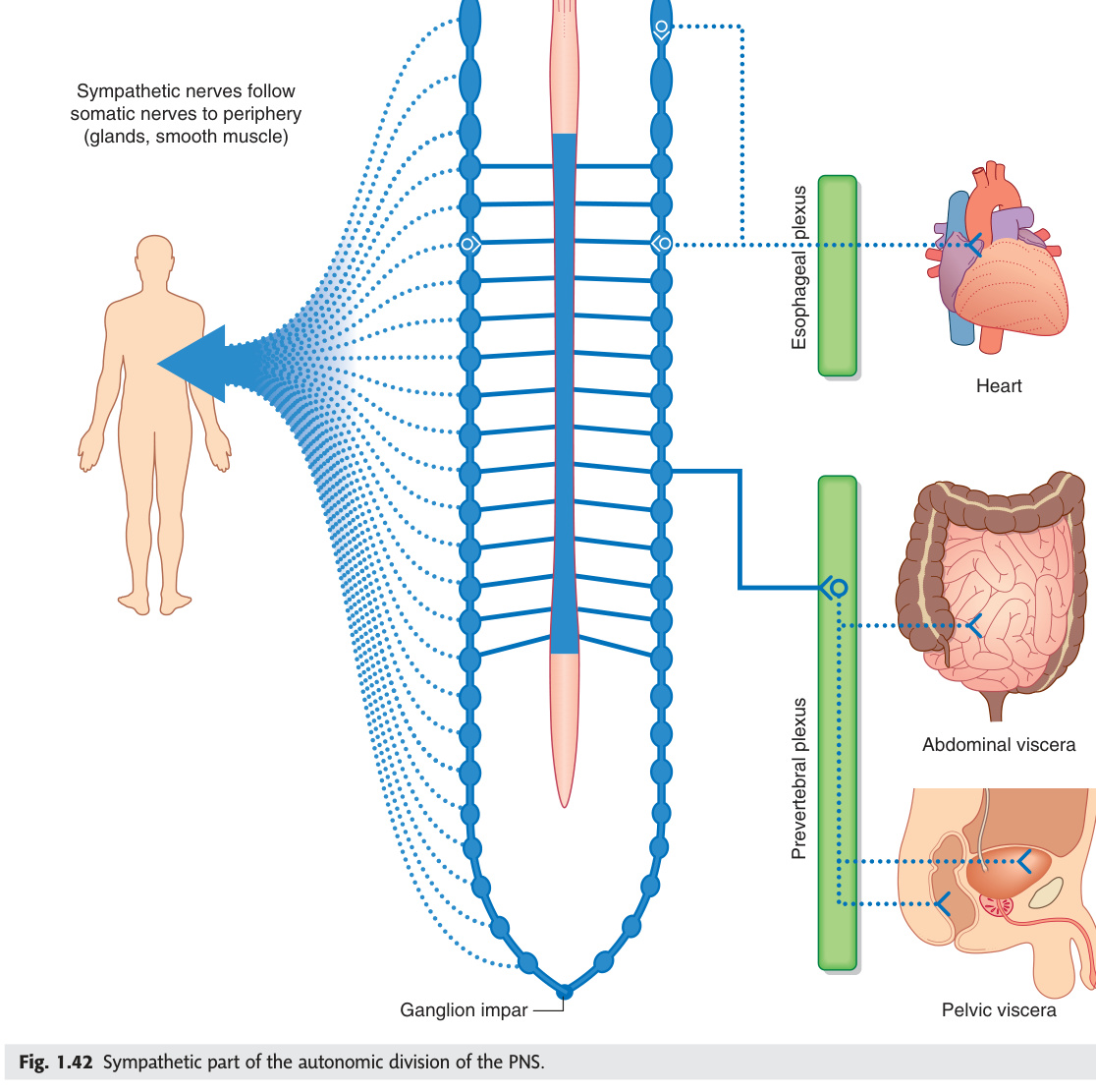
The autonomic nervous system (ANS) runs the body's background machinery — heart rate, blood pressure, digestion, bladder, pupils — without conscious input. It has two divisions that oppose each other: the sympathetic ("fight-or-flight," thoracolumbar T1–L2) and the parasympathetic ("rest-and-digest," craniosacral CN III/VII/IX/X + S2–S4). The rule that locks the anatomy into place: sympathetic preganglionic fibres are short and synapse in paravertebral ganglia far from the target organ, while parasympathetic preganglionic fibres are long and synapse in ganglia near or inside the target organ. Both systems use acetylcholine at the preganglionic synapse (nicotinic receptors) — but at the postganglionic synapse, sympathetic releases noradrenaline (adrenergic receptors) while parasympathetic releases acetylcholine again (muscarinic receptors). The one exception to memorise: sympathetic sweat gland fibres are cholinergic postganglionic, not adrenergic.
| Feature | Sympathetic ("fight or flight") | Parasympathetic ("rest and digest") |
|---|---|---|
| Origin | Thoracolumbar: T1–L2 lateral horn of spinal cord | Craniosacral: CN III, VII, IX, X (brain) + S2–S4 (sacral) |
| Pre-ganglionic fibre | Short myelinated (B fibre) | Long myelinated (B fibre) |
| Ganglion location | Paravertebral (sympathetic chain) or prevertebral (coeliac, superior mesenteric, inferior mesenteric ganglia) | In or near target organ (intramural or close ganglia: ciliary, pterygopalatine, submandibular, otic) |
| Post-ganglionic fibre | Long unmyelinated (C fibre) | Short unmyelinated (C fibre) |
| Neurotransmitters | Pre: ACh (nicotinic). Post: noradrenaline (α + β receptors). Exception: sweat glands + some vasodilator vessels → ACh (muscarinic). Adrenal medulla: ACh → chromaffin cells release adrenaline/noradrenaline directly into blood | Pre: ACh (nicotinic). Post: ACh (muscarinic). All parasympathetic post-ganglionic = cholinergic |
- What are the origins of the sympathetic and parasympathetic systems? Sympathetic: thoracolumbar (T1–L2 lateral horn). Parasympathetic: craniosacral — CN III, VII, IX, X from brainstem + S2–S4 sacral cord. No autonomic output from cervical or lower lumbar/sacral levels for sympathetic.
- Where do sympathetic vs parasympathetic preganglionic fibres synapse? Sympathetic: in paravertebral chain ganglia or prevertebral ganglia (far from target — short pre, long post). Parasympathetic: in terminal ganglia near or inside the target organ (long pre, short post).
- What neurotransmitters are used at each ANS synapse? Both systems: ACh at preganglionic synapse (nicotinic N2 receptors). Sympathetic postganglionic: noradrenaline (α/β adrenergic receptors). Parasympathetic postganglionic: ACh (muscarinic M1–M5). Exception: sympathetic postganglionic to sweat glands = ACh (muscarinic).
- What is the adrenal medulla in ANS terms? Modified postganglionic sympathetic neurons (chromaffin cells). Preganglionic (T8–L1 via greater splanchnic nerve) synapses directly on chromaffin cells → releases adrenaline (80%) + noradrenaline into bloodstream. No separate postganglionic neuron.
- What does the divergence ratio mean for sympathetic vs parasympathetic? Sympathetic: 1 preganglionic → 20 postganglionic (1:20 ratio) = widespread diffuse responses. Parasympathetic: 1:1 ratio = precise, organ-specific responses. Explains why "fight-or-flight" activates the whole body while parasympathetic effects are localised.
Sympathetic System — Outflow
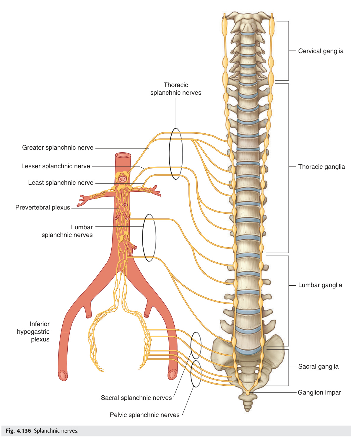
The sympathetic system is entirely thoracolumbar (T1–L2) — every sympathetic preganglionic originates from the lateral horn of the spinal cord at these levels, exits via the ventral root, and passes via white rami communicantes to the sympathetic chain. From there, the preganglionic fibres can synapse at the same level, travel up or down the chain and synapse at another level, or pass through the chain without synapsing to reach prevertebral ganglia via splanchnic nerves. The splanchnic nerves are the key visceral pathways: the greater (T5–T9) reaches the coeliac ganglion, the lesser (T10–T11) the superior mesenteric, and the lumbar splanchnics (L1–L2) the inferior mesenteric and hypogastric plexus. The adrenal medulla is the one target that receives preganglionic fibres directly (T8–L1 via greater splanchnic) without a postganglionic relay — because chromaffin cells are embryologically modified postganglionic neurons.
| Target organ | Pre-ganglionic level | Sympathetic effect |
|---|---|---|
| Head (pupil, salivary glands, blood vessels, sweat) | T1–T2 → superior cervical ganglion → follow arteries | Pupil dilation (dilator pupillae) + vasoconstriction + reduced salivation (thicker saliva) |
| Heart | T1–T5 → cardiac accelerator nerves | Increased HR + force (β1) |
| Lungs | T2–T5 → pulmonary plexus | Bronchodilation (β2) + reduced secretions |
| GI tract | T5–L2 → coeliac/superior/inferior mesenteric ganglia via splanchnic nerves | Reduced peristalsis + constrict sphincters + reduce secretions. Splanchnic vasodilation → blood diverted to muscles in fight/flight |
| Adrenal medulla | T8–L1 → greater splanchnic nerve → adrenal medulla directly (no postganglionic — chromaffin cells are modified postganglionic neurons) | Adrenaline (80%) + noradrenaline secreted into bloodstream |
| Bladder/genitalia | T10–L2 → hypogastric plexus | Bladder relaxation (internal sphincter contracts — prevents retrograde ejaculation) + ejaculation (sympathetic) |
| Limbs | T1–L2 → sympathetic chain → grey rami → spinal nerves → limb vessels + sweat glands + arrector pili | Vasoconstriction + sweating + piloerection |
Greater splanchnic: T5–T9 → coeliac ganglion (foregut + adrenal medulla). Lesser splanchnic: T10–T11 → superior mesenteric ganglion (midgut). Least splanchnic: T12 → renal plexus. Lumbar splanchnic: L1–L2 → inferior mesenteric ganglion + hypogastric plexus (hindgut + pelvic organs).
- What are white and grey rami communicantes? White rami: myelinated preganglionic fibres from T1–L2 ventral roots → sympathetic chain (only at these levels). Grey rami: unmyelinated postganglionic fibres from every sympathetic ganglion → every spinal nerve (distribute to skin: sweat glands, arrector pili, blood vessels).
- Name the three splanchnic nerves and their targets. Greater (T5–T9) → coeliac ganglion (foregut) + adrenal medulla. Lesser (T10–T11) → superior mesenteric ganglion (midgut). Least (T12) → renal plexus. Lumbar splanchnic (L1–L2) → inferior mesenteric + hypogastric plexus (hindgut + pelvic).
- Why does sympathetic activation cause ejaculation but parasympathetic causes erection? Erection: pelvic splanchnic (S2–S4) → vasodilation of helicine arteries → corpora fill. Ejaculation: hypogastric plexus (T10–L2) → smooth muscle contraction of vas deferens/seminal vesicles/prostate. "Point" (para = erection), "Shoot" (symp = ejaculation).
- What sympathetic level supplies the heart and what is the effect? T1–T5 → cardiac accelerator nerves → β1 receptors → ↑HR + ↑contractility + ↑conduction velocity. Blocked by β1-selective blockers (metoprolol, atenolol).
- Why is the adrenal medulla a special case in sympathetic anatomy? Chromaffin cells are modified postganglionic neurons — they receive preganglionic fibres directly (no separate postganglionic relay). They release adrenaline (80%) + noradrenaline (20%) directly into the bloodstream, acting as a circulating hormone rather than a local neurotransmitter.
Sympathetic Chain & Ganglia
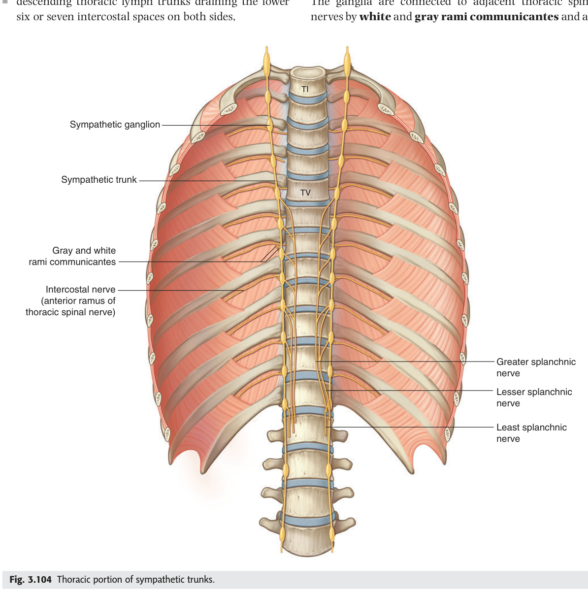
The sympathetic trunk is a paired chain of ganglia running from the base of the skull to the coccyx, just lateral to the vertebral bodies. Despite the cord only outputting sympathetic fibres from T1–L2, the chain extends the full length of the spine because preganglionic fibres travel up and down within it. The three cervical ganglia are the most clinically tested: the superior cervical ganglion (behind the ICA at C1–C3) supplies the head — its postganglionic fibres travel on the ICA to reach the dilator pupillae, Müller's muscle, and forehead sweat glands, so any lesion from the hypothalamus to the superior cervical ganglion (1st or 2nd order) can cause Horner's syndrome with facial anhidrosis. The stellate ganglion (inferior cervical + T1, behind the subclavian artery) supplies the upper limb and heart — it can be blocked for refractory ventricular tachycardia or complex regional pain syndrome.
The sympathetic trunk (paravertebral chain) runs from the base of skull to the coccyx, flanking the vertebral column (slightly anterior). 22–23 ganglia (3 cervical + 11–12 thoracic + 4 lumbar + 4–5 sacral + 1 ganglion impar). Grey rami communicantes (unmyelinated post-ganglionic): from every sympathetic ganglion to every spinal nerve. White rami communicantes (myelinated pre-ganglionic): only from T1–L2 to sympathetic trunk.
| Cervical ganglion | Location | Supplies |
|---|---|---|
| Superior cervical | C1–C3, behind internal carotid | Head (via ICA): pupil + upper eyelid (Müller's) + forehead sweat. Horner's syndrome if damaged |
| Middle cervical | C6 level (sometimes absent) | Heart + thyroid |
| Inferior cervical / stellate ganglion | Fused inferior cervical + 1st thoracic (stellate). C7/T1, behind subclavian artery | Upper limb + heart + lung. Stellate ganglion block: used for refractory VT, complex regional pain syndrome, Raynaud's |
- How many ganglia does the sympathetic trunk have and how are they distributed? ~22–23 ganglia: 3 cervical + 11–12 thoracic + 4 lumbar + 4–5 sacral + 1 ganglion impar (coccygeal). Despite preganglionic output only from T1–L2, the chain extends full length because fibres travel up/down within the trunk.
- What does the superior cervical ganglion supply and where is it located? Located at C1–C3, behind the internal carotid artery. Postganglionic fibres travel on the ICA → dilator pupillae + Müller's upper eyelid muscle + forehead sweating. Damage (1st or 2nd order lesion) → Horner's syndrome with ipsilateral facial anhidrosis.
- What is the stellate ganglion and what are its clinical uses? Fusion of inferior cervical + 1st thoracic ganglia, located at C7/T1 behind the subclavian artery. Supplies upper limb + heart + lung. Stellate block: used for refractory VT, complex regional pain syndrome (CRPS), Raynaud's. Complication: Horner's syndrome (blocks T1 preganglionic to superior cervical ganglion pathway).
- What are white rami communicantes and at which levels do they exist? Myelinated preganglionic sympathetic fibres from spinal nerve → sympathetic chain. Only at T1–L2 levels (where sympathetic cell bodies are). All other levels receive only grey rami (postganglionic) from the chain back to the spinal nerve.
- What are prevertebral ganglia (give examples) and how do they differ from paravertebral? Prevertebral = unpaired ganglia in front of vertebral column, within the abdominal/pelvic plexuses. Examples: coeliac ganglion, superior mesenteric ganglion, inferior mesenteric ganglion, aorticorenal ganglion. They receive preganglionic fibres via splanchnic nerves that pass THROUGH the paravertebral chain without synapsing.
Parasympathetic System — Outflow
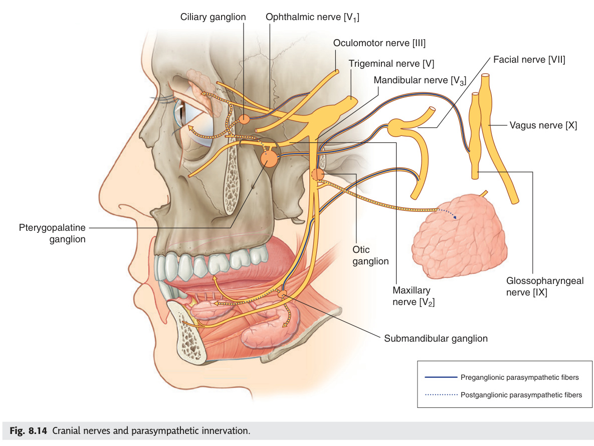
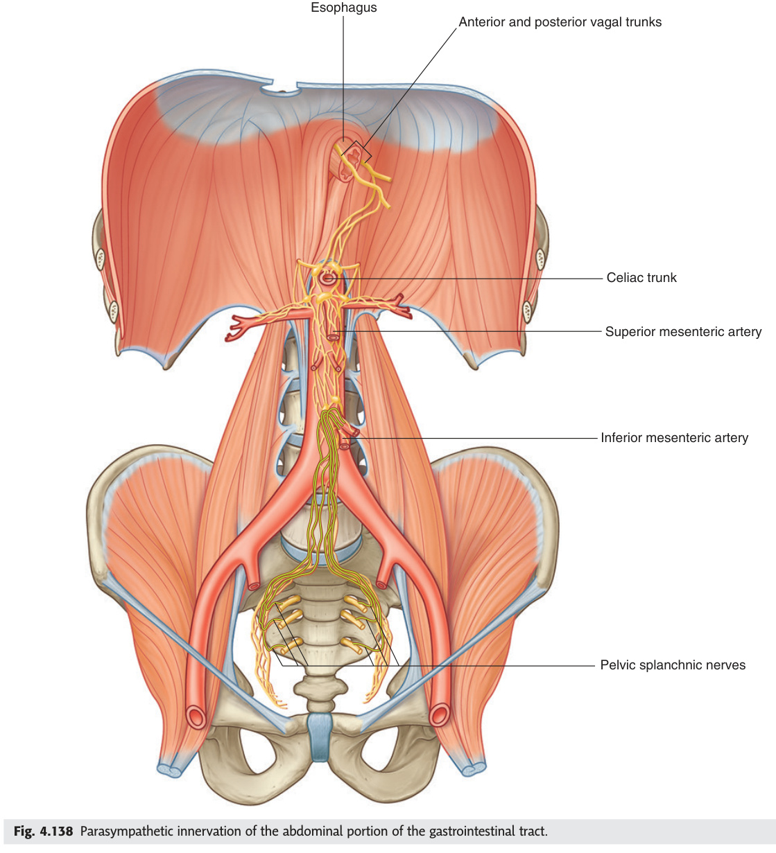
The parasympathetic outflow comes from two widely separated sources: the cranial nerves (CN III, VII, IX, X) and the sacral cord (S2–S4). CN X (vagus) is by far the dominant nerve, carrying 75% of all parasympathetic output — it supplies the heart, lungs, and the entire GI tract from oesophagus to the left colic flexure (splenic flexure). From the splenic flexure down to the pelvic organs, the pelvic splanchnic nerves (S2–S4) take over. There is a strict anatomical rule: the vagus does NOT supply pelvic organs, and the sacral parasympathetics do NOT supply above the splenic flexure. For the head, each of the four cranial ganglia (ciliary, pterygopalatine, submandibular, otic) receives fibres from a specific cranial nerve — the TMU exam has asked about CN IX → otic ganglion → parotid as a definition question ("Ganglion" in 2019 paper).
| Cranial nerve | Pre-ganglionic nucleus | Ganglion | Distribution |
|---|---|---|---|
| CN III (oculomotor) | Edinger-Westphal nucleus (midbrain) | Ciliary ganglion (orbit) | Sphincter pupillae (miosis) + ciliary muscle (accommodation) |
| CN VII (facial) | Superior salivatory nucleus (pons) | Pterygopalatine ganglion (via greater petrosal nerve): lacrimal gland + nasal + palatine glands. Submandibular ganglion (via chorda tympani → lingual nerve): submandibular + sublingual glands | Lacrimation + nasal secretion + submandibular/sublingual salivation |
| CN IX (glossopharyngeal) | Inferior salivatory nucleus (medulla) | Otic ganglion (via lesser petrosal nerve → auriculotemporal nerve → parotid gland) | Parotid salivation. Frey's syndrome (gustatory sweating): reinnervation of sweat glands by parasympathetic fibres after parotidectomy |
| CN X (vagus) | Dorsal motor nucleus of vagus (medulla) + nucleus ambiguus | Intramural ganglia within target organs | Heart (SA node — bradycardia; AV node — slows conduction) + lungs (bronchoconstriction + secretion) + GI tract (T-O junction to left colic flexure — motility + secretion). Does NOT supply pelvic organs |
| S2–S4 (pelvic splanchnic nerves) | Sacral parasympathetic nucleus | Intramural ganglia in pelvic organs | Descending colon + rectum + bladder (detrusor — micturition) + genitalia (erection — "S2–S4 keeps the penis off the floor"). Does NOT supply adrenal medulla |
Erection: Parasympathetic (pelvic splanchnic, S2–S4) → vasodilation of helicine arteries → filling of corpora cavernosa. Ejaculation: Sympathetic (hypogastric plexus, T10–L2) → smooth muscle contraction of vas deferens + seminal vesicles + prostate → emission; then somatic pudendal nerve (S2–S4) → bulbospongiosus contraction → propulsion. "Point" = parasympathetic erection. "Shoot" = sympathetic ejaculation. Both needed for normal sexual function.
- What does CN III (Edinger-Westphal nucleus) supply via the ciliary ganglion? Sphincter pupillae (miosis/pupillary constriction) + ciliary muscle (accommodation/near vision). CN III palsy → fixed dilated pupil (sphincter pupillae paralysed + sympathetic dilator unopposed) + loss of accommodation.
- What does CN VII supply parasympathetically and via which ganglia? Superior salivatory nucleus (pons). Two pathways: (1) Greater petrosal nerve → pterygopalatine ganglion → lacrimal gland + nasal/palate glands. (2) Chorda tympani → joins lingual nerve → submandibular ganglion → submandibular + sublingual salivary glands.
- CN IX → which ganglion → which gland? (TMU 2019 definition question) CN IX inferior salivatory nucleus (medulla) → lesser petrosal nerve → otic ganglion (just below foramen ovale) → postganglionic fibres travel with auriculotemporal nerve (CN V3) → parotid gland. Frey's syndrome: misdirected regeneration after parotidectomy → sweating on eating.
- What is the strict anatomical boundary of CN X (vagus) parasympathetic supply? Oesophagus → stomach → small intestine → ascending colon → transverse colon → up to the left colic (splenic) flexure. Below the splenic flexure: pelvic splanchnic nerves (S2–S4) take over. Vagus does NOT supply pelvic organs, bladder, or genitalia.
- "Point and Shoot" — explain erection vs ejaculation neural control. Erection: parasympathetic (pelvic splanchnic S2–S4) → NO-mediated vasodilation of helicine arteries → corpora fill. Ejaculation: sympathetic (hypogastric plexus T10–L2) → smooth muscle contraction → emission; somatic pudendal nerve → bulbospongiosus → propulsion. Para = Point; Symp = Shoot.
Cranial Parasympathetic Ganglia
The four cranial parasympathetic ganglia are relay stations where the long preganglionic fibre synapses before the short postganglionic fibre reaches the target. Each ganglion has three roots: a parasympathetic (motor) root that actually synapses, a sympathetic root that passes through without synapsing, and a sensory root from a branch of CN V that also passes through without synapsing. This means the ciliary ganglion, for example, carries sympathetic fibres to the dilator pupillae through it even though those fibres do not synapse there — a common exam trap. The otic ganglion (below foramen ovale, medial to V3) is the one most commonly tested because it is the relay for CN IX → parotid — and Frey's syndrome (gustatory sweating after parotidectomy, caused by misdirected parasympathetic regeneration into sweat gland territory) is a classic viva question.
| Ganglion | Location | Pre-ganglionic nerve | Post-ganglionic target |
|---|---|---|---|
| Ciliary | In orbit (behind eyeball, lateral to optic nerve) | CN III (Edinger-Westphal nucleus → inferior division → nerve to inferior oblique → ciliary ganglion) | Sphincter pupillae + ciliary muscle. Sensory root: nasociliary (CN V1) passes through without synapsing. Sympathetic root: also passes through (for dilator pupillae) without synapsing — "sympathetic root doesn't synapse in ciliary ganglion" |
| Pterygopalatine (sphenopalatine) | Pterygopalatine fossa (behind maxillary sinus) | CN VII → greater petrosal nerve → nerve of pterygoid canal → pterygopalatine ganglion | Lacrimal gland (via zygomatic → lacrimal nerve) + nasal mucosa + palate glands. Used for "cluster headache" sphenopalatine ganglion block |
| Submandibular | Hung from lingual nerve (CN V3 branch) near submandibular gland | CN VII → chorda tympani → lingual nerve → submandibular ganglion | Submandibular + sublingual salivary glands |
| Otic | Just below foramen ovale, medial to mandibular nerve | CN IX → lesser petrosal nerve → otic ganglion | Parotid salivary gland (via auriculotemporal nerve CN V3). Frey's syndrome after parotidectomy: misdirected regeneration of secretomotor fibres → sweating on eating |
- Name the four cranial parasympathetic ganglia and their CN input. Ciliary (CN III), Pterygopalatine (CN VII via greater petrosal nerve), Submandibular (CN VII via chorda tympani), Otic (CN IX via lesser petrosal nerve). Mnemonic: "Cats Play Some Music" — Ciliary, Pterygopalatine, Submandibular, Otic.
- What is the key rule about sympathetic and sensory roots of these ganglia? All four ganglia have three roots: parasympathetic (synapses here), sympathetic (passes through without synapsing), and sensory/CN V branch (passes through without synapsing). The ganglion is only a relay for the parasympathetic fibres.
- Ciliary ganglion: location, input, and what it controls. Located in the orbit, lateral to the optic nerve. Input: CN III inferior division → nerve to inferior oblique → ciliary ganglion. Output: short ciliary nerves → sphincter pupillae (miosis) + ciliary muscle (accommodation). Sympathetics to dilator pupillae pass through without synapsing.
- Pterygopalatine ganglion: where is it and what does it supply? Pterygopalatine fossa. CN VII → greater petrosal nerve → nerve of pterygoid canal → ganglion. Supplies: lacrimal gland (via zygomatic → lacrimal nerve), nasal mucosa, palate glands. Used for sphenopalatine ganglion block in cluster headache.
- What is Frey's syndrome and which ganglion does it involve? After parotidectomy, regenerating auriculotemporal nerve (CN V3) fibres from the otic ganglion misdirect into sweat gland territory instead of parotid. Result: gustatory sweating (sweating over the cheek on eating). Involves the otic ganglion (CN IX → lesser petrosal → otic → auriculotemporal → parotid).
Enteric Nervous System
The enteric nervous system (ENS) is the "second brain" — 100–500 million neurons in the gut wall that can coordinate peristalsis, secretion, and blood flow entirely independently of the brain or spinal cord. It has two plexuses: Auerbach's myenteric plexus (between the circular and longitudinal muscle layers — controls motility) and Meissner's submucosal plexus (controls secretion and absorption). The exam-critical disease here is Hirschsprung's disease: failure of neural crest cells to migrate to the distal gut → aganglionosis of the rectum (always) ± variable colon length → the aganglionic segment is permanently contracted (no inhibitory ENS neurons to relax it) → functional obstruction, proximal megacolon. The gold-standard diagnosis is rectal suction biopsy showing absent ganglion cells with increased acetylcholinesterase staining.
The "second brain" — 100–500 million neurons in the GI wall, capable of functioning independently of CNS. Two main plexuses:
| Plexus | Location | Function |
|---|---|---|
| Myenteric plexus (Auerbach's) | Between circular + longitudinal muscle layers of gut wall | Controls motility (peristalsis, segmentation). Contains predominantly excitatory (ACh + substance P) + inhibitory (NO + VIP) neurons |
| Submucosal plexus (Meissner's) | In submucosa | Controls secretion + absorption + local blood flow |
Hirschsprung's disease (congenital aganglionic megacolon): failure of neural crest cell migration → absence of ganglion cells (both Auerbach's + Meissner's plexuses) in the distal rectum (always) ± variable length of colon. Aganglionic segment: tonic contracted (no inhibitory ENS neurons) → functional obstruction. Proximal bowel: massively dilated (normal ganglion cells respond to obstruction). Presentation: neonates — failure to pass meconium within 48 hours + abdominal distension; chronic constipation in older children. Diagnosis: rectal suction biopsy (absent ganglion cells + increased acetylcholinesterase). Treat: resection of aganglionic segment + pull-through procedure. Associated: Down syndrome (5%), MEN2A (RET proto-oncogene).
- Name the two ENS plexuses, their locations, and functions. Myenteric (Auerbach's): between circular + longitudinal muscle layers → controls motility (peristalsis + segmentation). Submucosal (Meissner's): in submucosa → controls secretion, absorption, local blood flow.
- What is Hirschsprung's disease and what always fails to develop? Congenital aganglionosis from failure of neural crest cell migration. The rectum is always aganglionic; length of involvement varies. Both Auerbach's + Meissner's plexuses absent in the affected segment.
- Why is the aganglionic segment contracted and the proximal bowel dilated? Aganglionic segment: no inhibitory ENS neurons → tonic contraction → functional obstruction. Proximal normal bowel: ganglion cells intact, responds to obstruction with dilatation → megacolon.
- How is Hirschsprung's diagnosed and treated? Diagnosis: rectal suction biopsy — absent ganglion cells + increased acetylcholinesterase staining (diagnostic hallmark). Treatment: resection of aganglionic segment + pull-through procedure (Swenson, Soave, or Duhamel technique).
- What genetic associations does Hirschsprung's have? Down syndrome (5% of Hirschsprung's cases have trisomy 21). MEN2A: RET proto-oncogene mutation (10q11.2) — same gene involved in both familial medullary thyroid cancer/phaeochromocytoma and Hirschsprung's. RET mutations cause failure of neural crest migration.
Autonomic Receptors
Autonomic receptors are the translation layer between neurotransmitter and tissue response — knowing the receptor tells you instantly what drug will block or mimic that effect. For adrenergic receptors: α1 on vascular smooth muscle causes vasoconstriction (blocked by prazosin/tamsulosin — used for BPH and hypertension), β1 on the heart increases rate and contractility (blocked by metoprolol — used for heart failure, AF), and β2 on bronchial smooth muscle causes bronchodilation (activated by salbutamol — used in asthma). For muscarinic receptors: M2 on the SA/AV node is the target of atropine (blocks vagal bradycardia — used in organophosphate poisoning), and M3 on smooth muscle and glands is blocked by oxybutynin (overactive bladder) and activated by pilocarpine (glaucoma — contracts ciliary muscle and sphincter pupillae). The α2 receptor is pre-synaptic autoreceptor — its agonism (clonidine) reduces NE release, lowering sympathetic tone, explaining its use in hypertension and ADHD.
| Receptor | Location | Response | Drug examples |
|---|---|---|---|
| α1 (post-synaptic) | Vascular smooth muscle + bladder + prostate | Vasoconstriction + bladder sphincter contraction | Prazosin/tamsulosin (α-blockers — BPH, hypertension). Phenylephrine (agonist — nasal decongestant) |
| α2 (pre-synaptic) | Pre-synaptic terminal (autoreceptor) | Inhibits NE release (negative feedback) | Clonidine (α2 agonist — HTN, ADHD) |
| β1 | Heart (SA + AV node + myocardium) + kidney (JGA — renin release) | Increased HR + contractility; renin release | Metoprolol, atenolol (selective β1 blockers — HTN, angina, AF). Dobutamine (β1 agonist — cardiogenic shock) |
| β2 | Bronchial + uterine + vascular smooth muscle | Bronchodilation + uterine relaxation + vasodilation | Salbutamol (β2 agonist — asthma, tocolysis). Non-selective β-blockers (propranolol) cause bronchospasm in asthmatics |
| M1 | CNS + gastric parietal cells | CNS effects + gastric acid secretion | Pirenzepine (M1 antagonist — peptic ulcer) |
| M2 | SA node + AV node | Bradycardia + slowed AV conduction (vagal) | Atropine (muscarinic antagonist — bradycardia, organophosphate poisoning) |
| M3 | Smooth muscle + glands + bladder detrusor | Contraction of smooth muscle; secretion; detrusor contraction | Oxybutynin (M3 antagonist — overactive bladder). Pilocarpine (M3 agonist — glaucoma) |
- A patient on propranolol develops bronchospasm — which receptor explains this? β2 on bronchial smooth muscle normally causes bronchodilation; propranolol (non-selective β-blocker) blocks β2 → bronchospasm. Use selective β1-blockers (metoprolol) in asthmatics.
- Atropine reverses organophosphate bradycardia — which receptor does it block? M2 (muscarinic) on SA/AV node; organophosphates inhibit AChE → excess ACh → vagal bradycardia; atropine blocks M2 to restore rate.
- Tamsulosin relieves BPH symptoms — which receptor and mechanism? α1-blocker; α1 on prostate smooth muscle and internal urethral sphincter → relaxation → improved urine flow without much BP effect.
- What is the α2 receptor and why does clonidine lower blood pressure? α2 is a pre-synaptic autoreceptor — its activation inhibits NE release (negative feedback). Clonidine (α2 agonist) reduces sympathetic outflow → lower HR and BP.
- Pilocarpine is used in glaucoma — which receptor and two effects? M3 agonist → contracts ciliary muscle (opens trabecular meshwork → increases aqueous drainage) AND contracts sphincter pupillae (miosis) → reduces intraocular pressure.
Clinical Applications
Clinical autonomic medicine is essentially applied receptor pharmacology — if you know which neurotransmitter is in excess or deficit and which receptor it acts on, you can predict the entire clinical picture. Diabetic autonomic neuropathy silently destroys unmyelinated C-fibres supplying every organ: resting tachycardia (cardiac vagal loss), orthostatic hypotension (no vasoconstriction on standing), gastroparesis (Auerbach's plexus damage), and anhidrosis — often discovered only when the patient presents with a painless MI. The toxidrome mnemonics are the classic exam shortcut: organophosphate poisoning (AChE inhibition) produces SLUDGE from muscarinic excess, while the anticholinergic toxidrome ("dry as a bone, red as a beet, hot as a hare, mad as a hatter") is the mirror image — every feature is the opposite. Horner's syndrome (ptosis, miosis, anhidrosis) marks a third-order sympathetic neuron lesion, classically painful in carotid dissection — the pain localises the level. Raynaud's phenomenon, where sympathetic vasoconstriction spirals out of control in cold, reminds us that the vasomotor tone is a sympathetic α1 reflex, which is why stellate ganglion block or surgical sympathectomy can be curative.
Diabetic autonomic neuropathy: commonest cause of autonomic neuropathy in developed world. Features: cardiovascular (resting tachycardia, orthostatic hypotension — no reflex vasoconstriction), GI (gastroparesis → erratic glucose control; diabetic diarrhoea → Meissner's plexus damage → impaired absorption control), genitourinary (atonic bladder → overflow incontinence; erectile dysfunction), sudomotor (anhidrosis distally → compensatory hyperhidrosis proximally). Horner's syndrome: see Unit 13 (3rd order neuron lesion = painful → carotid dissection). Raynaud's phenomenon: excessive sympathetic vasoconstriction → digits go white (ischaemia) → blue (cyanosis) → red (reperfusion) on cold exposure. Primary (no cause) or secondary (systemic sclerosis, SLE, vibration white finger). Treat: CCBs (nifedipine), sympathectomy (stellate ganglion block or surgical) in severe cases.
Muscarinic (SLUDGE/DUMBELS): Salivation (excessive), Lacrimation, Urination (incontinence), Defaecation/Diarrhoea, GI upset/vomiting, Emesis/Bradycardia/Bronchoconstriction + bronchorrhoea. Eyes: miosis (constriction) + blurred vision.
Nicotinic (excess): Muscle fasciculations → weakness → paralysis (respiratory failure → death). Tachycardia (may mask muscarinic bradycardia), hypertension.
CNS: anxiety, seizures, coma.
Treatment: atropine (blocks muscarinic — high doses needed, titrate until secretions dry) + pralidoxime (2-PAM — reactivates cholinesterase if given early, before "ageing"). Maintain airway + benzodiazepines for seizures.
Anticholinergic toxidrome (atropine, tricyclics, antihistamines, hyoscine, Jimson weed): blocks muscarinic receptors → opposite of SLUDGE:
"Mad as a hatter" (CNS) + "Dry as a bone" (no secretions, dry mouth) + "Red as a beet" (vasodilation, flushing) + "Hot as a hare" (hyperthermia — no sweating) + "Blind as a bat" (mydriasis + cycloplegia) + "Full as a flask" (urinary retention) + tachycardia.
Treatment: physostigmine (cholinesterase inhibitor — crosses BBB; for severe CNS toxicity); benzodiazepines for agitation; cooling; catheter for retention. Avoid physostigmine in TCA overdose (risk of cardiac arrest).
A fall in systolic BP ≥20 mmHg OR diastolic BP ≥10 mmHg within 3 minutes of standing (or head-up tilt to 60°), documented on at least two separate occasions. Caused by failure of the normal sympathetic reflex vasoconstriction that compensates for gravitational venous pooling on standing.
Standing → ~500–700 mL blood pools in lower limbs + splanchnic bed → ↓ venous return → ↓ cardiac output → baroreceptor reflex (carotid sinus + aortic arch) detects ↓ BP → sympathetic outflow ↑ → (1) arterial vasoconstriction (α1) restores BP; (2) venoconstriction ↑ venous return; (3) ↑ HR (β1); (4) renin-angiotensin activated. In autonomic failure, one or more of these steps fails → BP falls → cerebral hypoperfusion → presyncope or syncope.
| Category | Causes | Distinguishing Features |
|---|---|---|
| Neurogenic (autonomic failure) |
Primary: Multiple system atrophy (MSA — Shy-Drager syndrome: cerebellar/parkinsonian + severe autonomic failure); Pure autonomic failure (PAF — isolated postganglionic degeneration); Parkinson's disease (late — Lewy body α-synuclein deposits in sympathetic ganglia) Secondary: Diabetic autonomic neuropathy (most common worldwide); amyloidosis; Guillain-Barré syndrome; HIV; paraneoplastic (anti-Hu antibodies, small-cell lung cancer) |
No compensatory tachycardia on standing (HR change <15 bpm) — baroreceptor reflex intact but efferent sympathetic limb damaged → cannot vasoconstrict. Supine hypertension often co-exists (loss of vasoconstrictor tone) |
| Hypovolaemia | Dehydration, haemorrhage, Addison's disease (aldosterone deficiency → sodium/water loss), diuretics, vomiting/diarrhoea | Compensatory tachycardia IS present (reflex intact, just insufficient venous return). Treat the volume deficit |
| Drug-induced | Anti-hypertensives (alpha-blockers — prazosin, tamsulosin; ACE inhibitors; calcium channel blockers), levodopa (dopamine → vasodilation), tricyclic antidepressants (alpha-blockade), antipsychotics (alpha-blockade), opioids, diuretics | Drug history essential. Most common preventable cause in elderly. Review and rationalise medications |
| Prolonged bed rest / deconditioning | ICU admission, post-surgery, elderly immobility | Baroreceptor reflex down-regulated by chronic supine position. Graded mobilisation programme |
Symptoms: dizziness, presyncope, visual greyout on standing; syncope; "coat-hanger" headache (neck/occipital pain on standing — due to ischaemia of neck/shoulder muscles in watershed territory); cognitive slowing; falls in elderly.
Investigations: lying + standing BP (3 minutes) — gold standard bedside test; 24-hour ambulatory BP (supine nocturnal hypertension); tilt-table test (for indeterminate cases); plasma noradrenaline lying vs standing (fails to rise in postganglionic failure — PAF, DAN; rises appropriately in hypovolaemia); HbA1c (DAN); serum cortisol/Synacthen test (Addison's).
Non-pharmacological treatment: avoid prolonged standing; rise slowly from lying; elevate head of bed 10–20° at night (reduces nocturnal hypertension + renal natriuresis → expands plasma volume by morning); compression stockings + abdominal binder (reduce venous pooling); increase salt + fluid intake (2–3 L water + 8–10 g NaCl/day); counter-pressure manoeuvres (leg crossing, squatting at prodrome).
Pharmacological: Fludrocortisone (mineralocorticoid → ↑ plasma volume — first-line in hypovolaemic/neurogenic); Midodrine (α1 agonist → vasoconstriction — avoid late evening to prevent nocturnal supine hypertension); Droxidopa (norepinephrine prodrug — for neurogenic OH in MSA/Parkinson's).
| Feature | Serotonin Syndrome (5-HT excess) | Neuroleptic Malignant Syndrome (NMS) |
|---|---|---|
| Mechanism | Excess serotonin (5-HT1A + 5-HT2A) at brainstem + spinal cord receptors | Dopamine (D2) receptor blockade in nigrostriatum + hypothalamus → loss of dopaminergic inhibition |
| Causative drugs | SSRIs + MAOIs (most dangerous — never combine); SSRIs + tramadol/triptans/linezolid/fentanyl/St John's Wort; serotonin release (MDMA/ecstasy); lithium toxicity; high-dose single SSRI | Antipsychotics (first-generation: haloperidol, chlorpromazine — highest risk; second-generation less common). Abrupt withdrawal of dopaminergic drugs (L-DOPA). Metoclopramide (anti-emetic D2 blocker) |
| Onset | Rapid — hours after drug change/addition | Insidious — days to weeks after starting/increasing antipsychotic |
| Hyperthermia | Yes — severe; due to excess motor activity (clonus + tremor → heat generation) + central 5-HT1A hypothalamic effects | Yes — severe; due to loss of dopaminergic inhibition of temperature regulation in hypothalamus + extreme rigidity → heat generation |
| Autonomic features | Diaphoresis, tachycardia, hypertension, hyperthermia, tachypnoea, diarrhoea (bowel hypermotility) | Diaphoresis, tachycardia, labile BP (hypo- or hypertension), tachypnoea. Diarrhoea NOT a feature |
| Neuromuscular — KEY differentiator | Clonus (spontaneous + inducible — elicited at ankles; ocular clonus) + hyperreflexia + tremor + agitation. Lower limbs more affected than upper. Clonus is the hallmark | "Lead pipe" rigidity (severe, generalised, uniform) + akinesia. Reflexes normal or ↓. NO clonus. Cogwheel rigidity may be present |
| Mental status | Agitation, confusion, anxiety (early); coma (severe) | Confusion, altered consciousness; stupor |
| Investigations | CK elevated (rhabdomyolysis from clonus). Metabolic acidosis. Leukocytosis | CK markedly elevated (severe rhabdomyolysis from rigidity — often >10,000 IU/L). Leukocytosis. Low serum iron (diagnostic clue). LFTs elevated |
| Treatment | (1) Remove offending agents. (2) Cyproheptadine (5-HT2A antagonist — 12 mg loading, then 2 mg 2-hourly; PO/NGT). (3) Benzodiazepines (agitation + muscle hyperactivity). (4) Cooling (ice packs, cooling blanket). (5) Intubation/paralysis for severe hyperthermia/rigidity. NOT bromocriptine or dantrolene primarily. | (1) Stop antipsychotic immediately. (2) Dantrolene (muscle relaxant — reduces rigidity + hyperthermia; 1 mg/kg IV, repeat up to 10 mg/kg/day). (3) Bromocriptine (D2 agonist — restores dopaminergic tone; 2.5 mg PO TID, titrate up). (4) Benzodiazepines. (5) Cooling. (6) IV fluids (rhabdomyolysis → renal protection). NB: dantrolene also used in malignant hyperthermia (RyR1 mutation + inhalational anaesthetic/suxamethonium). |
| Duration | 24–72 hours with treatment (rapid resolution once offending drug cleared) | Days–weeks (slow resolution even after stopping antipsychotic) |
Serotonin Syndrome = CLONUS (lower limb hyperreflexia + clonus + tremor; rapid onset; cyproheptadine). NMS = RIGIDITY (lead pipe; slow onset; days after antipsychotic; dantrolene + bromocriptine). Both: hyperthermia + diaphoresis + tachycardia + elevated CK. The neuro exam distinguishes them: tap the ankle → clonus = serotonin syndrome; brick wall resistance = NMS.
Test Unit 17 knowledge
Autonomic pathways, ganglia, receptors, and clinical ANS disorders.
- Name four organ-system features of diabetic autonomic neuropathy. Cardiovascular (resting tachycardia, orthostatic hypotension); GI (gastroparesis, diabetic diarrhoea); genitourinary (atonic bladder → overflow incontinence, erectile dysfunction); sudomotor (distal anhidrosis → proximal compensatory hyperhidrosis).
- What is the classic toxidrome mnemonic for organophosphate poisoning? SLUDGE: Salivation, Lacrimation, Urination, Defaecation, GI upset, Emesis — plus bradycardia and bronchoconstriction/bronchorrhoea. Also nicotinic: fasciculations → paralysis. Treat with atropine + pralidoxime.
- What key clinical finding distinguishes serotonin syndrome from NMS? Serotonin syndrome = clonus (spontaneous, inducible ankle clonus) + hyperreflexia; rapid onset; treat with cyproheptadine. NMS = "lead pipe" rigidity; slow onset over days; treat with dantrolene + bromocriptine. Both cause hyperthermia and elevated CK.
- Describe the classic triad of Horner's syndrome and name one painful cause. Ptosis (superior tarsal muscle paralysis) + miosis (dilator pupillae denervation) + anhidrosis (ipsilateral facial); painful Horner's = carotid artery dissection (3rd-order neuron runs along ICA).
- Why does Raynaud's phenomenon respond to calcium channel blockers? Excessive α1-mediated sympathetic vasoconstriction in digital vessels; nifedipine (CCB) relaxes vascular smooth muscle by blocking voltage-gated Ca²⁺ entry, reducing the sympathetically-driven spasm.
Micturition Reflex & Neurogenic Bladder ★★★
The bladder is one of medicine's best illustrations of dual autonomic control — two opposing systems held in perfect balance, with the cortex as the social override. During storage, sympathetic T10–L2 fibres relax the detrusor (β3) and clamp the internal sphincter (α1), while the pudendal nerve voluntarily contracts the external sphincter; the pontine storage centre reinforces continence. The social decision to void flips the switch: the pontine micturition centre (PMC) activates pelvic splanchnics (parasympathetic S2–S4) to contract the detrusor via M3 and simultaneously withdraws sphincter tone — a beautifully coordinated sequence that only works if the PMC is connected to the sacral cord. Cut the connection above the pons (stroke, cortical lesion) and urgency results — the PMC is intact but no longer socially inhibited. Cut it between the pons and the sacral cord (cervical/thoracic SCI) and you get detrusor-sphincter dyssynergia: the detrusor contracts but the external sphincter also fires — high-pressure voiding that destroys kidneys. Damage the sacral cord itself (S2–S4 — cauda equina) and everything goes flaccid: atonic bladder → overflow incontinence.
| Phase | Neural Control | Mechanism |
|---|---|---|
| Storage phase | Sympathetic (T10–L2, hypogastric nerve): β3 → detrusor relaxation; α1 → internal urethral sphincter (IUS) contraction. Somatic (pudendal S2–S4): external urethral sphincter (EUS) contracted voluntarily. Pontine storage centre: inhibits voiding reflex | Urine accumulates: bladder fills → stretch receptors in wall signal to pons. Continence maintained by IUS + EUS tone |
| Voiding phase | Parasympathetic (S2–S4, pelvic splanchnic): M3 → detrusor contracts (detrusor overactivity). Sympathetic inhibited (IUS relaxes). Somatic (pudendal): EUS relaxes (voluntary relaxation). Pontine micturition centre (PMC): coordinates | PMC activated by cortex (social decision to void) → pelvic splanchnic → detrusor contracts + simultaneous IUS/EUS relaxation → voiding. PMC also inhibits EUS (via Onuf's nucleus, S2–S4) |
| Lesion level | Type | Features | Complications |
|---|---|---|---|
| Suprapontine (cortex/internal capsule/basal ganglia) — stroke, MS, Parkinson's, frontal lobe tumour | UMN — overactive (detrusor hyperreflexia) | Urgency + frequency + urge incontinence. Bladder contracts involuntarily (PMC intact, just disinhibited). Small-capacity high-pressure bladder. EUS coordination preserved (PMC intact → coordinated voiding) | Urge incontinence. No upper tract damage (because coordinated) |
| Suprasacral spinal cord (above S2–S4 — cervical/thoracic SCI, MS) | UMN — overactive + DYSSYNERGIA | Detrusor-sphincter dyssynergia (DSD): detrusor contracts but EUS also contracts simultaneously (PMC disconnected → no coordination). High voiding pressure + incomplete emptying. Autonomic dysreflexia risk (above T6) | High-pressure voiding → hydronephrosis + renal failure if untreated. Recurrent UTIs. Life-threatening autonomic dysreflexia |
| Sacral cord/conus/cauda equina (S2–S4 — disc prolapse, tumour, sacral injury) | LMN — areflexic (acontractile) | Flaccid atonic bladder. No detrusor contraction → overflow incontinence (continuous dribbling when full). Large-capacity low-pressure bladder. EUS flaccid (may leak passively) | Overflow incontinence. Recurrent UTIs. Upper tract may be preserved if low pressure |
Life-threatening emergency in SCI above T6. Trigger: any noxious stimulus below lesion (full bladder/bowel most common, pressure sore, DVT, tight clothing). Mechanism: mass sympathetic discharge below lesion (T6 and above → no supraspinal inhibition) → severe vasoconstriction → hypertension (SBP can reach 250–300 mmHg). Above T6 level: baroreceptor reflex activates → bradycardia + vasodilation above lesion (facial flushing + sweating + pounding headache), but cannot override sublesional vasoconstriction.
Symptoms: "pounding" headache (severe) + sweating + flushing above level + piloerection below + hypertension + bradycardia. Emergency management: sit patient upright (↓ venous return → ↓ BP) + identify + remove trigger immediately (catheterise, check bowel, remove tight clothing) + antihypertensive (GTN spray under tongue, nifedipine bite + swallow) + monitor BP until resolved. If untreated → intracerebral haemorrhage + death.
- During bladder storage, which nerve/receptor keeps the detrusor relaxed and which keeps the internal sphincter closed? Sympathetic T10–L2 (hypogastric nerve): β3 receptors on detrusor → relaxation; α1 receptors on internal urethral sphincter → contraction. Pudendal nerve keeps external sphincter closed voluntarily.
- What is the role of the pontine micturition centre (PMC) in voiding? PMC coordinates simultaneous detrusor contraction (via pelvic splanchnics, M3) and sphincter relaxation (inhibits Onuf's nucleus → EUS relaxes). Without intact cord connection between PMC and S2–S4, this coordination is lost → detrusor-sphincter dyssynergia.
- A patient with a T4 spinal cord injury has urge incontinence without sphincter dyssynergia — explain. Suprapontine lesion type: above T6 → above the autonomic dysreflexia threshold; PMC intact but cortical inhibition lost → detrusor hyperreflexia (urgency) but PMC coordination preserved → no dyssynergia. This contrasts with suprasacral (below pons but above S2) SCI where PMC is disconnected → DSD.
- What is autonomic dysreflexia and what is the immediate management? Life-threatening mass sympathetic discharge in SCI ≥ T6 triggered by noxious stimulus below lesion (most commonly full bladder). Presents with hypertension, bradycardia, headache, sweating/flushing above level. Management: sit upright, identify and remove trigger (catheterise, evacuate bowel), sublingual GTN or nifedipine for BP control.
- A cauda equina injury at S2–S4 causes what type of bladder and why? LMN (areflexic) bladder: damage to the sacral reflex arc → no detrusor contraction → large flaccid bladder fills continuously → overflow incontinence (continuous dribble). Low pressure so upper tracts usually preserved, but UTIs common. Managed with clean intermittent self-catheterisation (CISC).
Referred Pain Pathways
Referred pain is the anatomical reason a heart attack feels like left arm pain, and why you should never diagnose "musculoskeletal shoulder pain" without ruling out a subphrenic abscess. The mechanism is convergence-projection: visceral afferents (C-fibres travelling with sympathetic nerves) enter the dorsal horn at T1–L2 and synapse on the exact same second-order neurons as somatic afferents from skin and muscle — the brain, inexperienced with visceral signals, defaults to interpreting it as coming from the surface it knows. The rule is simple: pain is referred to the dermatome of the spinal level where the organ's afferents enter. Cardiac afferents enter at T1–T5 → left arm and chest wall; gastric/duodenal afferents at T5–T9 → epigastrium; appendix at T10 → periumbilical (classic early appendicitis pain, before parietal peritoneum is involved and pain localises to RIF). The important exception is phrenic nerve referral (C3–C5) — diaphragmatic irritation from blood, pus, or perforated viscus refers to the shoulder tip because the phrenic nerve itself carries the pain to C3–C5, not via sympathetic convergence.
Convergence-projection theory: visceral pain afferents (C-fibres, unmyelinated) travel with sympathetic fibres → enter cord at T1–L2 (pelvic organs also via S2–4 parasympathetics). At the dorsal horn, visceral afferents synapse on the same second-order neurons as somatic afferents from a given dermatome. The brain, which rarely receives direct visceral pain, interprets the signal as coming from the more familiar somatic territory → pain is "projected" (referred) to that dermatome.
Key rule: referred pain is felt in the dermatomal territory of the segmental spinal level where visceral afferents enter the cord — NOT necessarily at the organ's surface location. Phrenic nerve referral (C3–C5) to the shoulder tip is an exception mediated by diaphragmatic irritation, not T-level convergence.
| Organ / Source | Visceral afferent level | Referred to | Classic scenario |
|---|---|---|---|
| Heart (ischaemia) | T1–T5 | Central chest + left arm inner aspect (T1–T2 = medial forearm → little finger, via ulnar nerve territory) + jaw/neck (C3–C5) | Myocardial infarction; occasionally right arm or back (posterior MI) |
| Oesophagus | T3–T7 | Central retrosternal chest | Oesophageal spasm mimics cardiac chest pain |
| Gallbladder / bile ducts | T7–T9 (right greater splanchnic) | Right hypochondrium + right shoulder tip (phrenic C3–C5, diaphragmatic irritation by bile) | Biliary colic; shoulder tip pain is phrenic referral — not T7–T9 |
| Liver capsule / subphrenic abscess (right) | Phrenic nerve (C3–C5) | Right shoulder tip | Right subphrenic collection; hepatic abscess |
| Spleen / left subphrenic | Phrenic nerve (C3–C5) | Left shoulder tip | Splenic rupture → Kehr's sign: left shoulder tip pain on Trendelenburg position (blood pools under left hemidiaphragm) |
| Stomach / pancreas | T6–T9 (coeliac plexus) + T10–L2 (pancreatic tail) | Epigastrium + boring pain through to back (L1–L2) | Acute pancreatitis: epigastric pain radiating to back, relieved by leaning forward |
| Appendix (early — visceral phase) | T10 | Periumbilical (central dull aching) | First 12–24 h of appendicitis before parietal peritoneum involved |
| Appendix (late — somatic phase) | Parietal peritoneum (direct somatic) | Right iliac fossa (McBurney's point) — sharp, precisely localised | Pain shifts central → RIF as inflammation reaches parietal peritoneum |
| Small intestine / colon (mid) | T9–T11 | Periumbilical / central abdomen | Small bowel obstruction, mesenteric ischaemia |
| Kidney / upper ureter | T10–L1 (lesser + least splanchnic) | Loin (T10–T11) + groin (L1) + testis/labia (T10 = testicular dermatome) | Renal colic: loin-to-groin pain; testicular pain in a male = always consider renal/ureteric origin |
| Lower ureter | L1 | Groin + medial thigh + tip of penis (via ilioinguinal + genitofemoral) | Ureteric stone in pelvic ureter |
| Testis / ovary | T10–T11 | Periumbilical / loin | Testicular torsion presenting as abdominal pain — especially in children |
| Uterus / cervix | T10–L1 | Lower abdomen + sacral/low back | Dysmenorrhoea, labour contractions |
| Rectum / pelvic organs | S2–S4 | Perineum, sacral region, inner thighs | Rectal pain, bladder neck pain |
| Diaphragm (central part — phrenic supply) | C3–C5 phrenic nerve | Ipsilateral shoulder tip | Sub-phrenic abscess, diaphragmatic pleurisy, splenic rupture (left), hepatic/gallbladder (right) |
Q: Why does acute cholecystitis cause right shoulder tip pain in addition to right hypochondrial pain?
A: Two separate mechanisms. (1) Right hypochondrial pain: gallbladder visceral afferents travel with the greater splanchnic nerve (T7–T9) → convergence at T7–T9 dorsal horn → referred to right upper quadrant dermatomes. (2) Right shoulder tip pain: inflamed gallbladder irritates the adjacent right hemidiaphragm → stimulates phrenic nerve (C3, C4, C5) → referred to C3–C5 dermatome = shoulder tip. This is phrenic nerve referral, independent of the T7–T9 pathway.
Q: A 16-year-old presents with central periumbilical pain for 12 hours that has now shifted to the right iliac fossa. Explain anatomically.
A: Early appendicitis: visceral inflammation → afferents travel at T10 → converge on T10 dorsal horn → pain perceived in T10 dermatome = umbilical region (central, dull, poorly localised). As inflammation progresses → parietal peritoneum of the right iliac fossa becomes involved → somatic parietal pain fibres (sharp, precisely localised, carried in ilioinguinal/anterior cutaneous branches of T10–T12) → pain localises to McBurney's point (2/3 along line from umbilicus to right ASIS). The shift from visceral (central) to somatic (localised) pain is diagnostically significant.
Q: Kehr's sign — describe and explain its mechanism.
A: Kehr's sign = left shoulder tip pain precipitated or exacerbated by placing the patient in the Trendelenburg position (head-down). It is a classic sign of splenic rupture or left subphrenic pathology. Mechanism: blood or fluid pools under the left hemidiaphragm in Trendelenburg position → irritates the central left diaphragm → stimulates the left phrenic nerve (C3, C4, C5) → referred pain to the left shoulder tip (C4 dermatome). Also described with left-sided ectopic pregnancy, splenic abscess, and pancreatic tail pathology.
- Explain the convergence-projection theory of referred pain. Visceral C-fibre afferents (travelling with sympathetics, entering T1–L2) synapse on the same dorsal horn second-order neurons as somatic afferents from a dermatome. The brain interprets the signal as somatic → pain is "referred" to the dermatomal territory of that spinal level, not the organ's surface.
- A patient has central periumbilical pain that shifts to the RIF over 12 hours — what is the anatomical explanation? Early: appendix visceral afferents enter at T10 → convergence → umbilical area pain (dull, poorly localised). Later: parietal peritoneum of RIF becomes inflamed → somatic parietal fibres → sharp, well-localised pain at McBurney's point (T10–T12 dermatome, RIF).
- Why does MI cause left arm pain (T1–T5) but a perforated duodenal ulcer initially causes epigastric pain (T5–T9)? Referred pain goes to the dermatomal level where visceral afferents enter the cord: cardiac = T1–T5 → left arm/chest; duodenal = T5–T9 → epigastrium. Once parietal peritoneum is involved, pain localises to the actual site.
- What is Kehr's sign, what causes it, and which nerve carries the pain? Left shoulder tip pain worsened by Trendelenburg position → classic sign of splenic rupture. Mechanism: blood under left hemidiaphragm → irritates left phrenic nerve (C3–C5) → referred to C4 dermatome (left shoulder tip). Phrenic referral is independent of T-level sympathetic convergence.
- Why does gallbladder disease cause both right hypochondrial AND right shoulder tip pain? Two pathways: (1) visceral afferents T7–T9 → right upper quadrant dermatomes (RHC pain); (2) inflamed gallbladder irritates right hemidiaphragm → right phrenic nerve C3–C5 → right shoulder tip. Both can coexist in the same patient.
Vasovagal Syncope
Vasovagal syncope is the common faint — the body's autonomic system briefly hitting an emergency stop — and understanding its circuit explains both why it happens and how to prevent it. The trigger (pain, fear, prolonged standing, blood draw) causes venous pooling → reduced preload → the left ventricle contracts vigorously on a near-empty chamber → C-fibre mechanoreceptors in the inferoposterior LV wall fire (the Bezold-Jarisch reflex) → paradoxical withdrawal of sympathetic tone and simultaneous vagal surge → sudden BP drop + bradycardia → cerebral hypoperfusion → collapse. The key clinical point is context and recovery: young patient, prodrome (nausea, greying vision, sweating), brief LOC, rapid full recovery on becoming supine — this pattern is almost never a serious arrhythmia. The tilt-table test reproduces it; treatment is volume loading, compression stockings, and salt supplementation, with fludrocortisone or midodrine in refractory cases. Distinguishing it from cardiac syncope (no prodrome, any posture, older patient, structural heart disease) is one of medicine's most important history-taking skills.
Vasovagal (neurocardiogenic / reflex) syncope: transient loss of consciousness (TLOC) caused by a self-limited neurally-mediated reflex that produces simultaneous sympathetic withdrawal and vagal surge → acute fall in BP and/or HR → cerebral hypoperfusion → TLOC. The most common cause of syncope in young adults. Recovery is typically rapid and complete on becoming supine.
- Trigger: pain, fear, blood/gore, prolonged standing (venous pooling), heat, micturition, defecation, Valsalva, venesection
- Venous pooling (upright posture) → ↓ venous return → ↓ end-diastolic volume → vigorous contraction of a relatively under-filled left ventricle
- C-fibre mechanoreceptors in the inferoposterior left ventricular wall stimulated by forceful contraction → afferent signals via vagus → nucleus tractus solitarius (NTS) in medulla
- Paradoxical central response (the brain interprets vigorous LV contraction as "too much pressure"):
- ↓ Sympathetic outflow → arteriolar vasodilation → ↓ SVR → ↓ BP (vasodepressor component)
- ↑ Vagal outflow → bradycardia ± transient asystole (cardioinhibitory component)
- ↓ Cerebral perfusion pressure → TLOC (usually <30 seconds)
- Recovery: becoming supine → ↑ venous return → cerebral perfusion restored → rapid recovery
Inferior MI variant: inferoposterior MI stimulates the same C-fibre receptors at rest → paradoxical bradycardia + hypotension (Bezold-Jarisch) instead of the expected tachycardia — treat with atropine, not sympathomimetics alone.
Types:
| Type | Dominant mechanism | HR pattern | Treatment relevance |
|---|---|---|---|
| Vasodepressor | Sympathetic withdrawal → vasodilation | Relatively preserved HR; BP falls | Counter-pressure manoeuvres, fluids |
| Cardioinhibitory | Vagal surge → bradycardia ± asystole (>3 s) | Marked bradycardia or asystole | Pacemaker considered if asystole >6 s documented |
| Mixed | Both vasodilation + bradycardia | Both components | Most common clinical pattern |
Prodromal symptoms (seconds before TLOC): nausea, pallor, sweating (sudomotor), yawning, visual greyout/blackout, lightheadedness, muffled hearing. Prodrome = sympathetic withdrawal + vagal surge before loss of consciousness. Patient can often abort TLOC by lying down or squatting during prodrome.
| Feature | Vasovagal syncope | Cardiac arrhythmia | Epileptic seizure |
|---|---|---|---|
| Posture at onset | Upright (standing/sitting) | Any posture | Any posture |
| Prodrome | Yes — nausea, pallor, sweating (seconds) | None or palpitations | Aura (sensory, motor, déjà vu) |
| Triggers | Heat, pain, blood, crowding, venepuncture | Exercise (structural), none | Sleep deprivation, flashing lights |
| Convulsive movements | Brief myoclonic jerks (anoxic) — irregular, start after TLOC | None | Tonic then rhythmic clonic — starts at onset |
| Colour | Pallor → flushing on recovery | Pallor / cyanosis | Cyanosis (tonic phase) |
| Duration of TLOC | <30 seconds typically | Seconds to minutes | Typically 1–3 minutes |
| Recovery | Rapid, immediate; feels well quickly | Rapid | Prolonged post-ictal confusion (minutes to hours) |
| Injury | Rare (slumps slowly) | Common (sudden drop) | Common (tongue biting, incontinence) |
Acute: lay patient supine + elevate legs (Trendelenburg) → restores venous return and cerebral perfusion → usually sufficient. Atropine IV (0.6 mg) for refractory bradycardia/asystole.
Long-term / prevention:
- Counter-pressure manoeuvres (first-line, non-pharmacological): leg crossing + tensing, squatting, hand-grip + arm-tensing at onset of prodrome → ↑ venous return + ↑ peripheral resistance → prevents or aborts TLOC
- Recognise and avoid triggers; adequate hydration + salt intake; tilt training
- Fludrocortisone (mineralocorticoid): ↑ plasma volume — useful in orthostatic/hypovolaemic subtype
- Pacemaker: only for cardioinhibitory type with documented asystole >6 seconds on tilt-table test — not for vasodepressor type (pacemaker corrects bradycardia but does nothing for vasodilation)
- Beta-blockers: no longer recommended (may be harmful)
Q: Explain the mechanism of vasovagal syncope in a medical student who faints during a surgical procedure.
A: Prolonged upright standing + emotional stress (pain, blood, anxiety) → venous pooling in lower limbs → reduced venous return → vigorous contraction of under-filled left ventricle → stimulation of C-fibre mechanoreceptors (Bezold-Jarisch receptors) in inferoposterior LV wall → vagal afferents to NTS → paradoxical reflex: (1) sympathetic withdrawal → vasodilation → ↓ BP [vasodepressor component] + (2) vagal surge → bradycardia [cardioinhibitory component] → ↓ cerebral perfusion pressure → TLOC. Prodrome (nausea, pallor, sweating, visual greying) occurs during the sympathetic withdrawal phase. Becomes supine on floor → cerebral perfusion restored → rapid recovery.
Q: A patient with an acute inferior MI develops unexpected bradycardia and hypotension. Explain the mechanism and management.
A: Inferior MI (RCA territory) → ischaemia/necrosis of the inferoposterior left ventricular wall → stimulation of C-fibre mechanoreceptors in that region at rest → Bezold-Jarisch reflex → vagal surge (bradycardia) + sympathetic withdrawal (hypotension). This is a vagally-mediated paradoxical response. Management: (1) Atropine IV 0.5–1 mg (blocks vagal bradycardia) — first-line; (2) Right heart-raising manoeuvres (elevate legs) to improve RV preload (especially if RV infarction co-exists); (3) Avoid GTN/morphine which worsen hypotension. Temporary pacing if atropine-refractory.
Q: Distinguish convulsive syncope from an epileptic tonic-clonic seizure.
A: Convulsive syncope: myoclonic jerks (irregular, brief) occur in 30–50% of vasovagal episodes when cerebral anoxia is severe; they begin after loss of consciousness (consequence of anoxia, not the cause); the context is typical of syncope (upright posture, trigger, pallor, prodrome, rapid recovery without post-ictal confusion). Epileptic tonic-clonic seizure: tonic phase (rigid stiffening) precedes clonic phase (rhythmic jerking); jerking begins at or near onset of unconsciousness; post-ictal confusion/drowsiness lasts minutes to hours; tongue biting and urinary incontinence more common; EEG ictal pattern. Key discriminator: post-ictal state — absent in syncope, present in epilepsy.
- Describe the Bezold-Jarisch reflex step by step. Trigger → venous pooling → ↓ preload → vigorous LV contraction on near-empty chamber → C-fibre mechanoreceptors in inferoposterior LV wall activated → afferents via vagus → NTS in medulla → paradoxical sympathetic withdrawal + vagal surge → ↓ BP + ↓ HR → cerebral hypoperfusion → LOC.
- Name four classic prodromal features of vasovagal syncope that help distinguish it from cardiac syncope. Nausea, greying/tunnelling of vision (grey-out), sweating/clamminess, pallor/light-headedness — all occurring seconds before LOC. Cardiac syncope (arrhythmia, structural) typically has no prodrome and can occur in any posture.
- An inferior MI patient develops unexpected bradycardia and hypotension — what is the mechanism? RCA ischaemia of the inferoposterior LV wall → C-fibre stimulation → Bezold-Jarisch reflex → vagal surge + sympathetic withdrawal. Treat with IV atropine (not GTN/morphine, which worsen hypotension); temporary pacing if refractory.
- What is the tilt-table test and what does it diagnose? Patient tilted 60–80° head-up for 20–45 minutes while BP and HR monitored. Positive if symptomatic hypotension + bradycardia reproduced. Diagnoses vasovagal/neurocardiogenic syncope and POTS (postural orthostatic tachycardia syndrome, in which HR rises ≥30 bpm on tilting without hypotension).
- How do you distinguish convulsive syncope from a true epileptic tonic-clonic seizure? Convulsive syncope: irregular myoclonic jerks begin AFTER LOC (consequence of cerebral anoxia); no post-ictal confusion; typical vasovagal context. Epileptic TC: tonic phase precedes clonic; jerks begin at/near onset; post-ictal confusion lasting minutes-hours; tongue biting; EEG ictal pattern.