Spinal Cord & Spinal Nerves
Spinal Cord — Transverse Section
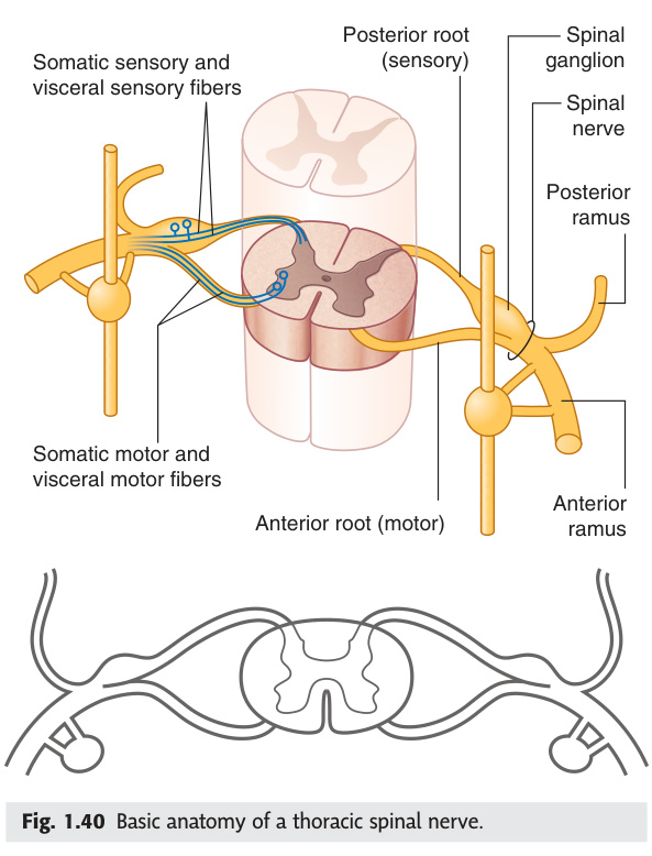
Grey matter = H-butterfly · White matter = tracts · Grey:White ratio highest in cervical enlargement (limb innervation)
Spinal Cord
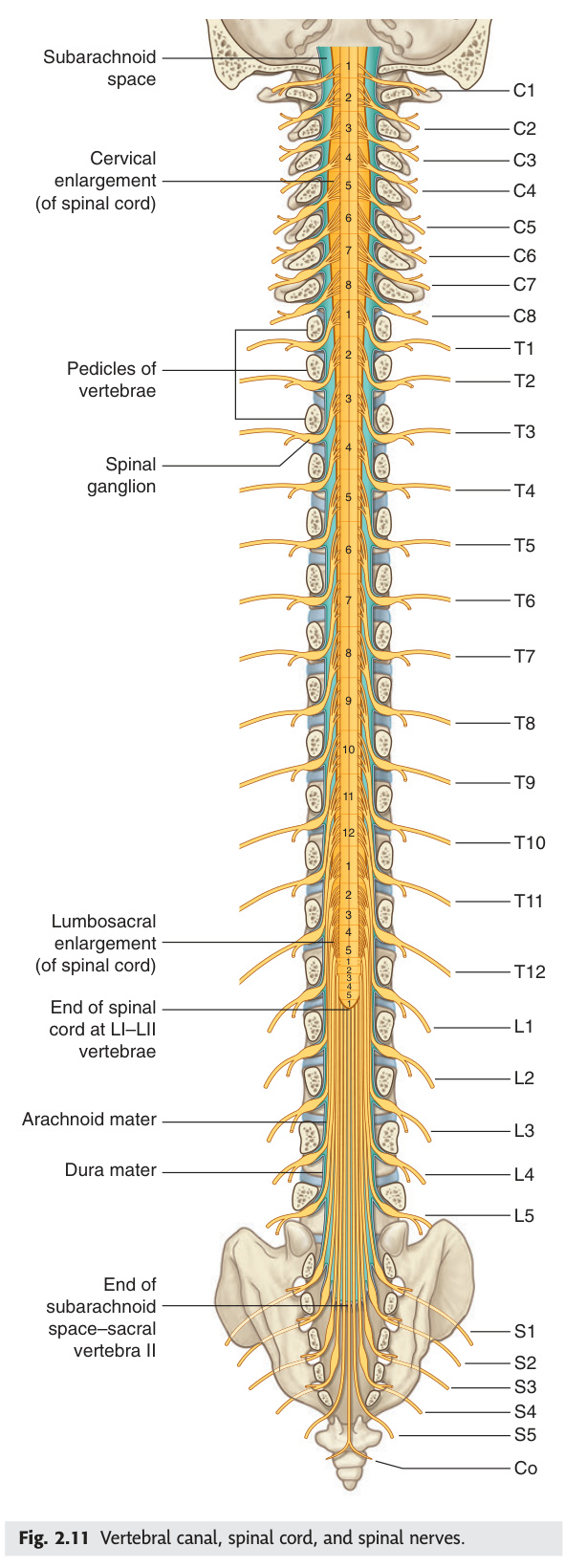
The spinal cord is the great cable of the nervous system — a finger-wide column that extends from the foramen magnum down to the L1/L2 vertebral level in adults, where it tapers into the conus medullaris. Think of it as a relay station: sensory signals race up, motor commands race down, and the H-shaped grey matter in the middle is where neurons actually live. Two enlargements — cervical (C4–T1) and lumbosacral (L1–S3) — exist because more neurons are needed to serve the limbs; the grey-to-white ratio is highest there. A single blood supply rule governs the most devastating strokes: the anterior spinal artery feeds the anterior two-thirds of the cord (motor + pain/temperature tracts), while two posterior spinal arteries supply only the posterior columns — knowing this one fact explains the entire clinical picture of anterior cord syndrome.
| Feature | Detail |
|---|---|
| Extent | Foramen magnum (medulla-cord junction, C1) → L1/L2 vertebral level (conus medullaris) in adults. At birth: L3. Filum terminale (pia mater strand) continues to coccyx |
| Enlargements | Cervical enlargement (C4–T1): brachial plexus. Lumbosacral enlargement (L1–S3): lumbosacral plexus. Both have more grey matter (more neurons for limb innervation) |
| Grey matter | H-shaped; dorsal (posterior) horns = sensory; ventral (anterior) horns = motor (alpha motor neurons). Lateral horn (T1–L2) = sympathetic preganglionic cell bodies. Rexed laminae I–X |
| White matter | Surrounds grey matter. Dorsal funiculus (posterior columns) + lateral funiculus + ventral funiculus. Each contains ascending + descending tracts |
| Blood supply | Anterior spinal artery (1): from vertebral arteries; supplies anterior 2/3 of cord (corticospinal + spinothalamic + anterior horn). Posterior spinal arteries (2): from PICAs; supply posterior 1/3 (dorsal columns). Artery of Adamkiewicz: main reinforcing artery to lower thoracic/lumbar cord (T9–L2, usually left side) — at risk during aortic surgery → paraplegia |
Occlusion of anterior spinal artery (aortic dissection, AAA repair, thromboembolism) → infarction of anterior 2/3 of spinal cord. Features: bilateral loss of motor function (corticospinal tracts) + bilateral loss of pain/temperature (spinothalamic tracts) BELOW the level, WITH PRESERVED vibration and proprioception (posterior columns spared — supplied by posterior spinal arteries). This is the classic "dissociated sensory loss" pattern for anterior cord syndrome. Distinguishes from complete cord transection (all modalities lost).
- Where does the adult spinal cord end and what continues below? Conus medullaris at L1/L2. Below it: cauda equina (lumbar + sacral nerve roots) + filum terminale (pia strand to coccyx). At birth the cord ends at L3.
- Why do cervical and lumbosacral enlargements exist? Greater number of neurons needed to serve the upper and lower limbs — grey-to-white ratio is highest here because of the dense motor and sensory neuron population for limb innervation.
- What is in the lateral horn of the spinal cord and at which levels? Sympathetic preganglionic cell bodies (T1–L2). The parasympathetic equivalent is the sacral intermediolateral cell column (S2–S4).
- Which artery supplies the anterior 2/3 of the cord and what are its clinical consequences if occluded? Anterior spinal artery (from vertebral arteries). Occlusion → anterior cord syndrome: bilateral motor loss + bilateral pain/temperature loss below, with preserved vibration/proprioception (posterior columns spared).
- What is the Artery of Adamkiewicz and why does it matter surgically? The main radicular reinforcing artery to the lower thoracic/lumbar cord (T9–L2, usually left side). At risk during aortic surgery — its occlusion causes paraplegia from anterior cord ischaemia.
Major Spinal Tracts
Two questions dominate spinal tract anatomy: where does each tract cross, and what modality does it carry? Pain and temperature fibres enter the cord, cross within 1–2 segments in the anterior white commissure, and travel up as the contralateral spinothalamic tract — that crossing within the cord is the key to Brown-Séquard syndrome. Fine touch, vibration, and proprioception travel ipsilaterally all the way to the medulla before crossing at the sensory decussation — which is why a unilateral cord lesion produces opposite-sided sensory deficits for different modalities. Motor commands from cortex cross at the medullary pyramids as the corticospinal decussation, then descend as the lateral corticospinal tract. The TMU 2019 exam asked for the full "superficial sensory pathway of trunk and limbs" (8 marks) — that is the spinothalamic tract in its entirety, from receptor to cortex.
| Tract | Location | Carries | Decussation |
|---|---|---|---|
| Lateral corticospinal tract (descending) | Lateral funiculus | Voluntary motor commands to contralateral limbs | At medullary pyramids (pyramidal decussation). 85–90% cross; 10–15% remain as anterior corticospinal tract (crosses in cord) |
| Lateral spinothalamic tract (ascending) | Lateral funiculus (anterior spinothalamic in ventral funiculus) | Pain + temperature (lateral); crude touch + pressure (anterior) | Crosses within 1–2 spinal cord segments of entry in anterior white commissure → contralateral |
| Dorsal columns (ascending): Fasciculus gracilis + cuneatus | Posterior funiculus | Fine touch, vibration, proprioception, 2-point discrimination. Gracilis (lower limb, medial) + cuneatus (upper limb, lateral) | Ascend ipsilaterally → cross in medulla (at sensory decussation in nucleus gracilis + cuneatus) → contralateral medial lemniscus → thalamus → somatosensory cortex |
| Rubrospinal tract | Lateral funiculus (anterior to corticospinal) | Motor coordination (from red nucleus). Minor in humans | Crosses at midbrain |
IPSILATERAL (right) below lesion: 1. Motor loss (UMN signs — corticospinal tract already crossed in medulla); 2. Loss of fine touch/vibration/proprioception (dorsal columns ascend ipsilaterally, cross later in medulla).
CONTRALATERAL (left) below lesion: loss of pain + temperature (spinothalamic tract crosses within 1–2 segments of entry → travels contralaterally).
AT the level of lesion: ipsilateral LMN signs (anterior horn cell/root damage) + ipsilateral dermatomal band of all sensory loss.
Clinical causes: penetrating trauma (knife), MS, tumour, epidural abscess, cord compression from disc.
- Where does the spinothalamic tract cross and what does it carry? Crosses within 1–2 spinal cord segments of entry, in the anterior white commissure → travels contralaterally. Lateral spinothalamic = pain + temperature; anterior spinothalamic = crude touch + pressure.
- Where do dorsal column fibres cross? Ascend ipsilaterally to the medulla → cross at the sensory decussation in the nucleus gracilis/cuneatus → contralateral medial lemniscus → thalamus → cortex. Carry: fine touch, vibration, proprioception, 2-point discrimination.
- What is the full superficial sensory pathway of the trunk and limbs? (TMU 2019 Q8) 1st neuron: receptor → dorsal root ganglion → enters cord. 2nd neuron: crosses in anterior white commissure → lateral spinothalamic tract → thalamus (VPL). 3rd neuron: thalamus → primary somatosensory cortex (postcentral gyrus, areas 3/1/2).
- In Brown-Séquard syndrome (right cord hemisection at T10), list all deficits. Right (ipsilateral) below: UMN signs + loss of fine touch/vibration/proprioception (dorsal columns). Left (contralateral) below: loss of pain/temperature (spinothalamic crossed 1–2 levels below). At lesion level right: LMN signs + band of all-modality sensory loss.
- Where does the lateral corticospinal tract cross? At the medullary pyramids (pyramidal decussation) — 85–90% of fibres. The remaining 10–15% descend as the anterior corticospinal tract and cross within the cord. Both ultimately reach contralateral anterior horn cells.
Spinal Nerve Formation
Every spinal nerve is a merger of two roots: the dorsal (sensory) root carrying signals in from the periphery via the dorsal root ganglion, and the ventral (motor) root carrying commands out from anterior horn cells. They unite in the intervertebral foramen and immediately divide into dorsal ramus (paravertebral muscles and back skin) and ventral ramus (limbs and anterior trunk). The key counting rule to memorise: there are 8 cervical nerves but only 7 cervical vertebrae, because C1–C7 exit above their vertebra while C8 exits below C7. Below that, every nerve exits below its vertebra — and this numbering gap is why C8 radiculopathy comes from the C7/T1 disc, not from a non-existent C8 vertebra.
31 pairs of spinal nerves: 8 cervical (C1–C8) + 12 thoracic (T1–T12) + 5 lumbar + 5 sacral + 1 coccygeal. Each formed by union of dorsal (posterior) root (sensory, DRG) + ventral (anterior) root (motor). Unite in intervertebral foramen → spinal nerve → immediately divides into dorsal ramus (posterior structures, paravertebral muscles + skin) + ventral ramus (limbs + anterior trunk). Note: C1 has a ventral root but usually no dorsal root (no sensory dermatome for C1).
C1–C7 nerves exit ABOVE their corresponding vertebra. C8 nerve exits BELOW C7 vertebra (no C8 vertebra). T1–S5 nerves exit BELOW their corresponding vertebra. Therefore: 8 cervical nerves but only 7 cervical vertebrae.
- How many spinal nerves are there and how are they distributed? 31 pairs: 8 cervical + 12 thoracic + 5 lumbar + 5 sacral + 1 coccygeal. C1 usually has no dorsal root and therefore no sensory dermatome.
- What are the two roots of a spinal nerve and what does each carry? Dorsal (posterior) root = sensory/afferent, with dorsal root ganglion containing cell bodies. Ventral (anterior) root = motor/efferent, with cell bodies in the anterior horn.
- What is the cervical nerve numbering rule and why does C8 exit below C7? C1–C7 exit above their vertebra; C8 exits below C7 (no C8 vertebra). T1–S5 exit below their vertebra. Result: 8 cervical nerves, 7 cervical vertebrae.
- What are dorsal and ventral rami and what do they supply? The spinal nerve immediately divides after exiting the foramen. Dorsal ramus → deep back muscles + posterior trunk skin. Ventral ramus → anterior trunk + all four limbs (forms the major plexuses).
- Where are dorsal root ganglion cell bodies located and why does this matter clinically? In the intervertebral foramen, just proximal to the nerve union. They house the 1st-order sensory neuron cell bodies — targeted by herpes zoster (shingles), which reactivates in the DRG and causes dermatomal vesicular rash.
Cervical Plexus (C1–C4)
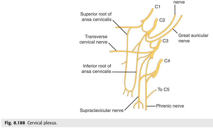
The cervical plexus (C1–C4) is clinically dominated by one nerve: the phrenic. "C3, 4, 5 keeps the diaphragm alive" is the most tested mnemonic in anatomy — damage above C3 (e.g., high cervical cord injury) stops breathing entirely, while damage below C5 spares the diaphragm. The phrenic nerve also has a sensory component to the central diaphragm and pericardium, which is why diaphragmatic irritation (subphrenic abscess, ectopic pregnancy rupture) refers pain to the shoulder tip via the C3/C4 supraclavicular dermatome. The great auricular nerve (C2/C3) is the most commonly injured cutaneous nerve in parotidectomy — numbness over the ear and parotid region is the expected post-operative finding surgeons warn patients about.
| Branch | Levels | Distribution |
|---|---|---|
| Phrenic nerve | C3, C4, C5 ("C3,4,5 keeps the diaphragm alive") | Motor to diaphragm + sensory to central diaphragm + pericardium + mediastinal pleura. Left phrenic: runs anterior to vagus over left subclavian artery. Injury (e.g., thyroid surgery, lung cancer) → ipsilateral hemiparalysis of diaphragm on CXR (elevated hemidiaphragm) |
| Great auricular nerve | C2, C3 | Skin over parotid + ear + angle of mandible. Most commonly injured in parotidectomy → numbness over ear/parotid region |
| Lesser occipital | C2 | Skin of lateral scalp behind auricle |
| Supraclavicular nerves | C3, C4 | Skin over shoulder + clavicle. Referred pain from diaphragm (C3,4 → shoulder tip pain) |
| Ansa cervicalis | C1–C3 | Motor to infrahyoid muscles (sternothyroid, sternohyoid, omohyoid) |
- What are the roots of the phrenic nerve and what does it supply? C3, C4, C5 ("C3,4,5 keeps the diaphragm alive"). Motor to the entire diaphragm. Sensory to central diaphragm, pericardium, and mediastinal pleura. Injury → ipsilateral elevated hemidiaphragm on CXR.
- Why does diaphragmatic irritation cause shoulder-tip pain? The phrenic nerve (C3/C4) also carries sensory fibres from the central diaphragm. Pain is referred to the C3/C4 dermatome = shoulder tip / supraclavicular region (convergence of somatic + visceral C3/C4 inputs at the same spinal level).
- Which cervical plexus nerve is most commonly injured in parotidectomy? Great auricular nerve (C2/C3) — runs superficially over the parotid gland. Injury → numbness over the ear, parotid region, and angle of mandible. Patients should be warned pre-operatively.
- What does the ansa cervicalis (C1–C3) supply? Motor to the infrahyoid (strap) muscles: sternothyroid, sternohyoid, omohyoid. It does NOT supply thyrohyoid (C1 via hypoglossal nerve) or geniohyoid.
- What is the clinical significance of supraclavicular nerves (C3/C4)? They supply skin over the shoulder and clavicle. Also form part of the referred pain pathway for diaphragm/pericardium irritation. In Pancoast tumour, supraclavicular lymph nodes (same level) may enlarge.
Brachial Plexus (C5–T1)
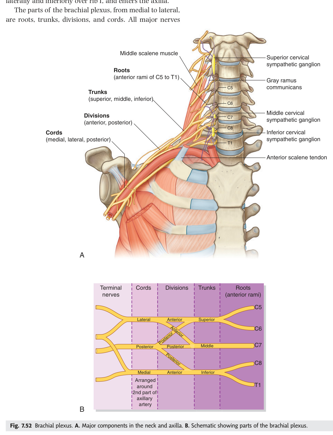
The brachial plexus (C5–T1) is the wiring harness for the entire upper limb — five roots reorganise into trunks, divisions, cords, and finally named branches. The mnemonic "Real Texans Drink Cold Beer" locks the order in. Clinically, two birth injury patterns dominate: Erb's palsy (C5/C6 upper trunk — "waiter's tip" arm from loss of deltoid, biceps, and supinators) and Klumpke's palsy (C8/T1 lower trunk — intrinsic hand wasting and claw hand, often with Horner's syndrome from T1 sympathetic damage). In the adult, the three most-tested nerve injuries are: radial nerve at the spiral groove (wrist drop, sensory loss at 1st web space), median nerve at the carpal tunnel (thenar wasting, LOAF muscles, night-time tingling), and ulnar nerve at the medial epicondyle (claw hand of ring/little fingers, hypothenar wasting). Each has a unique autonomous sensory zone that confirms the diagnosis at the bedside.
Roots: C5, C6, C7, C8, T1. Trunks: Upper (C5+C6) + Middle (C7) + Lower (C8+T1). Divisions: Each trunk → anterior + posterior. Cords: Lateral (anterior upper + anterior middle) + Posterior (all 3 posterior) + Medial (anterior lower). Branches from cords (lateral → medial → posterior).
| Injury | Roots | Mechanism | Deficit |
|---|---|---|---|
| Erb's palsy (upper trunk injury) | C5, C6 | Birth trauma — shoulder dystocia; fall on shoulder widening neck-shoulder angle | "Waiter's tip": arm adducted + medially rotated + elbow extended + forearm pronated. Deltoid, biceps, brachialis, brachioradialis weak. Absent biceps + brachioradialis reflex |
| Klumpke's palsy (lower trunk) | C8, T1 | Pulling on abducted arm; Pancoast tumour; cervical rib | Intrinsic hand muscles weak → claw hand. Ulnar nerve distribution predominantly. Horner's syndrome if T1 sympathetic preganglionic damaged (ptosis + miosis + anhidrosis) |
| Long thoracic nerve (nerve to serratus anterior) | C5, C6, C7 | Mastectomy, axillary surgery, carrying heavy loads | Winged scapula (serratus anterior paralysed → medial scapular border + inferior angle protrude when pushing against wall) |
| Axillary nerve | C5, C6 (posterior cord) | Shoulder dislocation, surgical neck of humerus fracture | Deltoid (shoulder abduction 15–90°) + teres minor paralysed. Sensory loss: regimental badge area (lateral arm, upper part) |
| Radial nerve | C5–T1 (posterior cord) | Midshaft humerus fracture (spiral groove), Saturday night palsy | Wrist drop (extensors paralysed), finger drop. Sensory: dorsum of 1st webspace (autonomous zone). Triceps spared if injured in spiral groove |
| Median nerve | C6–T1 (lateral + medial cord) | Carpal tunnel, supracondylar fracture (children), pronator teres compression | Ape hand (thenar wasting + cannot oppose). LOAF muscles in hand (Lumbricals 1+2, Opponens pollicis, Abductor pollicis brevis, Flexor pollicis brevis). Sensory: lateral 3.5 fingers palmar. Carpal tunnel: tingling in thumb + index + middle finger, worse at night, Tinel's + Phalen's signs |
| Ulnar nerve | C8, T1 (medial cord) | Medial epicondyle fracture, cubital tunnel, Guyon's canal | Claw hand (4th + 5th fingers, interossei weak → MCP hyperextension). Hypothenar wasting. Sensory: medial 1.5 fingers (both sides). "Ulnar paradox": more proximal = less claw (flexor digitorum profundus to ring/little also paralysed) |
- Give the order of brachial plexus organisation (mnemonic). Roots (C5–T1) → Trunks (upper C5/C6, middle C7, lower C8/T1) → Divisions (each trunk splits anterior + posterior) → Cords (lateral, posterior, medial) → Branches. "Real Texans Drink Cold Beer."
- Erb's palsy: roots, mechanism, and classic posture. C5, C6 (upper trunk). Birth trauma — shoulder dystocia widens neck-shoulder angle. "Waiter's tip": arm adducted + medially rotated + elbow extended + forearm pronated. Biceps + brachioradialis reflexes absent.
- Klumpke's palsy: roots, cause, and associated syndrome. C8, T1 (lower trunk). Pulling on abducted arm; Pancoast tumour; cervical rib. Intrinsic hand wasting + claw hand. Horner's syndrome if T1 sympathetic preganglionic fibres damaged (ptosis + miosis + anhidrosis).
- Radial nerve injury at the spiral groove: what is lost and what is spared? Lost: wrist + finger extension (wrist drop), sensory loss at dorsum of 1st web space. Spared: triceps (branch given off above spiral groove) + elbow extension. Cause: midshaft humerus fracture, Saturday night palsy.
- What are the LOAF muscles and which nerve supplies them? Median nerve (C6–T1) in the hand: Lumbricals 1+2, Opponens pollicis, Abductor pollicis brevis, Flexor pollicis brevis. Loss → ape hand (thenar wasting, cannot oppose thumb). Carpal tunnel syndrome is the most common cause.
Lumbar & Sacral Plexus
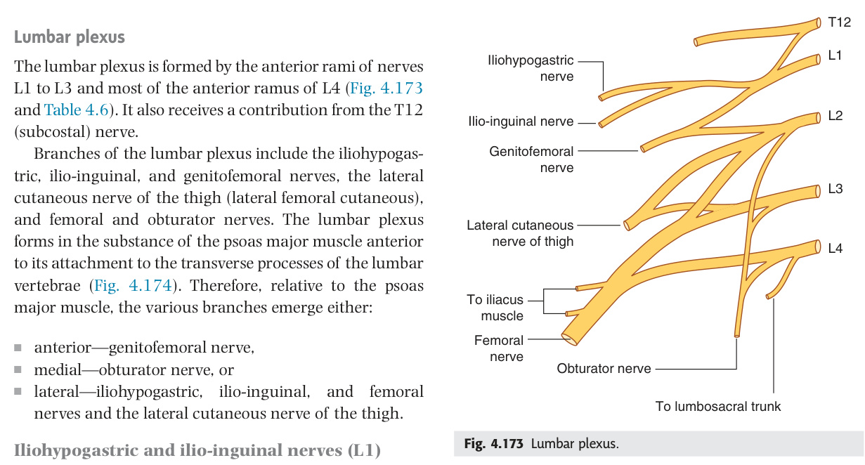
The lower limb is served by two overlapping plexuses: lumbar (L1–L4) and sacral (L4–S3). Three nerves dominate clinically. The femoral nerve (L2–L4) extends the knee via quadriceps and is tested by the knee jerk (L3/L4) — damage in the femoral triangle or from psoas abscess causes inability to climb stairs. The sciatic nerve (L4–S3) is the largest nerve in the body and supplies everything below the knee; its safe injection zone is the upper outer quadrant of the buttock. The common peroneal nerve, as it winds around the fibular neck, is the most vulnerable peripheral nerve in the lower limb — a simple leg cross or fibular neck fracture causes foot drop, and the key bedside test is whether inversion is preserved (tibial nerve intact → pure peroneal lesion, not sciatic or L5 root). The pudendal nerve (S2/S3/S4) is the nerve of the perineum — "S2, 3, 4 keeps the penis off the floor."
| Nerve | Roots | Motor | Sensory / Reflex |
|---|---|---|---|
| Femoral nerve | L2, L3, L4 | Quadriceps (knee extension) + iliopsoas (hip flexion) + sartorius + pectineus | Anterior thigh + medial leg (saphenous nerve = longest branch, runs with great saphenous vein). Knee jerk (L3, L4). Damaged in femoral triangle surgery, psoas abscess |
| Obturator nerve | L2, L3, L4 | Adductors (hip adduction) + obturator externus | Medial thigh. Obturator hernia → pressure on nerve → Howship-Romberg sign (medial thigh pain on hip internal rotation) |
| Sciatic nerve | L4, L5, S1, S2, S3 | Hamstrings (knee flexion) + all muscles below knee. Divides into common peroneal (fibular) + tibial above or at popliteal fossa | Back of thigh (posterior cutaneous nerve), whole leg below knee except medial strip (saphenous). Ankle jerk (S1, S2) |
| Common peroneal (fibular) | L4, L5, S1 | Dorsiflexion + eversion of foot (peroneus longus + brevis). Deep branch: toe extension | Dorsum of foot. Foot drop: fibular neck fracture (winds around neck of fibula) or leg crossing |
| Tibial nerve | L4, L5, S1, S2, S3 | Plantarflexion + inversion + toe flexors + intrinsic foot muscles | Sole of foot (sural nerve). Tarsal tunnel syndrome: compression under flexor retinaculum → burning pain in sole |
| Superior gluteal | L4, L5, S1 | Gluteus medius + minimus + tensor fasciae latae (hip abduction). Safe injection zone: upper outer quadrant of buttock | Trendelenburg sign if damaged: pelvis drops on opposite side during single-leg stance |
| Pudendal nerve | S2, S3, S4 | External anal + urethral sphincters + perineal muscles | Perineum + genitalia. Pudendal canal (Alcock's canal). Pudendal nerve block: ischial spine landmark |
- Femoral nerve: roots, key motor, reflex, and common injury site. L2, L3, L4. Motor: quadriceps (knee extension) + iliopsoas (hip flexion). Knee jerk (L3/L4). Injured in femoral triangle surgery, psoas abscess, or pelvic haematoma. Saphenous nerve = longest cutaneous branch (medial leg to foot).
- Sciatic nerve: roots, what it supplies, and safe injection zone. L4, L5, S1, S2, S3. Supplies all hamstrings + all muscles below knee (via common peroneal + tibial divisions). Safe gluteal injection: upper outer quadrant (avoids sciatic nerve, which exits below piriformis at mid-buttock).
- Common peroneal nerve: where is it most vulnerable and what is the classic deficit? Winds around the neck of fibula — most vulnerable peripheral nerve in the lower limb. Foot drop (tibialis anterior) + weak eversion (peroneus longus/brevis) + sensory loss dorsum of foot. Ankle jerk and inversion are preserved.
- Trendelenburg sign: which nerve and which muscle? Superior gluteal nerve (L4/L5/S1) → gluteus medius + minimus. Damage → pelvis drops to opposite (unaffected) side during single-leg stance because ipsilateral hip abductors cannot hold the pelvis level.
- Pudendal nerve: roots, supply, and surgical landmark. S2, S3, S4. Supplies external anal + urethral sphincters + perineal muscles + perineal skin + genitalia. Travels in Alcock's canal (pudendal canal). Block performed at ischial spine landmark — used in obstetric perineal anaesthesia.
Key Dermatomes & Myotomes
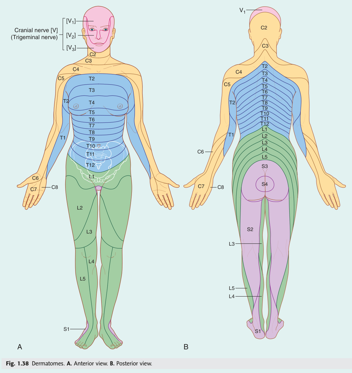
Dermatomes are the body's postcode system — each patch of skin reports back to a specific spinal level, and knowing the landmarks lets you localise a cord or root lesion precisely. The trunk landmarks confirmed in the TMU anatomy notes: T2 = sternal angle, T4 = nipple, T6 = xiphoid process, T10 = umbilicus (also where appendicitis pain begins as referred visceral pain), T12 = anterior superior iliac spine. For the limbs, the C6 autonomous zone is the tip of the thumb (carpal tunnel tingling), C7 is the middle finger (most common cervical disc level), L5 is the big toe and dorsum of foot (L4/5 disc), and S1 is the lateral foot and sole (L5/S1 disc, ankle jerk). Myotomes give you the reflex ladder: biceps = C5/C6, triceps = C7, knee = L3/L4, ankle = S1/S2 — one absent reflex places the lesion precisely.
| Level | Dermatome landmark | Myotome / Reflex |
|---|---|---|
| C3, C4 | Shoulder/clavicle (cape distribution) | Diaphragm (C3,4,5) |
| C5 | Lateral arm (regimental badge) | Shoulder abduction (deltoid); biceps jerk |
| C6 | Lateral forearm + thumb + index finger | Elbow flexion (biceps); brachioradialis jerk; wrist extension |
| C7 | Middle finger (longest finger) | Elbow + wrist extension (triceps); triceps jerk |
| C8 | Little finger + medial forearm | Finger flexion; intrinsic hand muscles |
| T4 | Nipple line | — |
| T10 | Umbilicus | Appendicitis referred pain; ovarian/testicular pain |
| L1 | Inguinal region | Cremasteric reflex (L1, L2) |
| L3, L4 | Medial thigh + knee | Knee extension (quadriceps); knee jerk (L3,L4) |
| L5 | Lateral lower leg + dorsum of foot + big toe | Foot dorsiflexion + big toe extension (L4, L5) |
| S1 | Lateral foot + sole | Plantarflexion + eversion; ankle jerk (S1, S2) |
| S2, S3, S4 | Perineum ("saddle area") | External sphincters; anal reflex (S3, S4) |
- List the trunk dermatome landmarks (TMU notes confirmed). T2 = sternal angle, T4 = nipple, T6 = xiphoid process, T8 = costal arch, T10 = umbilicus, T12 = anterior superior iliac spine. These are tested in fill-in-the-blank format in TMU exams.
- Which dermatome covers the umbilicus and what is its clinical relevance? T10. Appendicitis begins as periumbilical pain (T10 referred visceral pain from the appendix) before localising to McBurney's point as parietal peritoneum becomes involved (L1 somatic).
- Give the reflex ladder: reflex → root levels. Biceps jerk = C5/C6. Brachioradialis jerk = C6. Triceps jerk = C7. Knee jerk = L3/L4. Ankle jerk = S1/S2. Anal reflex = S3/S4. Cremasteric reflex = L1/L2.
- What is the autonomous sensory zone of C6, C7, L5, and S1? C6 = tip of thumb. C7 = middle finger (tip). L5 = dorsum of big toe / first web space. S1 = lateral border of foot / little toe. These zones remain sensory even when neighbouring dermatomes compensate, confirming the level of injury.
- L5 myotome: what movement tests it and which reflex is associated? Big toe extension (extensor hallucis longus — the key L5 myotome test) + foot dorsiflexion + hip abduction (gluteus medius). No reliable reflex for L5 in most patients (occasionally a reduced hamstring jerk).
Spinal Cord Lesion Patterns
Identifying a spinal cord lesion pattern is one of the highest-yield clinical skills in anatomy — the TMU 2019 exam devoted 9 marks to a single question about the internal capsule alone. The key is always: which tracts are hit, and how do they decussate? UMN lesions (above the anterior horn) give spasticity + hyperreflexia + Babinski; LMN lesions (at or below the anterior horn) give flaccidity + wasting + fasciculations + areflexia. Cauda equina syndrome (L2–S5 roots) is a surgical emergency — saddle anaesthesia + urinary retention + bilateral flaccid leg weakness demands urgent MRI and decompression within 48 hours. Subacute combined degeneration (B12 deficiency) is the classic "combined" lesion: dorsal columns + corticospinal tracts simultaneously, producing the paradox of absent ankle jerks (peripheral neuropathy) with extensor plantar responses (UMN cord involvement) — virtually pathognomonic.
| Feature | UMN lesion (above anterior horn cell) | LMN lesion (anterior horn cell, root, or nerve) |
|---|---|---|
| Tone | ↑ Spasticity (velocity-dependent) — clasp-knife | ↓ Flaccidity + hypotonia |
| Power | Weak — pyramidal distribution: extensors weak in arm, flexors weak in leg | Weak — distribution of root/nerve territory |
| Reflexes | ↑↑ Hyperreflexia + clonus (>3 beats) | ↓ Hyporeflexia or areflexia |
| Plantar reflex | Extensor (Babinski sign — hallux dorsiflexes + fanning of toes) | Flexor (normal) or absent |
| Wasting | Disuse atrophy only (mild, late) | Denervation atrophy (early, severe) |
| Fasciculations | Absent | Present (spontaneous LMN discharge) |
| Examples | Stroke, MS, spinal cord compression above lesion level, motor neuron disease (corticospinal component) | Disc prolapse (radiculopathy), peripheral neuropathy, Guillain-Barré, motor neuron disease (anterior horn component), polio |
| Syndrome | Structures damaged | Findings |
|---|---|---|
| Complete transection | All tracts | Bilateral motor + ALL sensory loss below level + autonomic (urinary retention, loss of sweating). Spinal shock initially (flaccid paralysis) → UMN signs weeks later |
| Brown-Séquard (hemisection) | Ipsilateral: corticospinal + dorsal columns. Contralateral: spinothalamic | Ipsilateral: UMN + loss of fine touch/proprioception/vibration. Contralateral: pain/temperature loss 1–2 levels below lesion |
| Central cord syndrome | Central grey + adjacent tracts (cervical cord) | Upper limbs weaker than lower (cervical cord central — arm fibres medial in corticospinal tract). Cape distribution of pain/temperature loss (arms > legs). Most common incomplete SCI. Cause: hyperextension injury in elderly cervical spondylosis |
| Posterior cord syndrome | Dorsal columns | Bilateral loss of fine touch/vibration/proprioception → sensory ataxia (positive Romberg). Motor intact. Causes: subacute combined degeneration (B12 deficiency), tabes dorsalis (tertiary syphilis), Friedrich's ataxia |
| Anterior cord syndrome | Anterior spinal artery territory | Bilateral motor loss + bilateral pain/temperature loss. Dorsal columns SPARED (vibration/proprioception intact) |
| Cauda equina syndrome | L2–S5 nerve roots in lumbar canal (below conus) | LMN signs bilaterally (flaccid, areflexic). Saddle anaesthesia (S3–S5). Bladder + bowel dysfunction (urinary retention + overflow incontinence). Sexual dysfunction. Bilateral sciatica. SURGICAL EMERGENCY → MRI spine urgently → decompression within 48 hours |
Degeneration of the dorsal columns (posterior funiculi) AND the lateral corticospinal tracts of the spinal cord due to vitamin B12 (cobalamin) deficiency. "Combined" = two separate tract systems involved simultaneously — a combination not explained by a single-level lesion.
Mechanism: B12 is essential for methylmalonyl-CoA mutase (myelin synthesis) and methionine synthase (DNA synthesis). Deficiency → abnormal fatty acid incorporation into myelin → demyelination of dorsal columns (posterior funiculi) + lateral corticospinal tracts. The dorsal columns are affected first and most severely; corticospinal tracts affected in moderate–severe deficiency.
Haematological features (often co-existing): megaloblastic anaemia (macrocytic, hypersegmented neutrophils >5 lobes), glossitis, angular cheilosis, jaundice (haemolysis). Note: neurological symptoms can occur without anaemia.
Neurological features — combined pattern:
• Posterior column signs (dorsal funiculi): bilateral loss of vibration sense (tuning fork test — most sensitive early sign) + proprioception loss → sensory ataxia + positive Romberg test + broad-based gait + pseudoathetosis of fingers (involuntary writhing when eyes closed — proprioception lost). Fine touch impaired. Pain and temperature preserved (spinothalamic tracts not primarily affected).
• Corticospinal tract signs (UMN below lesion): bilateral leg spasticity + hyperreflexia + extensor plantar responses (Babinski). Weakness of limbs.
• Paradox: ankle jerk may be absent (LMN, peripheral neuropathy from B12 deficiency affecting peripheral nerves simultaneously) + extensor plantar (UMN from cord). Absent ankle jerk + extensor plantar = subacute combined degeneration until proven otherwise.
• Peripheral neuropathy: burning/tingling in feet (glove-and-stocking distribution) from simultaneous peripheral nerve demyelination.
• Neuropsychiatric: cognitive impairment, depression, megaloblastic madness (psychosis in severe cases).
Causes of B12 deficiency: pernicious anaemia (autoimmune — anti-intrinsic factor antibodies; most common in adults; associated with other autoimmune diseases — Graves, Hashimoto, vitiligo, Addison's); strict vegans/vegetarians (B12 only in animal products); gastrectomy (loss of parietal cells → no intrinsic factor); terminal ileum disease/resection (B12 absorbs at terminal ileum); blind loop syndrome; nitrous oxide abuse (oxidises cobalamin → inactivation — can precipitate acute neurological decompensation in subclinical B12 deficiency during anaesthesia).
| Investigation | Finding | Note |
|---|---|---|
| Serum B12 | <200 pg/mL = deficient; 200–300 = borderline | Can be falsely normal in ~25% of neurologically affected patients (functional deficiency) |
| Methylmalonic acid (MMA) | Raised in B12 deficiency (most sensitive + specific functional marker) | Rises before serum B12 falls; better indicator of true tissue deficiency than serum B12 alone |
| Homocysteine | Raised in B12 AND folate deficiency | Not specific to B12; also elevated in renal failure, hypothyroidism |
| Full blood count | Macrocytic anaemia (MCV >100), hypersegmented neutrophils, oval macrocytes | May be absent in early/pure neurological B12 deficiency |
| Anti-intrinsic factor antibodies | Positive = pernicious anaemia (specific ~95%, sensitive ~50%) | Anti-parietal cell antibodies more sensitive but less specific |
| MRI spine | T2 hyperintensity in posterior columns (dorsal columns) ± lateral columns of cervical/thoracic cord | Characteristic "inverted V" sign on axial T2 (posterior column hyperintensity). May be normal early. |
Miosis (constricted pupil — sympathetic dilator pupillae paralysed) + partial ptosis (drooping of upper lid — superior tarsal/Müller's muscle paralysed; also lower lid elevation = "upside down ptosis"/reverse ptosis) + anhidrosis (absence of sweating on ipsilateral face — only with first- or second-order neuron lesions above the bifurcation of the common carotid). Enophthalmos is apparent (not real) — due to lid changes reducing palpebral aperture.
| Order | Pathway | Location of Lesion | Causes |
|---|---|---|---|
| 1st-order neuron (central) | Hypothalamus → descends through lateral tegmentum of brainstem + lateral funiculus of spinal cord → ciliospinal centre of Budge (C8–T2 lateral horn) | Hypothalamus, brainstem, cervical/upper thoracic spinal cord | Lateral medullary (Wallenberg) syndrome, MS, syringomyelia (C8–T2 lateral horn), Brown-Séquard at C8–T2, spinal cord tumour, stroke |
| 2nd-order neuron (preganglionic) | Exits cord at T1 → over apex of lung → over subclavian artery → ascends with common carotid artery → synapses in superior cervical ganglion (at C2/C3) | T1 root, superior pulmonary sulcus, subclavian, cervical sympathetic chain | Pancoast tumour (apex of lung — compresses T1 root; associated with Klumpke's palsy + Horner's = pathognomonic combination), cervical rib, thyroid goitre, neck surgery, lymph node metastasis, subclavian artery aneurysm |
| 3rd-order neuron (postganglionic) | Superior cervical ganglion → travels with internal carotid artery → enters cavernous sinus → joins ophthalmic division of CN V → ciliary nerves to dilator pupillae + superior tarsal muscle. Facial sweating fibres travel with external carotid artery (diverge at bifurcation) | Internal carotid artery, cavernous sinus | Internal carotid artery dissection (painful Horner's = emergency — often with neck pain + ipsilateral face pain + contralateral stroke symptoms), cavernous sinus thrombosis, cluster headache (due to perivascular inflammation around ICA — painful Horner's during attack), nasopharyngeal carcinoma |
Cocaine drops (4–10%): blocks noradrenaline reuptake → dilates normal pupil; fails to dilate Horner's pupil regardless of the order (blocks NA at neuroeffector junction — confirms Horner's, does not localise it). Now replaced by apraclonidine.
Apraclonidine (0.5% drops) — modern test: weak alpha-1 agonist; in denervation supersensitivity of Horner's iris → both miosis resolves + ptosis reverses (Horner's pupil dilates more than normal pupil → "reversal" of anisocoria confirms Horner's diagnosis). Safe alternative to cocaine.
Hydroxyamphetamine (1%) — order differentiation: releases stored noradrenaline from postganglionic terminal. 1st/2nd order lesion: 3rd-order neuron intact → noradrenaline stores present → pupil DILATES. 3rd-order lesion: postganglionic neuron degenerated → no stored NA → pupil FAILS to dilate. Thus: hydroxyamphetamine differentiates preganglionic (1st/2nd) from postganglionic (3rd order). Note: anhidrosis involving face = 1st or 2nd order (above ICA/ECA bifurcation); no anhidrosis = 3rd order (facial sweating fibres diverge at bifurcation).
Test Unit 16 knowledge
Spinal tracts, plexus injuries, dermatomes, and cord lesion MCQs.
- Give 4 UMN signs and 4 LMN signs. UMN: spasticity (clasp-knife), hyperreflexia + clonus, extensor plantar (Babinski), disuse atrophy only. LMN: flaccidity, areflexia, fasciculations, early severe denervation wasting.
- Cauda equina syndrome: what is it, presentation, and management? Compression of L2–S5 roots below conus medullaris. Presentation: saddle anaesthesia (S3–S5), urinary retention + overflow incontinence, bilateral flaccid leg weakness, sexual dysfunction. Surgical emergency: urgent MRI spine → decompression within 48 hours.
- What is the paradoxical finding in subacute combined degeneration and why does it occur? Absent ankle jerks (peripheral neuropathy from B12 deficiency demyelinating peripheral nerves) + extensor plantar responses (Babinski, from corticospinal tract UMN involvement in the cord). Two simultaneous pathologies at different levels of the nervous system.
- Central cord syndrome: which limbs are weaker and why? Upper limbs weaker than lower limbs. The corticospinal tract is somatotopically organised: cervical (arm) fibres run medially (closest to the central lesion) while lumbar (leg) fibres run laterally — the central cavity damages arm fibres preferentially. Cause: hyperextension in elderly with cervical spondylosis.
- Horner's syndrome triad and the order of neurons. Which order causes anhidrosis of the face? Miosis + partial ptosis + (apparent) enophthalmos. 1st-order (hypothalamus→cord), 2nd-order (cord→superior cervical ganglion, travels over lung apex), 3rd-order (along ICA). Anhidrosis of face = 1st or 2nd order (facial sweat fibres diverge at ECA at the common carotid bifurcation, proximal to the 3rd-order neuron).
Disc Prolapse & Radiculopathy ★★★
An intervertebral disc is a hydraulic shock absorber — gelatinous nucleus pulposus contained by the annulus fibrosus — and when the annulus tears, the nucleus herniates posterolaterally (because the posterior longitudinal ligament is strongest centrally) to compress the exiting nerve root. The L+1 rule for the lumbar spine is the clinical shortcut: the L4/L5 disc compresses the L5 root, L5/S1 compresses S1. For the cervical spine: C5/C6 disc → C6 root (thumb tingling, weak biceps), C6/C7 disc → C7 root (middle finger, weak triceps — the most common cervical level). The straight leg raise (SLR) test stretches the sciatic nerve; pain below 60° of elevation is positive for L4/L5/S1 radiculopathy. Red flags — saddle anaesthesia, bilateral symptoms, urinary/bowel dysfunction — mean central disc prolapse causing cauda equina syndrome until proven otherwise: MRI spine urgently.
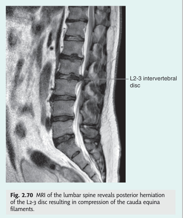
Disc structure: Nucleus pulposus (gelatinous, high water content in youth — 90%, decreases with age → disc degeneration) surrounded by annulus fibrosus (concentric rings of fibrocartilage). Disc receives nutrition by diffusion (avascular). Prolapse types: Protrusion (nucleus herniates but annulus intact) → extrusion (nucleus herniates through annulus) → sequestration (free fragment in spinal canal). Most common direction: posterolateral (posterior longitudinal ligament strongest centrally → disc bulges to side) → compresses exiting nerve root.
In the lumbar spine: the L4/L5 disc (between L4 and L5 vertebral bodies) compresses the L5 root (the traversing root at that level). The L5/S1 disc compresses the S1 root. In the cervical spine: the C5/C6 disc compresses C6 root; C6/C7 disc compresses C7 root. Exception: central disc prolapse → cauda equina syndrome (bilateral).
| Root | Disc | Pain radiation | Weakness | Sensory loss | Reflex lost |
|---|---|---|---|---|---|
| L3 | L2/L3 | Anterior thigh to medial knee | Hip flexion + knee extension (quad weakness) | Anterior/medial thigh | Knee jerk (L3,L4) |
| L4 | L3/L4 | Anterior thigh → medial lower leg → medial foot | Knee extension (quad) + foot dorsiflexion | Medial lower leg + medial foot | Knee jerk (L3,L4) |
| L5 | L4/L5 | Posterior thigh → lateral lower leg → dorsum of foot + big toe | Big toe extension (extensor hallucis longus — key L5 test) + foot dorsiflexion + eversion | Lateral lower leg + dorsum foot + big toe (L5 autonomous zone) | No specific reflex (sometimes ↓ hamstring) |
| S1 | L5/S1 | Posterior thigh → posterior lower leg → lateral foot + heel | Plantarflexion (can't stand on tiptoe) + eversion; gluteus maximus | Lateral foot + sole (S1 autonomous zone) | Ankle jerk (S1, S2) |
Straight leg raise (SLR) test: leg raised with knee extended → stretches sciatic nerve + L4/L5/S1 roots. Positive if sciatic pain (NOT just hamstring tightness) at <60° of elevation = radiculopathy. Crossed SLR: raising the UNAFFECTED leg causes pain in the AFFECTED leg = large central disc prolapse. More specific for disc herniation.
Femoral stretch test: prone, knee flexed → stretches femoral nerve + L2/L3/L4 roots. Positive (anterior thigh pain) = upper lumbar disc prolapse.
Red flags (must exclude serious pathology): saddle anaesthesia + urinary/bowel dysfunction → cauda equina syndrome (emergency); bilateral symptoms; age >50; night pain; unexplained weight loss; cancer history; progressive neurological deficit → MRI spine + urgent surgical referral.
| Root | Disc | Pain | Weakness | Sensory | Reflex |
|---|---|---|---|---|---|
| C5 | C4/C5 | Neck → shoulder → lateral arm | Deltoid + biceps (shoulder abduction + external rotation) | Lateral arm (regimental badge) | Biceps jerk ↓ |
| C6 | C5/C6 | Neck → lateral forearm → thumb + index finger | Biceps + wrist extensors (ECRL/ECRB) | Thumb + index finger (C6 autonomous zone = tip of thumb) | Biceps + brachioradialis ↓ |
| C7 | C6/C7 (most common) | Neck → posterior arm → middle finger | Triceps + wrist flexors + finger extensors | Middle finger (C7 autonomous zone) | Triceps jerk ↓ |
| C8 | C7/T1 | Neck → medial forearm → ring + little finger | Finger flexors + intrinsic hand muscles | Ring + little finger + medial forearm | None reliable |
Inability to dorsiflex the foot, causing the toes and forefoot to hang down during the swing phase of walking → steppage gait (patient lifts knee excessively to clear the dropped foot from the ground). Due to weakness of tibialis anterior (dorsiflexion) and toe extensors (extensor digitorum longus + extensor hallucis longus).
| Cause | Lesion Site | Distinguishing Features |
|---|---|---|
| Common peroneal (fibular) nerve palsy | Fibular head / neck (most vulnerable site — winds around lateral neck of fibula) |
Mechanism: fibular neck fracture; plaster cast pressure; leg crossing (habitual); prolonged squatting; prolonged bed rest; Baker's cyst. Motor: foot drop (tibialis anterior) + inability to evert foot (peroneus longus + brevis — deep + superficial peroneal branches). Sensory: dorsum of foot + first web space (autonomous zone of deep peroneal nerve). Preserved: ankle jerk intact (tibial nerve, S1). Plantarflexion intact. Inversion intact (tibialis posterior = tibial nerve). Reflexes: knee jerk + ankle jerk both normal. |
| L5 root lesion (radiculopathy) | L4/L5 disc prolapse compressing L5 root |
Motor: foot drop + weak big toe extension (extensor hallucis longus — key L5 test) + weak foot eversion. Also weak hip abduction (gluteus medius, L5) + weak knee flexion (hamstrings, L5 component). Sensory: lateral lower leg + dorsum of foot + big toe + first web space (L5 dermatome — broader than peroneal autonomous zone). Reflexes: no specific reflex lost (L5 has no reliable reflex in most patients; occasionally ↓ hamstring jerk). Back pain / sciatica: usually present (posterolateral L4/5 disc). SLR positive. Inversion: weakened (tibialis anterior = L4/L5 → weak; tibialis posterior also L5 component) — distinguishes from isolated peroneal where inversion preserved. |
| Sciatic nerve injury | Gluteal region, posterior thigh |
Both divisions affected (common peroneal + tibial): foot drop + weak plantarflexion + weak inversion. Motor: all muscles below knee weak + hamstrings (knee flexion weak). Sensory: entire leg below knee (except medial strip = saphenous nerve/femoral). Sole + dorsum of foot all affected. Reflexes: ankle jerk lost (tibial nerve component, S1). Knee jerk intact (femoral nerve). Cause: misplaced gluteal injection (safest site: upper outer quadrant), posterior hip dislocation, piriformis syndrome, pelvic tumour. |
| Central (UMN) cause | Contralateral leg motor cortex / corticospinal tract (stroke, tumour, MS) |
Key difference: UMN pattern — spasticity + hyperreflexia + extensor plantar (Babinski) + circumduction gait (not steppage). Foot may drag but tone is increased. Sensory: cortical sensory loss pattern (may be dissociated or absent). Other UMN signs: clonus, brisk knee/ankle jerk — distinguishes immediately from all peripheral causes. |
Test inversion (tibialis posterior = tibial nerve, L4/L5/S1): If inversion is PRESERVED → lesion is confined to common peroneal nerve or superficial peroneal only (tibial nerve intact). If inversion is WEAK → L5 root lesion or sciatic nerve (both supply tibialis posterior via tibial nerve's L5 contribution). Test ankle jerk (S1, tibial nerve): If ankle jerk is LOST with foot drop → sciatic nerve lesion (not isolated peroneal). Big toe extension (EHL — L5 hallmark): weakness suggests L5 root involvement rather than isolated peroneal nerve.
- Which disc compresses which root in the lumbar spine (L+1 rule)? L3/L4 disc → L4 root; L4/L5 disc → L5 root; L5/S1 disc → S1 root. Direction: posterolateral herniation (PLL weakest laterally) compresses the traversing root at that level, not the exiting root.
- L5 root compression: pain, weakness, sensory loss, reflex. Pain: posterior thigh → lateral lower leg → dorsum of foot + big toe. Weakness: big toe extension (EHL — key L5 test) + foot dorsiflexion. Sensory: lateral lower leg + dorsum foot + big toe. Reflex: none reliable (no L5 reflex in most patients).
- S1 root compression: weakness, sensory loss, reflex lost. Weakness: plantarflexion (cannot stand on tiptoe) + eversion + gluteus maximus. Sensory: lateral foot + sole + heel. Reflex: ankle jerk (S1/S2) lost — the most reliable reflex for S1 lesion.
- SLR test: what does a positive result indicate and what angle? Pain in sciatic distribution (not just hamstring tightness) at <60° of straight leg elevation = L4/L5/S1 radiculopathy from disc herniation. Crossed SLR (raising unaffected leg causes pain in affected leg) = large central disc prolapse, more specific.
- C6/C7 disc (most common cervical level): root, deficit, reflex. C7 root. Pain: neck → posterior arm → middle finger. Weakness: triceps + wrist flexors + finger extensors. Sensory: middle finger (C7 autonomous zone). Reflex: triceps jerk lost.
Syringomyelia ★★
Syringomyelia is the cord destroying itself from within — a fluid-filled cavity (syrinx) expands outward from the central grey matter, hitting structures in a predictable inside-out sequence. First to go: the anterior white commissure, where pain and temperature fibres from both sides are crossing. This produces the signature bilateral cape distribution of dissociated sensory loss (pain/temperature gone bilaterally over arms and shoulders, fine touch preserved). Then the anterior horn cells are damaged (LMN signs in arms — wasting, fasciculations, areflexia), then the corticospinal tracts (UMN signs in legs — spasticity, hyperreflexia). The commonest cause is Arnold-Chiari type I malformation (cerebellar tonsillar herniation >5 mm below foramen magnum), and the treatment — posterior fossa decompression — often causes the syrinx to collapse. The key MRI finding: a sausage-shaped T2-hyperintense central cavity on sagittal imaging.
A pathological fluid-filled cavity (syrinx) within the substance of the spinal cord, most commonly in the cervical cord, lined by glial cells (ependyma-lined central canal dilatation = hydromyelia; true syrinx = paracentral cavity lined by glia). The cavity progressively destroys the cord from the inside out.
| Cause | Mechanism | Notes |
|---|---|---|
| Arnold-Chiari type I (most common) | Cerebellar tonsillar herniation >5 mm below foramen magnum → impaired CSF flow at craniovertebral junction → CSF pulsations forced into cord parenchyma → syrinx | Presents in adults 20–40 years; often symptomatic only when syrinx develops. MRI craniovertebral junction diagnostic |
| Post-traumatic | Spinal cord injury → arachnoid scarring → impaired CSF dynamics → syrinx months to years post-injury | Progressive neurological deterioration after initial stabilisation → syrinx until proven otherwise |
| Post-infective / post-SAH | Arachnoiditis (meningitis, SAH) → fibrosis → CSF flow obstruction | LP may show xanthochromic or inflammatory CSF from remote event |
| Intramedullary tumour | Ependymoma (most common intramedullary tumour in adults), haemangioblastoma → secretion / local CSF obstruction → rostral/caudal syrinx | Ependymoma: central cord, bleeds (haemosiderin cap sign); haemangioblastoma: associated with von Hippel-Lindau syndrome |
| Idiopathic | No identifiable cause | Diagnosis of exclusion after MRI brain + full spine |
Pathophysiology of signs: Syrinx expands from central cord outward. First structure destroyed: anterior white commissure (decussating spinothalamic fibres for pain/temperature at that level) → then anterior horn cells (LMN at cord level) → then corticospinal tracts (UMN below) → then posterior columns (late).
Classic triad:
1. Cape distribution of dissociated sensory loss (bilateral pain + temperature lost over both arms, shoulders, upper back = like a cape) with preserved fine touch/vibration/proprioception (posterior columns intact — only cross in medulla).
2. LMN signs in upper limbs at the level of the syrinx (anterior horn cell damage): wasting of small hand muscles + fasciculations + areflexia in arms.
3. UMN signs in lower limbs below the syrinx (corticospinal tract compression): spasticity + hyperreflexia + extensor plantar responses.
Additional features: Horner's syndrome (ciliospinal centre C8–T2 affected) — miosis + partial ptosis + anhidrosis. Neuropathic (Charcot's) arthropathy of shoulder/elbow/wrist (pain sensation lost → repetitive microtrauma). Painless cuts/burns on hands (common presenting complaint). Scoliosis in children (paraspinal muscle imbalance).
Investigation: MRI spine T2 — hyperintense elongated central cavity (sausage-shaped). MRI brain — identify Chiari malformation. MRI whole spine — exclude tumour at both ends of cavity.
Most common cause: Arnold-Chiari type I malformation (cerebellar tonsillar herniation >5 mm below foramen magnum). MRI spine T2 = elongated hyperintense central cavity. Treatment: posterior fossa decompression (foramen magnum decompression + duraplasty) → syrinx often collapses.
Syringomyelia: bilateral loss of pain/temperature at the affected levels (anterior white commissure destroyed symmetrically) with preserved fine touch/vibration bilaterally (posterior columns intact). Symmetric cape distribution. Both are "dissociated" sensory patterns but in opposite configurations.
- What is the inside-out sequence of syrinx expansion and resulting deficits? 1. Anterior white commissure → bilateral loss of pain/temperature at affected levels (cape distribution). 2. Anterior horn cells → LMN signs in arms (wasting, fasciculations, areflexia). 3. Corticospinal tracts → UMN signs in legs (spasticity, hyperreflexia, Babinski). 4. Posterior columns (late) → vibration/proprioception loss.
- Why is the sensory loss "dissociated" in syringomyelia? Pain/temperature fibres (spinothalamic) cross in the anterior white commissure at cord level — the syrinx destroys them bilaterally there. Fine touch/vibration/proprioception (dorsal columns) travel ipsilaterally and only cross in the medulla — unaffected by a central cord lesion.
- What is the most common cause of syringomyelia and its treatment? Arnold-Chiari type I malformation — cerebellar tonsillar herniation >5 mm below the foramen magnum impairs CSF flow → CSF pulsations driven into cord parenchyma → syrinx. Treatment: posterior fossa decompression + duraplasty → syrinx often regresses.
- What additional features suggest syringomyelia at C8–T2 levels? Horner's syndrome (ciliospinal centre C8–T2 affected → miosis + ptosis + anhidrosis). Neuropathic (Charcot) joints of shoulder/elbow from painless microtrauma. Painless burns/cuts on hands (common presenting complaint). Scoliosis in children.
- How does syringomyelia differ from Brown-Séquard in its sensory pattern? Syringomyelia: bilateral pain/temperature loss at affected levels + preserved fine touch/vibration bilaterally (symmetric, cape). Brown-Séquard: ipsilateral fine touch/vibration loss + contralateral pain/temperature loss (asymmetric, hemicord). Both are "dissociated" but in opposite configurations.
Meralgia Paraesthetica (LFCN Entrapment) ★
Meralgia paraesthetica is a purely sensory mononeuropathy — no motor weakness, no reflex change — caused by entrapment of the lateral femoral cutaneous nerve (LFCN, L2/L3) under the inguinal ligament near the ASIS. The name itself is the diagnosis: "meralgia" = thigh pain, "paraesthetica" = abnormal sensation. The key bedside differentiator is that quadriceps strength and the knee jerk are completely normal — the moment you find motor weakness or a depressed knee jerk, the diagnosis switches to L3 radiculopathy or femoral nerve compression. Obesity is the most common risk factor (abdominal girth increases the angulation of the nerve at the inguinal ligament), and weight loss alone often resolves symptoms. The treatment ladder is conservative first: loose clothing, weight loss, NSAIDs/gabapentin; then steroid + LA injection at the ASIS; surgical decompression or neurectomy only for refractory cases.
Entrapment neuropathy of the lateral femoral cutaneous nerve (LFCN) at the inguinal ligament, causing burning pain, tingling, and numbness in the anterolateral thigh. A purely sensory mononeuropathy — no motor weakness, no reflex change.
| Feature | Detail |
|---|---|
| Nerve anatomy | LFCN arises from posterior divisions of L2 and L3 ventral rami → passes under (or through) the inguinal ligament just medial to the ASIS (anterior superior iliac spine) → supplies anterolateral thigh skin. Purely sensory — no muscular branches |
| Entrapment site | At the inguinal ligament near the ASIS — passes through a tight fascial tunnel. Variable anatomy (in 25%: passes through the inguinal ligament itself — more vulnerable) |
| Risk factors | Obesity (↑ abdominal girth → ↑ tension on inguinal ligament and nerve angulation); pregnancy; tight clothing (tight belt, corset, compression garment); prolonged standing; rapid weight gain; retroperitoneal haematoma/mass; hip surgery (anterior approach) |
| Symptoms | Burning, tingling, numbness on the anterolateral thigh (NOT medial thigh — medial thigh = obturator nerve). Worse on prolonged standing/walking/hip extension; relieved by sitting/hip flexion (slackens inguinal ligament). Hypersensitivity to light touch in the distribution |
| Signs | Tenderness at ASIS; Tinel's sign at ASIS. Sensory loss confined to anterolateral thigh. Normal quadriceps power + knee jerk (distinguishes from femoral nerve/L3 root lesion) |
| Diagnosis | Clinical diagnosis. Nerve conduction study (LFCN sensory conduction velocity) confirms but rarely required. USS/MRI if structural cause suspected (hernia, haematoma) |
| Treatment | Conservative: weight loss, loose-fitting clothing, avoid aggravating positions; NSAIDs/gabapentin for neuropathic pain. Local steroid ± LA injection at ASIS (diagnostic + therapeutic). Surgical decompression/neurectomy for refractory cases |
| Feature | Meralgia (LFCN) | L3 Radiculopathy | Femoral Nerve (L2–L4) |
|---|---|---|---|
| Sensory loss | Anterolateral thigh only | Anterior thigh + medial knee | Anterior thigh + medial lower leg (saphenous) |
| Motor weakness | None | Quad weakness (knee extension) | Quad weakness + hip flexion |
| Knee jerk | Normal | ↓ or absent | ↓ or absent |
| Medial thigh sensory | Normal | Affected | Affected (saphenous) |
| Back pain | Absent | Often present | Usually absent |
Test Unit 16 knowledge
Spinal tracts, plexus injuries, cord lesion patterns, and radiculopathy MCQs.
- Which nerve, which roots, and where is it entrapped? Lateral femoral cutaneous nerve (LFCN), L2/L3 ventral rami. Entrapped under (or through) the inguinal ligament just medial to the ASIS. Purely sensory — no muscular branches.
- What symptoms does it cause and what relieves them? Burning, tingling, numbness over the anterolateral thigh. Worse on prolonged standing/walking/hip extension (stretches the nerve). Relieved by sitting/hip flexion (slackens inguinal ligament).
- What single bedside finding rules out meralgia and points to L3 root or femoral nerve instead? Any motor weakness (quadriceps weakness) or a depressed/absent knee jerk. Meralgia = purely sensory, normal power, normal reflexes throughout.
- List three risk factors for meralgia paraesthetica. Obesity (increased abdominal girth), pregnancy, tight belts/clothing/compression garments, rapid weight gain, prolonged standing, anterior hip surgery. All increase tension or direct pressure on the nerve at the inguinal ligament.
- What is the treatment ladder? 1. Conservative: weight loss, loose clothing, NSAIDs/gabapentin. 2. Local steroid + LA injection near ASIS (diagnostic + therapeutic). 3. Surgical decompression or neurectomy for refractory cases. Most resolve with conservative measures.