Neuro Intro: Meninges, CSF & BBB
CSF Circulation — Production to Absorption
CNS vs PNS — Overview
The nervous system divides into the CNS (brain + spinal cord) and PNS (everything outside), but the more clinically important distinction is in how each handles damage. In the PNS, Schwann cells myelinate a single axon segment each and actively guide regeneration along endoneurial tubes after Wallerian degeneration at ~1 mm/day; in the CNS, oligodendrocytes myelinate multiple axons but the surrounding environment is actively inhibitory to regrowth (MAG, Nogo, CSPGs from reactive astrocytes and oligodendrocytes). This regeneration gap explains why a severed peripheral nerve can recover over months while a spinal cord injury rarely does. Grey matter in the CNS (cortex and nuclei — cell bodies, dendrites, synapses) corresponds to ganglia in the PNS, while white matter in both systems consists of myelinated axon tracts.
| Feature | CNS | PNS |
|---|---|---|
| Components | Brain + spinal cord | Cranial nerves (III–XII, outside brainstem) + spinal nerves + ganglia + peripheral nerves + autonomic nervous system |
| Myelination | Oligodendrocytes (one cell myelinates many axons) | Schwann cells (one cell myelinates one axon segment); unmyelinated = Schwann cell wraps but does not spiral) |
| Regeneration | Very limited (inhibitory environment: MAG, Nogo, CSPGs from astrocytes + oligodendrocytes) | Good regeneration potential (Wallerian degeneration then regrowth along endoneurial tubes at ~1 mm/day) |
| Support cells | Astrocytes, oligodendrocytes, microglia, ependymal cells | Schwann cells, satellite cells (in ganglia) |
| Grey matter | Cell bodies + dendrites + synapses (cortex + nuclei) | Ganglia = grey matter equivalent |
| White matter | Myelinated axon tracts (internal capsule, corpus callosum, spinal tracts) | Peripheral nerves = white matter equivalent |
- Which cell myelinates CNS axons vs PNS axons, and what is the key difference? CNS: oligodendrocytes (one cell → many axons); PNS: Schwann cells (one cell → one axon segment). Schwann cells guide PNS regeneration; CNS environment is inhibitory to regrowth (MAG, Nogo, CSPGs).
- At what rate does a PNS axon regenerate after injury? ~1 mm/day along endoneurial tubes after Wallerian degeneration (distal axon degeneration).
- What is the PNS equivalent of grey matter? Ganglia — collections of cell bodies outside the CNS.
- Name the four CNS support cells. Astrocytes, oligodendrocytes, microglia, ependymal cells.
- Why does CNS regeneration fail compared to PNS? Inhibitory molecular environment from reactive astrocytes and oligodendrocytes (MAG, Nogo, chondroitin sulfate proteoglycans) blocks axon regrowth; no guiding endoneurial tubes.
Neuroglia
Neurons make up only about 10% of CNS cells — the remaining 90% are glia, each with a distinct role and, importantly, a distinct tumour. Astrocytes (GFAP-positive, most abundant) maintain the BBB through capillary end-feet, buffer extracellular K⁺, and clear glutamate; their injury response (reactive gliosis) is the dominant CNS repair mechanism but also inhibits axon regeneration. Oligodendrocytes myelinate CNS axons (one cell to many axons) and are the autoimmune target in multiple sclerosis — their destruction unmyelinates central tracts, slowing or blocking conduction. Microglia are the brain's resident macrophages, of mesodermal origin (uniquely non-neural), activated in HIV encephalitis, neurodegeneration, and injury. Ependymal cells lining the ventricles produce and circulate CSF; ependymoma in the fourth ventricle is the classical obstructive childhood CNS tumour.
| Cell | Location | Function / Pathology |
|---|---|---|
| Astrocytes | CNS (most abundant glial cell) | BBB maintenance (end-feet on capillaries), metabolic support to neurons, K⁺ buffering, glutamate uptake, scar formation (gliosis) after injury. GFAP (glial fibrillary acidic protein) = marker. Reactive gliosis: main CNS response to injury (inhibits regeneration). Astrocytoma/glioblastoma = most common primary brain tumour |
| Oligodendrocytes | CNS white matter | Form myelin in CNS (one cell → multiple axons). MBP (myelin basic protein) = marker. Destruction in multiple sclerosis (MS) = autoimmune demyelination. Oligodendroglioma: slow-growing CNS tumour, "fried egg" cells, calcification on CT |
| Microglia | CNS (resident immune cells) | Mesodermal origin (not neural crest). CNS macrophages: phagocytose debris + pathogens. Activated in infection, trauma, neurodegeneration. HIV infects microglia → HIV encephalitis |
| Ependymal cells | Lining of ventricles + central canal of spinal cord | Ciliated cuboidal epithelium → circulate CSF. Choroid plexus ependymal cells → produce CSF. Ependymoma: tumour of ependymal cells; 4th ventricle commonest site in children → obstructive hydrocephalus |
| Schwann cells | PNS | Myelinate PNS axons + guide regeneration. Neural crest origin. Acoustic neuroma (vestibular schwannoma) = benign Schwann cell tumour of CN VIII |
- What is the most abundant glial cell and what are its key functions? Astrocyte (GFAP+): BBB maintenance via capillary end-feet, K⁺ buffering, glutamate uptake, reactive gliosis (scar formation after injury — inhibits regeneration).
- Which glial cell is the target of autoimmune attack in multiple sclerosis? Oligodendrocytes — demyelination of CNS white matter tracts slows/blocks axon conduction.
- What is unusual about the embryological origin of microglia? Mesodermal origin — the only CNS cell not from neuroectoderm; they are tissue-resident macrophages (phagocytose debris + pathogens; activated in HIV encephalitis).
- Where are ependymal cells found and what tumour do they give rise to? Line the ventricles and central canal of spinal cord; ependymoma (commonest in the 4th ventricle in children → obstructive hydrocephalus).
- What immunohistochemical marker identifies astrocytes? GFAP (glial fibrillary acidic protein). Glioblastoma (WHO grade IV astrocytoma) is the most common primary malignant brain tumour in adults.
Meninges
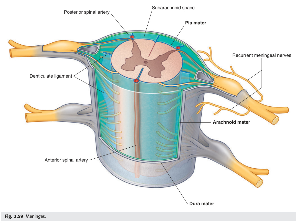
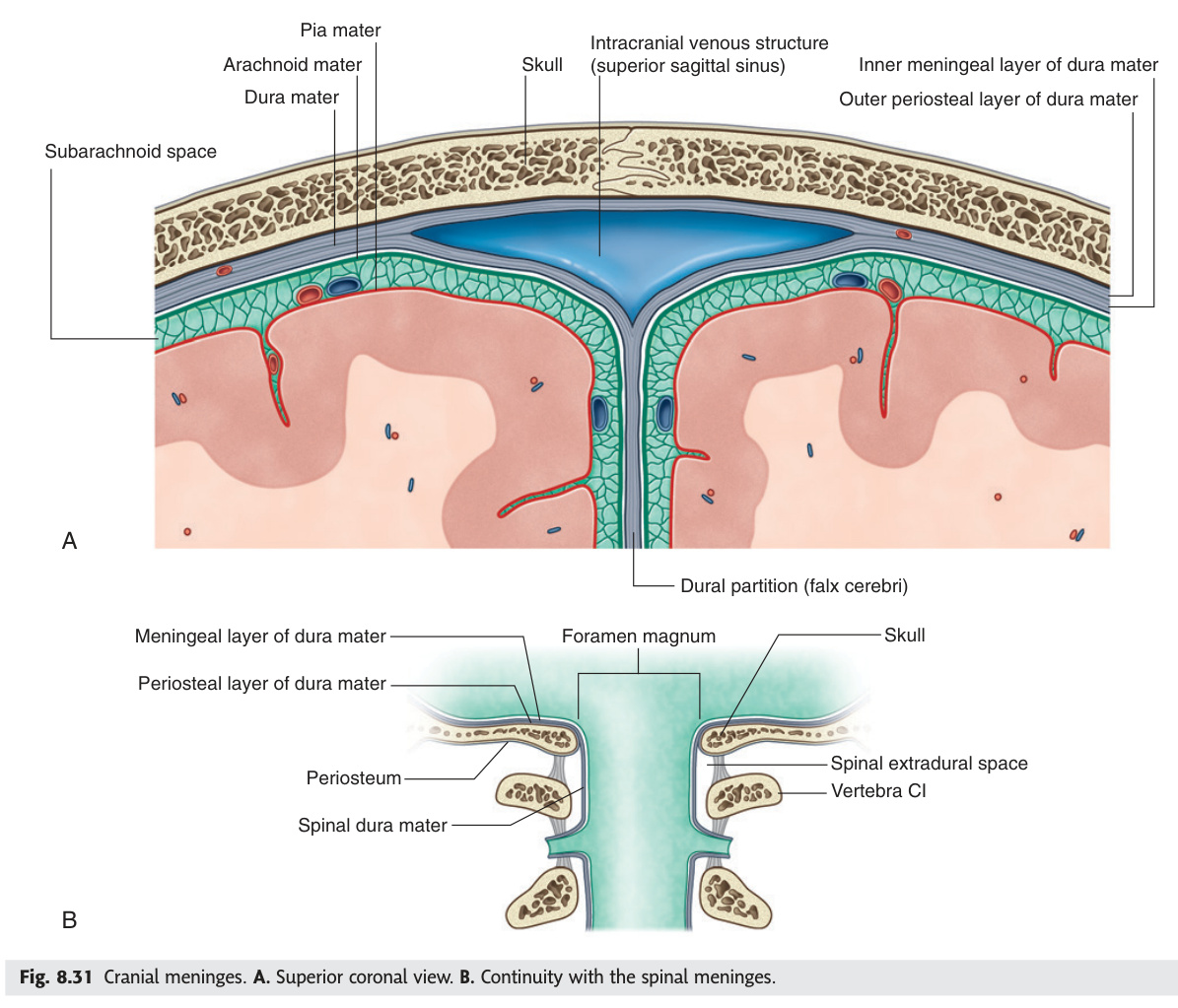
The three meningeal layers — dura, arachnoid, and pia — form concentric coverings around the brain, each defining a distinct bleeding compartment. The tough outer dura mater creates four folds (falx cerebri between hemispheres, tentorium cerebelli between cerebrum and cerebellum with its notch for the brainstem, falx cerebelli, diaphragma sellae) and houses the venous sinuses between its two intracranial layers. Arachnoid granulations projecting from the arachnoid into the superior sagittal sinus are the main CSF absorption site. Three traumatic haemorrhages map precisely to these spaces: extradural (middle meningeal artery rupture at the pterion → biconvex CT lesion with classic lucid interval, does not cross sutures); subdural (bridging vein tears → crescent CT lesion crossing sutures, commonest in elderly/anticoagulated); subarachnoid (berry aneurysm rupture → thunderclap headache, xanthochromic CSF after 2 hours, hyperattenuating cisterns on CT).
| Layer (outer → inner) | Structure | Space below |
|---|---|---|
| Dura mater | Tough outer fibrous layer. Intracranial: two layers (periosteal + meningeal). Spinal: only meningeal layer (no periosteal — epidural space between dura + vertebral periosteum). Dural folds: falx cerebri (between hemispheres), tentorium cerebelli (between cerebrum + cerebellum, has tentorial notch for brainstem), falx cerebelli, diaphragma sellae | Epidural (extradural) space: potential space between dura + bone (intracranially); real space (fat + epidural veins) spinally |
| Arachnoid mater | Avascular membranous layer; closely applied to dura but not attached (allows subdural space). Arachnoid trabeculae bridge to pia. Arachnoid granulations (Pacchioni's granulations): project into dural venous sinuses → absorb CSF | Subdural space: between dura + arachnoid (bridging veins cross this space). Subarachnoid space: between arachnoid + pia; contains CSF + major cerebral arteries + CN roots |
| Pia mater | Innermost, thin, vascular; closely adherent to brain + spinal cord surface. Follows all sulci and gyri. Denticulate ligaments: lateral projections from pia → dura → anchor spinal cord | — |
Subdural haematoma: tearing of bridging veins (crossing subdural space) — often minimal trauma in elderly/alcoholics/anticoagulated. Crescent-shaped hyperdensity on CT (acute — hyperdense) / hypodense (chronic >3 weeks) following brain surface; CROSSES suture lines but stopped by falx/tentorium. Acute: surgical drainage. Chronic: burr holes.
Subarachnoid haemorrhage (SAH): berry aneurysm rupture (Circle of Willis, most common: anterior communicating artery junction). Thunderclap headache ("worst headache of my life"). Blood in subarachnoid space = diffuse on CT (hyperattenuating cisterns). LP: xanthochromia (yellow CSF from haemoglobin breakdown, present 2 hours → 2 weeks after bleed). CT angiogram → identify aneurysm → coiling or clipping.
- Name the four dural folds and what each separates. Falx cerebri (between cerebral hemispheres); tentorium cerebelli (between cerebrum and cerebellum); falx cerebelli (between cerebellar hemispheres); diaphragma sellae (covers the pituitary fossa).
- Compare CT appearances of extradural vs subdural haematoma. Extradural: biconvex (lens-shaped), does NOT cross suture lines; subdural: crescent-shaped, crosses suture lines but stopped by falx/tentorium.
- Which artery ruptures in extradural haematoma and at which skull point? Anterior division of the middle meningeal artery; at the pterion — thinnest point of the skull (junction of frontal, parietal, temporal, and sphenoid bones).
- What is xanthochromia and when does it appear after SAH? Yellow discolouration of CSF from haemoglobin breakdown products; appears 2 hours after bleed, persists up to 2 weeks — used to confirm SAH when CT is normal.
- What is the most common site of berry aneurysm causing SAH? Anterior communicating artery (AComm) junction — presents with thunderclap headache ("worst headache of my life").
CSF Circulation
CSF is produced at ~500 mL/day by the choroid plexus (mainly in the lateral ventricles) and follows a one-way highway: lateral ventricles → foramen of Monro → third ventricle → aqueduct of Sylvius (the narrowest segment, most vulnerable to obstruction) → fourth ventricle → foramina of Luschka (two lateral apertures) and Magendie (one midline aperture) → subarachnoid space → absorbed at arachnoid granulations into the superior sagittal sinus. Obstruction within the ventricular system causes non-communicating hydrocephalus (commonest site: aqueduct stenosis); impaired reabsorption after meningitis or SAH causes communicating hydrocephalus. Normal pressure hydrocephalus (NPH) in the elderly demonstrates the classic "Wet, Wacky, Wobbly" triad (urinary incontinence, dementia, gait apraxia) — the gait prominence distinguishes it from Alzheimer's, and temporary LP drainage confirms the diagnosis before VP shunting.
Clear colourless fluid; 120–150 mL total (intracranial + spinal); produced at ~500 mL/day; normal pressure 5–15 cmH₂O (lumbar). Composition: protein 15–45 mg/dL, glucose ~2/3 serum glucose, <5 WBC/mm³ (all lymphocytes), no RBCs.
2. Flows from lateral ventricles → 3rd ventricle via foramen of Monro (interventricular foramen) (one on each side).
3. From 3rd ventricle → 4th ventricle via cerebral aqueduct of Sylvius (narrowest part — most vulnerable to obstruction → obstructive hydrocephalus).
4. From 4th ventricle → subarachnoid space via: foramina of Luschka (two lateral apertures) + foramen of Magendie (one midline/median aperture).
5. Circulates in subarachnoid space around brain + spinal cord.
6. Absorbed by arachnoid granulations projecting into dural venous sinuses (mainly superior sagittal sinus) → venous blood. Minor drainage also via spinal nerve root sleeves + cribriform plate lymphatics.
Mnemonic: Monro → Sylvius → Luschka + Magendie → subarachnoid → arachnoid granulations.
Non-communicating (obstructive): block within ventricular system. Most common site: aqueduct of Sylvius (stenosis — congenital or from tumour/haemorrhage). 4th ventricle outlet obstruction: Arnold-Chiari malformation, posterior fossa tumour. Features: headache (worse in morning), papilloedema, nausea, sunset sign (eyes downward deviated). Treat: ventriculoperitoneal (VP) shunt or endoscopic third ventriculostomy (ETV — makes hole in floor of 3rd ventricle).
Communicating: no block in ventricles; impaired reabsorption at arachnoid granulations (post-meningitis, post-SAH). Normal pressure hydrocephalus (NPH): triad = Wet (urinary incontinence) + Wacky (dementia) + Wobbly (gait apraxia) in elderly; LP drainage test → temporary improvement → VP shunt.
- Trace the CSF pathway from production to absorption. Choroid plexus (lateral ventricles mostly) → foramen of Monro → 3rd ventricle → aqueduct of Sylvius → 4th ventricle → foramina of Luschka (×2 lateral) + Magendie (×1 midline) → subarachnoid space → arachnoid granulations → superior sagittal sinus.
- Which is the narrowest CSF pathway segment and what does its obstruction cause? Aqueduct of Sylvius — obstruction causes non-communicating (obstructive) hydrocephalus.
- What is normal CSF composition (protein, glucose, cells)? Protein 15–45 mg/dL; glucose ~2/3 serum glucose; WBC <5/mm³ (lymphocytes only); no RBCs.
- Describe the "Wet, Wacky, Wobbly" triad and its diagnosis. Normal pressure hydrocephalus (NPH) — urinary incontinence + dementia + gait apraxia in the elderly; communicating hydrocephalus with normal LP pressure; treated with VP shunt after LP drainage test confirms improvement.
- What is endoscopic third ventriculostomy (ETV)? A hole made in the floor of the third ventricle to bypass aqueduct obstruction — alternative to VP shunt for non-communicating hydrocephalus.
Blood-Brain Barrier (BBB)
The blood-brain barrier is formed by tight junctions between cerebral capillary endothelial cells, maintained by astrocyte end-feet and pericytes, acting as a selective gateway that protects the brain while making CNS infections hard to treat. Lipid-soluble molecules (O₂, CO₂, ethanol, steroids, most anaesthetics) cross freely; hydrophilic drugs cannot — penicillin G only reaches therapeutic CNS levels when the BBB is inflamed by meningitis, justifying high-dose IV therapy. Dopamine cannot cross the BBB, so Parkinson's disease is treated with its precursor L-DOPA (crosses via amino acid transporters, then converted to dopamine inside the brain). The circumventricular organs — area postrema (chemoreceptor trigger zone), subfornical organ, pineal gland, neurohypophysis — deliberately lack a BBB to sample blood composition; the area postrema detects toxins and triggers vomiting, explaining why ondansetron (5-HT3 antagonist) acts there.
The BBB is formed by tight junctions between cerebral capillary endothelial cells, supported by astrocyte end-feet + pericytes. Restricts passage of molecules from blood to CNS.
| Freely crosses BBB | Requires transport / does NOT cross |
|---|---|
| Lipid-soluble molecules (O₂, CO₂, ethanol, most anaesthetics, steroid hormones) | Large molecules (proteins, antibodies — explain why CNS infections are hard to treat) |
| Small lipophilic drugs (chloramphenicol, rifampicin — used for meningitis) | Hydrophilic drugs (penicillin G only crosses inflamed BBB — use high dose in meningitis) |
| Water (via aquaporin-4 channels in astrocyte end-feet) | Dopamine (does NOT cross — hence L-DOPA used for Parkinson's, crosses then converted) |
Area postrema (vomiting centre in medulla — chemoreceptor trigger zone for antiemetics), subfornical organ, organum vasculosum of lamina terminalis (OVLT), pineal gland, neurohypophysis. These areas must "sample" the blood: area postrema detects toxins/emetics → triggers vomiting. Ondansetron (5-HT3 antagonist) acts at area postrema.
- What forms the structural basis of the BBB? Tight junctions between cerebral capillary endothelial cells, supported by astrocyte end-feet and pericytes.
- Why is L-DOPA used in Parkinson's rather than dopamine itself? Dopamine cannot cross the BBB; L-DOPA crosses via large neutral amino acid transporters and is converted to dopamine inside the brain by DOPA decarboxylase.
- Which antibiotic only penetrates the BBB when it is inflamed? Penicillin G — only reaches therapeutic CNS levels in bacterial meningitis (inflamed BBB); justifies high-dose IV therapy.
- Name two antibiotics that reliably cross the intact BBB. Chloramphenicol and rifampicin — both lipophilic, penetrating well regardless of inflammation.
- What is the area postrema and why is it clinically relevant? A circumventricular organ in the medulla without a BBB — acts as the chemoreceptor trigger zone (CTZ), detecting bloodborne toxins and triggering vomiting. Target of ondansetron (5-HT3 antagonist).
Dural Venous Sinuses
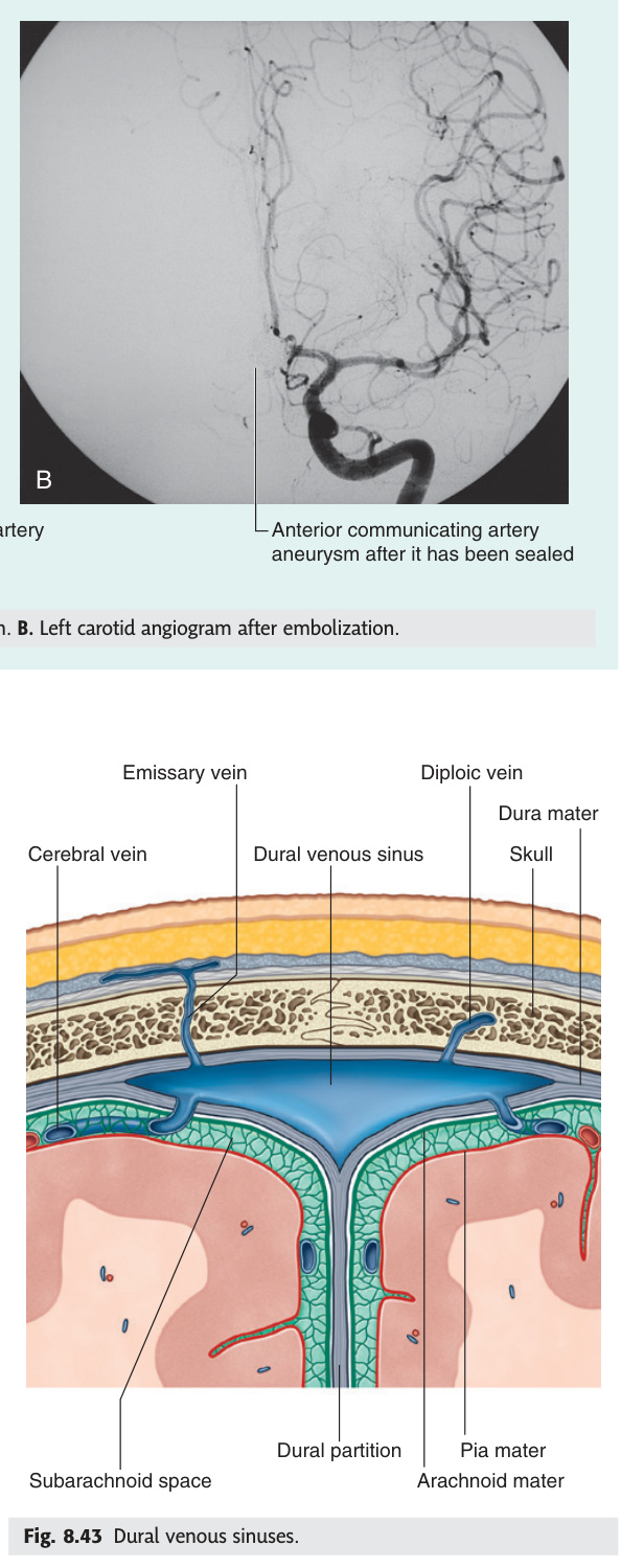
The dural venous sinuses are blood-filled channels formed between the two layers of the dura mater, draining cerebral venous blood toward the internal jugular veins. The superior sagittal sinus (SSS), running along the top of the falx, receives cortical bridging veins and houses arachnoid granulations for CSF absorption; SSS thrombosis causes headache, papilloedema, and parasagittal cortical infarction (bilateral leg weakness), with the "empty delta sign" on contrast CT. The cavernous sinus flanks the sella turcica and is anatomically unique — CN III, IV, V1, V2 run in its lateral wall (mnemonic O TOM CAT: Oculomotor, Trochlear, Ophthalmic, Maxillary), while CN VI and the internal carotid artery float freely within the sinus itself; cavernous sinus thrombosis from facial infection causes proptosis, chemosis, and complete ophthalmoplegia. The transverse-sigmoid system drains laterally to the jugular foramen and is at greatest risk from mastoiditis (lateral sinus thrombosis — picket-fence fever).
| Sinus | Location / Formation | Clinical |
|---|---|---|
| Superior sagittal sinus (SSS) | Upper edge of falx cerebri; from foramen caecum → internal occipital protuberance → confluens sinuum. Arachnoid granulations project into it. Bridging veins drain into it | SSS thrombosis: headache + papilloedema + seizures + focal deficits (parasagittal cortex infarction). Risk: OCP, dehydration, pregnancy, hypercoagulable states. MRI/MRV: "empty delta sign" on contrast CT (filling defect in SSS) |
| Cavernous sinus | On either side of sella turcica. Structures within: CN III + IV + V1 + V2 in lateral wall (mnemonic: O TOM CAT — Oculomotor, Trochlear, Ophthalmic, Maxillary). CN VI + internal carotid artery within the sinus itself (floating free) | Cavernous sinus thrombosis (CST): facial infection → angular vein → cavernous sinus. Features: proptosis + chemosis + ophthalmoplegia (all EOMs + V1/V2 sensory loss) + fever. Treat: IV antibiotics + anticoagulation. Carotid-cavernous fistula: direct (trauma) or indirect (spontaneous dural AVM) → pulsatile exophthalmos + bruit over eye + engorgement of conjunctival veins |
| Transverse + sigmoid sinuses | Transverse: in tentorial edge. Sigmoid: continues transverse → IJV at jugular foramen | Lateral sinus thrombosis: complication of mastoiditis. Otitic hydrocephalus (benign intracranial hypertension with lateral sinus thrombosis) |
- Which structures pass in the lateral wall of the cavernous sinus vs within it? Lateral wall (O TOM CAT): CN III (Oculomotor), CN IV (Trochlear), CN V1 (Ophthalmic), CN V2 (Maxillary); within the sinus: CN VI + internal carotid artery (floating freely).
- What are the clinical features of cavernous sinus thrombosis? Proptosis + chemosis + complete ophthalmoplegia (all EOMs paralysed) + V1/V2 sensory loss + fever — from facial infection spreading via the angular vein.
- What is the "empty delta sign" on contrast CT? A filling defect (non-enhancing centre surrounded by enhancing dura) in the superior sagittal sinus — indicates SSS thrombosis.
- Where do arachnoid granulations project and what is their function? Into the superior sagittal sinus — absorb CSF into the venous blood at a rate matching CSF production (~500 mL/day).
- Which sinus is most at risk from mastoiditis? Transverse/sigmoid sinus — lateral sinus thrombosis complicates coalescent mastoiditis; causes picket-fence fever + headache + papilloedema.
Lumbar Puncture (LP)
LP is performed at L3/L4 or L4/L5 because the spinal cord ends at L1/L2 (conus medullaris) in adults, leaving only freely floating cauda equina roots below — identified using Tuffier's line (connecting the iliac crests crosses L4). Layers traversed from skin to CSF: supraspinous ligament → interspinous ligament → ligamentum flavum → epidural space → dura → arachnoid → subarachnoid space. CSF analysis then cleanly separates meningitis causes: bacterial (turbid, neutrophils >1000, glucose ↓↓, protein ↑↑↑); viral (clear, lymphocytes 10–1000, glucose normal); TB (clear/fibrin web, lymphocytes, glucose ↓, protein ↑↑). In suspected meningococcal septicaemia (non-blanching rash) or raised ICP, antibiotics must be given before LP — never delay treatment for a diagnostic procedure. Meningism signs (Kernig's, Brudzinski's, nuchal rigidity) confirm meningeal irritation clinically but have only ~50–60% sensitivity.
Layers traversed (skin → CSF): skin → subcutaneous fat → supraspinous ligament → interspinous ligament → ligamentum flavum → epidural space (fat + epidural veins) → dura mater → arachnoid mater → subarachnoid space (CSF). "Some Say Lovers Try Positions That They Can't Handle"
Contraindications: raised ICP (risk of transtentorial herniation — always check fundoscopy/CT before LP if symptoms suggest mass lesion); local infection at puncture site; coagulopathy (INR >1.5, platelets <50×10⁹/L); spinal cord compression at lumbar level.
| CSF finding | Bacterial meningitis | Viral meningitis | TB meningitis | SAH |
|---|---|---|---|---|
| Appearance | Turbid/cloudy | Clear | Fibrin web/clear | Bloody/xanthochromic |
| WBC | >1000 PMN (neutrophils) | 10–1000 lymphocytes | 10–500 lymphocytes | RBCs (uniform all 3 tubes) |
| Protein | ↑↑↑ (>1 g/L) | Normal or mildly ↑ | ↑↑ (0.5–3 g/L) | Elevated (by lysis) |
| Glucose | ↓↓ (<2.2 mmol/L; CSF:serum <0.4) | Normal | ↓ (0.4–2.2) | Normal |
Nuchal rigidity: involuntary neck stiffness on passive flexion — resistance to chin-to-chest movement. Due to meningeal irritation/inflammation. Absent in coma, immunocompromised, very early disease.
Kernig's sign: patient supine + hip flexed 90° → attempts to extend the knee → pain and resistance at <135° of extension (hamstring spasm from lumbosacral nerve root irritation by inflamed meninges).
Brudzinski's sign: passive flexion of the neck → involuntary flexion of both hips and knees (reflex attempt to relieve meningeal stretch). Two forms: neck sign (above) and contralateral leg sign (pressing one flexed leg → other leg flexes reflexively).
Jolt accentuation: existing headache worsens on horizontal head rotation at 2–3 Hz — sensitive screen for meningitis when nuchal rigidity is absent.
Sensitivity of Kernig's + Brudzinski's: ~50–60% — absence does NOT rule out meningitis, especially in immunocompromised or elderly.
| Age Group | Most Likely Organisms | Empirical Antibiotic Cover |
|---|---|---|
| Neonates (<3 months) | Group B Streptococcus (GBS, S. agalactiae) — most common. Escherichia coli. Listeria monocytogenes. (GBS + E. coli account for ~70%) | Ampicillin + cefotaxime (or gentamicin). Ampicillin covers Listeria (cephalosporins do NOT cover Listeria — critical) |
| Infants & children (3 months – 18 years) | Neisseria meningitidis (meningococcus — serogroups B + C in UK; A in belt of Africa). Streptococcus pneumoniae. Haemophilus influenzae type b (now rare — HiB vaccine) | Ceftriaxone (3rd gen cephalosporin covers all three). Add dexamethasone before/with 1st dose (reduces hearing loss complication of H. influenzae + pneumococcal meningitis) |
| Adults (18–60 years) | S. pneumoniae (most common overall; highest mortality ~20–30%). N. meningitidis (purpuric non-blanching rash = meningococcal septicaemia — treat before LP; penicillin G or ceftriaxone) | Ceftriaxone ± dexamethasone. Add amoxicillin/ampicillin if Listeria risk (immunosuppressed, alcoholic, diabetic, elderly) |
| Elderly & immunocompromised (>60 years) | S. pneumoniae, Listeria monocytogenes (increased risk — T-cell immunity wanes). Gram-negative bacilli (E. coli, Klebsiella in nosocomial/neonatal) | Ceftriaxone + ampicillin (Listeria cover). Consider vancomycin if MRSA or penicillin-resistant pneumococcus |
| Post-surgical / post-traumatic | Staphylococcus aureus (including MRSA). Coagulase-negative staphylococci. Gram-negative rods (Pseudomonas, Klebsiella) | Vancomycin + ceftazidime/meropenem (broad gram-negative + MRSA cover) |
| Aseptic (viral) meningitis — organisms | Enteroviruses (most common cause of viral meningitis: echovirus, coxsackievirus, poliovirus). HSV-2 (genital herpes recurrence). Mumps. HIV seroconversion. EBV | Supportive; acyclovir if HSV suspected; no antibiotics required if definitively viral |
Aetiology: HSV-1 (in adults and children >3 months — reactivation from trigeminal ganglion tracking along olfactory tract to limbic system → temporal lobes + orbitofrontal cortex). HSV-2 (neonatal encephalitis via birth canal; also HSV-2 meningitis in adults as benign Mollaret's meningitis).
Presentation: subacute onset (days) fever + headache + altered consciousness + temporal lobe features — psychiatric symptoms (agitation, hallucinations, personality change), olfactory/gustatory hallucinations, temporal lobe seizures (lip-smacking, automatisms), memory impairment (hippocampal involvement), aphasia if dominant temporal lobe involved. Focal neurological signs appear as disease progresses.
Triad: fever + temporal lobe features + altered consciousness = HSE until proven otherwise → start acyclovir immediately.
| Investigation | Finding in HSE | Notes |
|---|---|---|
| MRI brain (gold standard imaging) | T2/FLAIR hyperintensity in bilateral (asymmetric) temporal lobes + insular cortex + orbitofrontal cortex. DWI restriction = cytotoxic oedema. May show haemorrhagic foci (haemorrhagic necrotising encephalitis). Ring enhancement ONLY in advanced necrotic lesions | CT is often normal early (days 1–3) — do not rely on CT. MRI may also be normal in first 24 hours. Repeat if high suspicion |
| CSF PCR for HSV-DNA | Positive HSV-1 DNA on PCR = diagnostic. Sensitivity 98%, specificity 99%. CSF: lymphocytic pleocytosis (10–200 WBC), mildly elevated protein, normal glucose, may contain RBCs (haemorrhagic component) | PCR may be negative in first 72 hours of illness — treat empirically and REPEAT LP at 3–7 days if initial PCR negative but clinical suspicion remains. CSF PCR becomes positive as disease progresses |
| EEG | Periodic lateralised epileptiform discharges (PLEDs) over temporal region — characteristic but not specific for HSE. Helps detect subclinical seizures | Useful adjunct when MRI non-diagnostic |
- Why is LP done at L3/L4 and not higher? The spinal cord ends at L1/L2 (conus medullaris) in adults; below this only cauda equina roots float freely. Tuffier's line (joining both iliac crests) crosses L4 spinous process.
- Name all layers traversed from skin to CSF during LP. Skin → subcutaneous fat → supraspinous ligament → interspinous ligament → ligamentum flavum → epidural space → dura mater → arachnoid mater → subarachnoid space.
- Compare CSF findings in bacterial vs viral vs TB meningitis. Bacterial: turbid, neutrophils >1000, glucose ↓↓, protein ↑↑↑; Viral: clear, lymphocytes 10–1000, glucose normal; TB: clear/fibrin web, lymphocytes 10–500, glucose ↓, protein ↑↑.
- Why must ampicillin be added to ceftriaxone in neonates and elderly? Listeria monocytogenes is intrinsically resistant to all cephalosporins; affects those with impaired T-cell immunity. Ampicillin (or co-trimoxazole) is the only standard cover for Listeria.
- What are the three meningism signs and their clinical limitations? Kernig's (resistance to knee extension with hip flexed 90°), Brudzinski's (neck flexion → involuntary hip/knee flexion), nuchal rigidity — all indicate meningeal irritation; sensitivity only ~50–60%, absent in immunocompromised and early disease.
Raised Intracranial Pressure (ICP)
The Monro-Kellie doctrine governs intracranial pressure: the skull is rigid, so any volume increase in brain, blood, or CSF must be compensated by a decrease in another compartment or ICP rises. Cerebral perfusion pressure (CPP = MAP − ICP) must be kept ≥60 mmHg; when ICP rises unchecked, Cushing's triad appears — hypertension with widened pulse pressure, bradycardia, and irregular respirations — a terminal sign of impending brainstem compression. Uncal herniation (temporal mass pushing the uncus over the tentorial edge) compresses CN III first (ipsilateral fixed dilated pupil before ophthalmoplegia) then the posterior cerebral artery. Management escalates through steps: head elevation and sedation → osmotherapy (mannitol or hypertonic saline) → brief hyperventilation (CO₂-driven vasoconstriction, temporary) → surgical decompression; dexamethasone reduces vasogenic oedema around tumours but worsens outcomes in TBI and stroke. IIH (obese women of childbearing age — headache, pulsatile tinnitus, papilloedema, normal CSF composition) is treated with weight loss + acetazolamide; the chief danger is permanent visual loss from optic nerve damage.
Normal ICP: 5–15 mmHg. Raised ICP (>20 mmHg sustained) = neurological emergency. Monro-Kellie doctrine: intracranial volume is fixed (skull rigid) → any increase in one compartment (brain, blood, CSF) must be compensated by decrease in another, or ICP rises.
Cushing's triad (terminal sign of brainstem compression): 1. Hypertension (widened pulse pressure — reflex response to maintain CPP) + 2. Bradycardia (vagal response to hypertension) + 3. Irregular respirations (Cheyne-Stokes, central neurogenic hyperventilation, ataxic breathing). Indicates imminent brainstem herniation — act immediately.
Uncal herniation: expanding temporal lobe lesion → uncus of temporal lobe herniates over tentorial edge → compresses CN III (ipsilateral fixed dilated pupil FIRST, then ophthalmoplegia + ptosis) + posterior cerebral artery (→ ipsilateral occipital infarct). Then ipsilateral hemiplegia (contralateral pyramidal tract at cerebral peduncle compresses against tent). Kernohan's notch phenomenon: contralateral cerebral peduncle compressed against opposite tentorial edge → "false localising" ipsilateral hemiplegia.
Tonsillar herniation (coning): cerebellar tonsils herniate through foramen magnum → brainstem compression → sudden death.
General (all patients): Head of bed 30°; avoid hypotension (MAP ≥80 mmHg); avoid hypoxia (O₂ sat ≥95%); avoid hyper/hyponatraemia; avoid pyrexia (temp control); avoid raised intrathoracic pressure.
Step 1 — Optimise sedation/analgesia: reduces agitation + pain which ↑ICP.
Step 2 — Osmotherapy: Mannitol 20% (0.25–1 g/kg IV bolus) — osmotic diuretic draws water from brain; avoid if hypovolaemic (intravascular depletion ↑↑ICP paradoxically). Or Hypertonic saline (3% NaCl) — may be preferred if haemodynamically unstable; ↑ serum Na to 145–155 mmol/L.
Step 3 — Controlled hyperventilation: reduce PaCO₂ to 30–35 mmHg → cerebral vasoconstriction → ↓ cerebral blood volume → ↓ ICP. Temporary bridge only (effects wear off in hours, rebound on normalisation).
Step 4 — Surgical decompression: Neurosurgical evacuation of haematoma/tumour, VP shunt for hydrocephalus, decompressive craniectomy (remove skull flap for malignant MCA infarction or refractory TBI).
Steroids: dexamethasone reduces oedema ONLY around tumours (vasogenic oedema) — NOT beneficial in TBI, stroke, or post-SAH (can worsen outcomes).
Raised intracranial pressure (>25 cmH₂O on LP) with normal CSF composition, normal brain parenchyma on MRI (no mass lesion, no ventricular dilatation), and no identifiable secondary cause. Modified Dandy criteria: symptoms/signs of raised ICP + elevated opening pressure on LP + normal CSF composition + no other cause found.
Classic patient: obese woman of childbearing age (15–44 years). Incidence rises steeply with obesity — adipose tissue may produce factors that increase CSF production or reduce absorption. Symptoms: daily positional headache (worse on lying down or Valsalva), pulsatile tinnitus (whooshing sound synchronous with heartbeat — almost pathognomonic), transient visual obscurations (seconds of greying/blurring, especially on postural change), horizontal diplopia (CN VI palsy — false localising sign from raised ICP stretching the long intracranial course of abducens).
Signs: bilateral papilloedema (virtually always present at diagnosis — graded by Frisén scale; visual field loss is the main risk). Visual field defects: enlarged blind spot first, then inferior nasal constriction, then generalised constriction → if untreated → permanent visual loss (the major morbidity). Normal consciousness (distinguishes from other causes of raised ICP — patient looks well but has papilloedema).
| Feature | IIH | Notes |
|---|---|---|
| MRI brain | Empty sella (flattening of pituitary gland by chronically raised CSF pressure), posterior globe flattening, distension of optic nerve sheath, transverse sinus stenosis (may be cause or effect) | Ventricles are normal or small (not enlarged — distinguishes from hydrocephalus) |
| LP opening pressure | >25 cmH₂O (some use >28 in obese); CSF composition entirely normal (protein, glucose, cells all normal) | LP is both diagnostic AND temporarily therapeutic (CSF removal gives hours of headache relief). Done in lateral decubitus position with legs extended to measure true opening pressure |
| Secondary causes to exclude | Venous sinus thrombosis (SSS thrombosis); venous outflow obstruction; medications: tetracyclines (minocycline, doxycycline), vitamin A excess (retinoids, isotretinoin for acne), anabolic steroids, oral contraceptives, growth hormone; hypoparathyroidism; Cushing's disease withdrawal | Always check for these before diagnosing IIH — it is a diagnosis of exclusion |
1st line: Weight loss (10% body weight → significant ICP reduction; most important long-term treatment) + acetazolamide (carbonic anhydrase inhibitor → reduces CSF production; standard first-line medication; side effects: paraesthesia, taste disturbance, renal stones).
2nd line: Topiramate (anti-epileptic with carbonic anhydrase activity + promotes weight loss). Loop diuretics (furosemide) — second agent.
Serial therapeutic LP: gives temporary relief (hours–days); used as bridge when vision threatened acutely.
Surgical (sight-threatening or medically refractory): (a) Optic nerve sheath fenestration (ONSF) — incision in optic nerve sheath behind the globe → CSF decompression → protects vision; primarily local effect; does not reduce ICP globally. (b) CSF diversion: lumboperitoneal shunt (LP shunt) or ventriculoperitoneal shunt (VP shunt); reduces ICP globally; high revision rate.
Test Unit 15 knowledge
Meninges, CSF pathway, LP anatomy, and intracranial haemorrhage MCQs.
- State the Monro-Kellie doctrine. The skull is a rigid box — any increase in one intracranial compartment (brain/blood/CSF) must be compensated by a decrease in another, or ICP rises.
- What is CPP and what is the minimum target? Cerebral Perfusion Pressure = MAP − ICP; target ≥60 mmHg. Normal ICP is 5–15 mmHg; treat if sustained >20 mmHg.
- What is Cushing's triad and what does it indicate? Hypertension (widened pulse pressure) + bradycardia + irregular respirations — a terminal sign of imminent brainstem compression; act immediately.
- In uncal herniation, which nerve is compressed first and what is the first clinical sign? CN III — first sign is ipsilateral fixed dilated (blown) pupil, as the pupilloconstrictor fibres run on the outside of CN III and are compressed before motor fibres.
- When is dexamethasone helpful vs harmful for raised ICP? Helpful: vasogenic oedema around brain tumours. Harmful (worsens outcomes): traumatic brain injury, ischaemic stroke, and haemorrhagic stroke.
Cerebral Arterial Territories & Stroke Syndromes ★★★
The cerebral circulation divides into anterior (ACA, MCA) and posterior (PCA, basilar, PICA, AICA) territories, each producing a recognisable syndrome when occluded. MCA is the most common stroke territory — contralateral face + arm weakness > leg, hemisensory loss, and homonymous hemianopia; dominant hemisphere involvement adds aphasia (Broca's for expression at the inferior frontal gyrus, Wernicke's for comprehension at the posterior superior temporal gyrus). PICA infarct (Wallenberg/lateral medullary syndrome) causes the classic crossed sensory loss — ipsilateral face pain/temperature loss + Horner's + dysphagia/hoarseness + contralateral body sensory loss, without hemiplegia (corticospinal tract is ventral and spared). Basilar artery occlusion causes locked-in syndrome: bilateral pontine infarct with bilateral hemiplegia and anarthria but preserved vertical eye movements and consciousness. Lacunar infarcts (small vessel lipohyalinosis in perforating arteries) produce pure motor, pure sensory, or ataxic-hemiparesis syndromes without cortical features; watershed infarcts from global hypoperfusion damage zones between adjacent territories.
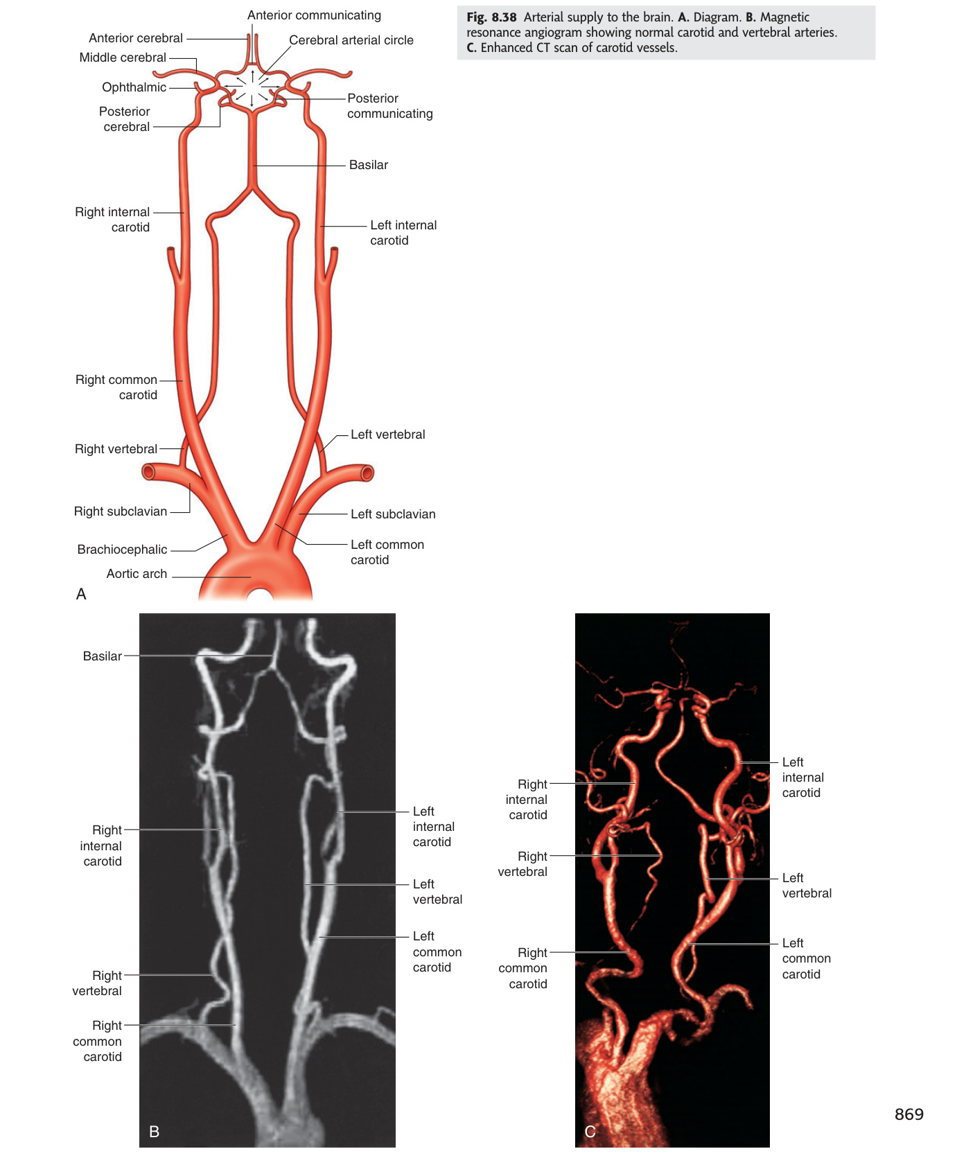
The Circle of Willis (circulus arteriosus) is an anastomotic polygon at the base of the brain formed by: bilateral ACAs (joined by AComm), bilateral MCAs, bilateral PCAs (joined to ICA by PComms). Complete circle in only 20–25% — frequent anatomical variants. Functions as collateral bypass when one vessel occluded.
| Artery | Territory | Stroke Syndrome |
|---|---|---|
| ACA (Anterior Cerebral) | Medial surface of frontal + parietal lobe; anterior corpus callosum. Leg area of motor + sensory cortex | Contralateral LEG > arm/face weakness + sensory loss. Urinary incontinence (medial frontal lobe). Behavioural changes (prefrontal cortex). Grasp reflex. Bilateral ACA infarct (from AComm aneurysm or ICA bifurcation occlusion) → akinetic mutism (alert but mute + immobile — bilateral supplementary motor cortex) |
| MCA (Middle Cerebral) | Lateral surface of frontal + parietal + temporal lobes; deep (lenticulostriate arteries) → putamen, caudate, internal capsule | Most common stroke territory. Face + arm >> leg weakness (contralateral). Hemisensory loss. Dominant hemisphere (L in R-handed): Broca's area (inferior frontal gyrus) → expressive aphasia; Wernicke's area (posterior STG) → receptive aphasia. Non-dominant: hemispatial neglect, anosognosia. Homonymous hemianopia (optic radiation). Internal capsule infarct (lenticulostriate) → pure motor hemiplegia or pure sensory stroke |
| PCA (Posterior Cerebral) | Occipital lobe (visual cortex) + posterior temporal + thalamus + midbrain | Contralateral homonymous hemianopia WITH macular sparing (dual MCA/PCA supply to macular cortex). Thalamic infarct: contralateral hemisensory loss ± thalamic pain (Déjérine-Roussy syndrome). Top-of-basilar syndrome: bilateral PCA occlusion → cortical blindness (Anton's syndrome — patient denies being blind) + altered consciousness |
| PICA (Post. Inferior Cerebellar) | Lateral medulla + inferior cerebellum | Wallenberg syndrome (lateral medullary syndrome): ipsilateral face pain/temperature loss (CN V nucleus) + Horner's syndrome (descending sympathetic) + dysphagia/hoarseness (CN IX/X nuclei) + ataxia (cerebellum/inf. cerebellar peduncle). Contralateral body pain/temperature loss (spinothalamic tract). No hemiplegia (corticospinal tract in ventral medulla, spared). "Crossed sensory loss" = lateral medullary infarct until proven otherwise |
| AICA (Ant. Inferior Cerebellar) | Lateral pons + inner ear (labyrinthine artery) + anterior cerebellum | Ipsilateral: facial sensory loss + CN VI (abducens) + CN VII palsy + sensorineural deafness + vertigo (inner ear ischaemia). Contralateral body sensory loss. Ipsilateral ataxia |
| Basilar artery | Pons + midbrain + supplies SCA + AICA | Basilar artery occlusion = locked-in syndrome: bilateral pontine infarct → bilateral hemiplegia + anarthria + dysphagia. Preserved: vertical eye movements + blinking (corticospinal fibres to CN III intact above). Patient conscious but cannot move; communicates by eye movement. Distinguish from vegetative state (vegetative = unconscious, no voluntary movement) |
| Type | Mechanism | Syndromes |
|---|---|---|
| Lacunar infarcts | Small vessel disease (lipohyalinosis from HTN/DM) → occlusion of perforating arteries (lenticulostriate, thalamoperforators) → small deep infarcts in basal ganglia, thalamus, internal capsule, pons | (1) Pure motor hemiplegia (internal capsule posterior limb or pons); (2) Pure sensory stroke (thalamus — VPL nucleus); (3) Sensorimotor stroke (posterior internal capsule + thalamus); (4) Ataxic hemiparesis (pons or posterior limb IC); (5) Dysarthria-clumsy hand (pons). NIHSS typically low. MRI DWI best for acute lacune |
| Watershed (borderzone) infarcts | Global hypoperfusion (cardiac arrest, severe hypotension, carotid occlusion) → ischaemia in zones between terminal territories of adjacent arteries | ACA/MCA borderzone: "man in a barrel" syndrome — proximal arm + leg weakness with face + hands spared (medial shoulder-hip cortex in borderzone). MCA/PCA borderzone: transcortical aphasia (speech preserved but repetition lost; Broca's/Wernicke's areas in MCA territory spared but association fibres in borderzone damaged) |
- What deficits distinguish dominant vs non-dominant MCA stroke? Dominant (usually left): Broca's aphasia (expressive, inferior frontal gyrus) + Wernicke's aphasia (receptive, posterior STG); Non-dominant: hemispatial neglect, anosognosia.
- Describe Wallenberg (lateral medullary) syndrome — artery, deficits. PICA infarct: ipsilateral face pain/temperature loss (CN V nucleus) + Horner's + dysphagia/hoarseness (CN IX/X) + ipsilateral ataxia + contralateral body pain/temperature loss (spinothalamic). No hemiplegia — corticospinal tract is ventral and spared.
- What is locked-in syndrome and which artery is occluded? Basilar artery occlusion → bilateral pontine infarct → bilateral hemiplegia + anarthria with preserved vertical eye movements + blinking + consciousness. Communicate by eye movement.
- What is a watershed infarct and when does it occur? Ischaemia in zones between adjacent arterial territories during global hypoperfusion (cardiac arrest, severe hypotension); ACA/MCA borderzone → "man in a barrel" (proximal arm + leg weakness with face + hands spared).
- Name five lacunar syndromes and their common sites. Pure motor hemiplegia (posterior IC/pons); pure sensory stroke (thalamus); sensorimotor stroke (IC + thalamus); ataxic hemiparesis (pons/IC); dysarthria-clumsy hand (pons) — all from small vessel lipohyalinosis.
Ring-Enhancing Lesions & Cerebral Abscess ★★★
A ring-enhancing lesion — central non-enhancement (necrosis, pus, or cyst) surrounded by a contrast-enhancing ring with oedema — is non-specific and always demands a differential with the MAGIC mnemonic. Metastases are the most common cause in adults over 40, typically multiple at the grey-white junction with disproportionate oedema. A cerebral abscess has a smooth, thin, regular ring with the critical distinguishing feature of restricted diffusion centrally on DWI-MRI — pus is viscous and cellular, restricting water movement, unlike the free diffusion of necrotic GBM. GBM shows a thick, irregular ring, may cross the corpus callosum as a "butterfly glioma", and has free central diffusion. In AIDS patients with CD4 <100, multiple lesions in the basal ganglia are treated empirically for toxoplasmosis (pyrimethamine + sulfadiazine) — failure to respond in 2 weeks mandates biopsy to exclude primary CNS lymphoma. LP is contraindicated in all these conditions given the risk of herniation — MRI is the investigation of choice.
A lesion on contrast CT/MRI showing a central area of no enhancement (necrosis, pus, or cystic change) surrounded by a ring of enhancement (breakdown of the blood-brain barrier at the lesion periphery) with surrounding low-density oedema. Always a differential diagnosis situation — the ring is non-specific.
M — Metastasis: most common cause of ring-enhancing lesion in adults >40 years. Location: grey-white junction (haematogenous spread; high flow at GM/WM interface). Features: multiple lesions = metastases until proven otherwise; disproportionate oedema for lesion size; primary tumours: lung (most common), breast, renal, melanoma, colorectal. Free diffusion centrally (necrotic debris).
A — Abscess (cerebral abscess): smooth, thin, regular ring (unlike the thick/irregular ring of GBM). Key distinguishing feature: restricted diffusion centrally on DWI-MRI (pus = high viscosity, high cellularity → bright DWI, dark ADC map). Also: satellite lesions, daughter abscesses. Clinical: fever + headache + focal deficit (classic triad in only ~50% of cases).
G — Glioblastoma multiforme (GBM, WHO grade IV): most common primary malignant brain tumour in adults (peak 55–65 years). Features: thick, irregular, nodular ring with satellite fingers; crossing corpus callosum = "butterfly glioma" (virtually diagnostic of GBM). No restricted diffusion centrally. MGMT promoter methylation predicts chemotherapy response. Treatment: maximal safe resection + radiotherapy + temozolomide (Stupp protocol). Median survival 14–16 months.
I — Infarct (subacute): ring enhancement at 1–4 weeks post-ischaemic infarct (luxury perfusion + BBB breakdown at periphery). Conforms to vascular territory; other features of infarct present (DWI restriction in acute phase).
C — Cysticercosis (neurocysticercosis): Taenia solium (pork tapeworm) larval cysts in brain. Endemic in SE Asia, Latin America, sub-Saharan Africa. "Pea-in-a-pod": scolex (white dot) visible within cyst = pathognomonic. Multiple lesions at grey-white junction. Presents with new-onset seizures. Treat: albendazole/praziquantel + steroids (for inflammatory reaction); antiepileptics.
Also consider: Toxoplasmosis (AIDS patients — CD4 <100: multiple ring-enhancing lesions often in basal ganglia; treat empirically pyrimethamine + sulfadiazine; failure to respond in 2 weeks → biopsy to exclude CNS lymphoma); Primary CNS lymphoma (periventricular, often homogeneous enhancement in immunocompetent but ring in AIDS; treat with methotrexate); Tumefactive MS (open ring sign — incomplete ring, open side toward grey matter = characteristic of demyelination).
| Source | Location in brain | Organism(s) |
|---|---|---|
| Sinusitis (frontal/ethmoid) | Frontal lobe | Streptococcus viridans, anaerobes, Staphylococcus |
| Otitis media / mastoiditis | Temporal lobe or cerebellum | Mixed (gram-negative, anaerobes, Streptococcus) |
| Dental abscess | Frontal lobe | Streptococcus, anaerobes |
| Haematogenous (endocarditis, lung abscess) | Multiple, grey-white junction; MCA territory | Staphylococcus aureus, Streptococcus, gram-negative rods |
| Cyanotic congenital heart disease (R→L shunt) | Multiple | Mixed — bypasses pulmonary capillary filter |
| Immunocompromised (AIDS, transplant) | Variable | Toxoplasma gondii, Nocardia, Aspergillus, Candida, Cryptococcus |
| Post-surgical / post-traumatic | At operative site | Staphylococcus aureus, gram-negative rods |
Stages (Britt & Enzmann):
(1) Early cerebritis (days 1–3): ill-defined area of necrosis + inflammation; no ring yet on imaging.
(2) Late cerebritis (days 4–9): central necrotic core expands; early ring formation; oedema peaks.
(3) Early capsule (days 10–13): collagen capsule forms; ring enhancement well-defined; begins to contain pus.
(4) Late capsule (day 14+): mature thick capsule. Capsule is thinner on the ventricular side (less vascular deep white matter) — at risk of rupture into ventricle → ventriculitis (catastrophic, 80% mortality).
Investigation: MRI gadolinium is gold standard. DWI restriction centrally = pus. LP is CONTRAINDICATED (risk of herniation if ICP raised). Blood cultures. Search for source (CXR for lung abscess, echo for endocarditis, dental OPG, sinus CT).
Treatment:
• Aspiration (stereotactic CT-guided) for lesions >2.5 cm, diagnostic + therapeutic. Excision if multiloculated or fungal. Send pus for microscopy + culture.
• Antibiotics: empirical — cefotaxime/ceftriaxone + metronidazole (broad spectrum + anaerobic cover) × 6–8 weeks IV then oral. Adjust on culture. Add anti-staphylococcal (flucloxacillin or vancomycin) if post-surgical/trauma.
• Dexamethasone: only if significant mass effect / herniation risk (reduces oedema but may slow capsule formation and reduce antibiotic penetration — use cautiously).
• Treat source (sinus drainage, dental extraction, endocarditis treatment).
- Give the MAGIC mnemonic for ring-enhancing lesions. Metastases (multiple, grey-white junction, disproportionate oedema); Abscess (smooth thin ring, restricted DWI centrally); GBM (thick irregular ring, may be butterfly); Infarct (subacute, vascular territory); Cysticercosis (scolex pea-in-pod, grey-white junction, seizures).
- What DWI-MRI finding distinguishes a cerebral abscess from GBM? Abscess: restricted diffusion centrally (bright DWI, dark ADC) — pus is viscous and cellular. GBM: free diffusion centrally (liquefactive necrosis = low viscosity). This distinction avoids unnecessary biopsy.
- In AIDS (CD4 <100) with multiple ring-enhancing lesions in the basal ganglia, what is the first treatment and when do you biopsy? Empirical pyrimethamine + sulfadiazine for toxoplasmosis; biopsy only if no radiological improvement in 2 weeks (to exclude primary CNS lymphoma).
- Which cerebral abscess source predicts frontal lobe location? Sinusitis (frontal/ethmoid) or dental abscess. Otitis media/mastoiditis → temporal lobe or cerebellum. Haematogenous (endocarditis, cyanotic CHD) → multiple at grey-white junction.
- Why is LP contraindicated in suspected cerebral abscess? Risk of transtentorial herniation if ICP is raised — MRI with gadolinium is the investigation of choice. LP is unsafe whenever a mass lesion is suspected.