The Ear
Ear Anatomy — Schematic Cross-Section
Purple = External ear · Teal = Middle ear cavity · Gold = Cochlea/Inner ear · Blue = Semicircular canals
External Ear
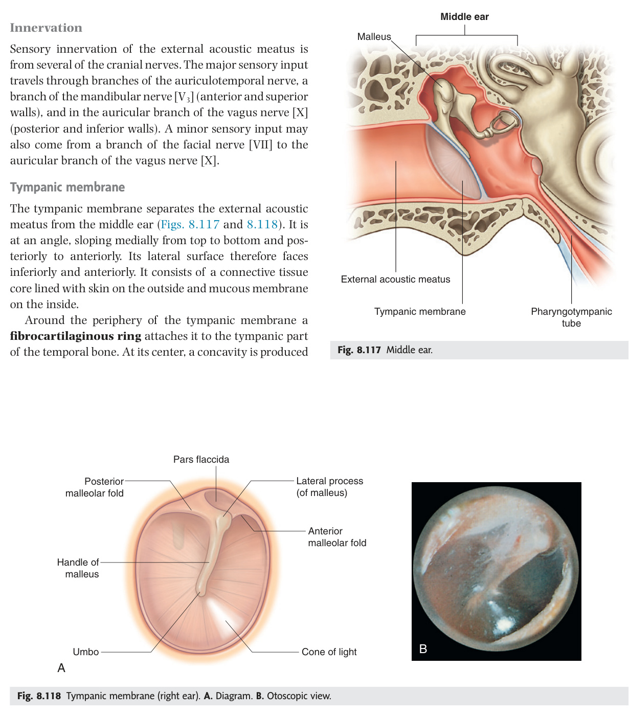
The external ear is a funnel system — the auricle collects sound and channels it through the S-shaped external auditory meatus (EAM) to the tympanic membrane, converting pressure waves into mechanical vibration. The EAM is cartilaginous in its outer third (with hair follicles and ceruminous glands) and bony in its inner two-thirds; its curve must be straightened by pulling the auricle upward and backward in adults — downward and backward in children — before otoscopy can reach the drum. The tympanic membrane has a large taut pars tensa and a small lax pars flaccida (Shrapnell's membrane) above the malleolar folds — cholesteatoma originates at the pars flaccida, while the safe anteroinferior quadrant of the pars tensa is where myringotomy incisions are placed to avoid the ossicles, chorda tympani, and facial nerve. Arnold's nerve (auricular branch of CN X) supplying part of the posterior EAC explains why syringing or otoscopy can trigger a vagal cough reflex or syncope.
| Structure | Details |
|---|---|
| Auricle (pinna) | Elastic cartilage covered by skin. Parts: helix + antihelix + tragus + antitragus + lobule (fibrofatty, no cartilage). Sensory supply: great auricular nerve (C2–C3), auriculotemporal nerve (CN V3), lesser occipital nerve (C2), Arnold's nerve (auricular branch of CN X) |
| External auditory meatus (EAM) | S-shaped canal, 2.5 cm long. Outer 1/3 = cartilaginous (skin with hair follicles + ceruminous glands → earwax). Inner 2/3 = bony (in temporal bone). Straighten canal for otoscopy: pull auricle UP and BACK in adults (DOWN and BACK in children) |
| Tympanic membrane (eardrum) | At end of EAM; obliquely placed (superior-posterior end deeper). Layers: outer squamous epithelium + middle fibrous layer + inner mucosal layer. Parts: pars tensa (lower 4/5, tight) + pars flaccida/Shrapnell's membrane (upper 1/5, above malleolar folds, lax) |
The auricular branch of CN X (Arnold's nerve) supplies part of the posterior EAM. Stimulation (syringing, otoscopy, earwax) can trigger vagal reflexes: cough reflex (most common — "ear-cough reflex"), bradycardia, syncope. This explains why some patients cough when their ears are cleaned. The cough reflex centre in the medulla is connected to CN X nuclei.
- How do you straighten the EAM for otoscopy in adults vs children? Adults: pull auricle upward and backward; children: downward and backward.
- Name the two parts of the tympanic membrane. Pars tensa (lower 4/5, taut); pars flaccida/Shrapnell's membrane (upper 1/5, lax, above malleolar folds — origin of acquired cholesteatoma).
- Which TM quadrant is safe for myringotomy and why? Anteroinferior — avoids the chorda tympani, ossicles, and facial nerve which all lie posterosuperiorly.
- Where is the normal cone of light reflex seen on the TM? Anteroinferior quadrant (5 o'clock in right ear; 7 o'clock in left ear).
- What nerve in the EAC triggers a cough reflex on syringing? Arnold's nerve (auricular branch of CN X) — stimulation activates vagal reflexes including cough, bradycardia, or syncope.
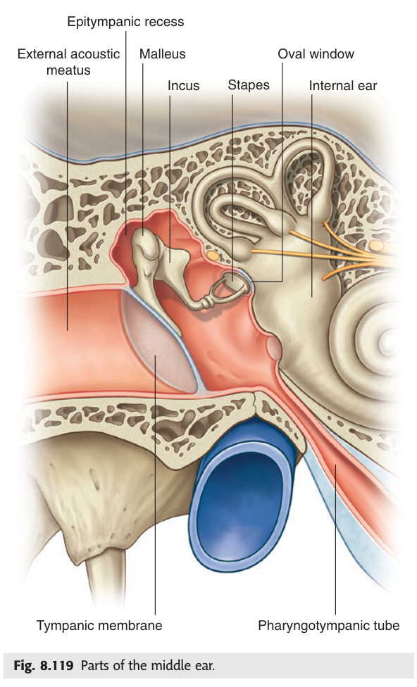
Middle Ear (Tympanic Cavity)
The middle ear is a box with six walls, each with its own clinical story: the thin tegmen above separates it from the middle cranial fossa (cholesteatoma erosion here causes meningitis or temporal lobe abscess), the jugular floor overlies the high-riding jugular bulb visible on otoscopy, and the anterior wall contains the auditory tube and the thin bone of the carotid canal. The medial wall is most event-rich — the oval window (sealed by the stapes footplate) transmits vibration into the perilymph, the round window provides a pressure-relief outlet, and the promontory of the basal cochlear turn carries the tympanic plexus of CN IX. Crossing the middle ear unsheathed between the malleus handle and the long process of the incus is the chorda tympani, carrying taste from the anterior two-thirds of the tongue and parasympathetic supply to the submandibular and sublingual glands — naked exposure to any middle ear pathology or surgery. Cholesteatoma — a locally destructive accumulation of desquamating keratin — is the most important disease of this space, eroding ossicles, the facial canal, the tegmen, and the sigmoid sinus plate through collagenase activity.
| Wall | Contents / Features | Clinical Significance |
|---|---|---|
| Lateral (membranous) | Tympanic membrane + bony wall of epitympanum (attic) above | Perforation here in acute otitis media (AOM); cholesteatoma in attic |
| Medial (labyrinthine) | Oval window (fenestra vestibuli — covered by stapes footplate) + round window (fenestra cochleae — covered by secondary tympanic membrane) + promontory (basal turn of cochlea, tympanic plexus CN IX) | Otosclerosis: bony fixation of stapes footplate → conductive deafness; stapedectomy + prosthesis |
| Roof (tegmental) | Tegmen tympani (thin plate of petrous temporal bone); separates from middle cranial fossa | Cholesteatoma erosion → meningitis, temporal lobe abscess |
| Floor (jugular) | Jugular fossa (internal jugular vein bulb) below | High-riding jugular bulb → visible in middle ear on otoscopy; at risk during myringotomy |
| Posterior (mastoid) | Aditus (opening to mastoid antrum) + pyramid (contains stapedius muscle, CN VII branch) | Mastoiditis: infection spreads from middle ear → mastoid antrum → air cells |
| Anterior (carotid) | Auditory tube opening + tensor tympani muscle (semicanal above tube) + carotid canal | Internal carotid artery separates from middle ear by thin bone; at risk in skull base surgery |
The chorda tympani (branch of CN VII) crosses the middle ear between the handle of malleus + long process of incus, medial to the TM. It carries: taste from anterior 2/3 tongue + parasympathetic to submandibular + sublingual glands. Crossed WITHOUT a sheath (not protected). Damaged in: otitis media, cholesteatoma, mastoid surgery, mandibular nerve blocks → altered taste ipsilaterally + dry mouth.
Types:
(1) Congenital: embryonic squamous cell rest behind an intact TM; presents in children as a white pearly mass in the anterosuperior quadrant (most common at age 4–7); no history of ear disease.
(2) Acquired — primary: retraction pocket of pars flaccida (Shrapnell's membrane) → invaginates into epitympanum (attic) → accumulates keratin debris. Associated with Eustachian tube dysfunction + chronic negative middle ear pressure.
(3) Acquired — secondary: squamous epithelium migrates through a marginal (posterosuperior) or attic perforation.
Presentation: Painless, persistent, foul-smelling otorrhoea (the keratin accumulates + gets infected) + conductive hearing loss. Otoscopy: attic retraction pocket or perforation with cheesy white debris. No cone of light.
Complications (bone erosion pathway): Ossicles (incus long process first → conductive HL) → lateral semicircular canal (perilymph fistula + vertigo) → cochlea (SNHL) → CN VII canal in tympanic segment (facial palsy) → tegmen (meningitis + temporal lobe abscess) → sigmoid sinus (thrombosis). Mnemonic: F A C E S = Facial nerve, Abscess, Cochlea, Erosion of ossicles, Sigmoid sinus.
Investigation: CT temporal bones (soft tissue mass + bone erosion + opacification of middle ear). MRI DWI (restricted diffusion = cholesteatoma — helps detect recurrence).
Treatment: Surgical — mastoidectomy. Canal wall up (CWU): preserves anatomy, better hearing, but higher recurrence (cavity difficult to clear). Canal wall down (CWD): creates a wide cavity (modified radical mastoidectomy), lower recurrence, needs life-long aural toilet. Regular follow-up to detect recurrence (second-look surgery at 12 months in CWU).
- What does the medial wall of the middle ear contain? Oval window (stapes footplate), round window, promontory (basal cochlear turn + tympanic plexus CN IX), and the facial nerve canal.
- What does the chorda tympani carry and where does it cross the middle ear? Taste from anterior 2/3 tongue + parasympathetics to submandibular and sublingual glands; crosses unsheathed between the malleus handle and long process of the incus.
- What is a cholesteatoma and where does acquired cholesteatoma originate? Locally destructive accumulation of desquamating keratinising squamous epithelium; originates from a retraction pocket of the pars flaccida.
- What does the tegmen tympani separate and what happens if it erodes? Separates the middle ear from the middle cranial fossa; erosion by cholesteatoma leads to meningitis or temporal lobe abscess.
- Give the FACES mnemonic for cholesteatoma complications. Facial nerve palsy, Abscess (temporal/cerebellar), Cochlea (SNHL), Erosion of ossicles (conductive HL), Sigmoid sinus thrombosis.
Ossicles & Middle Ear Muscles
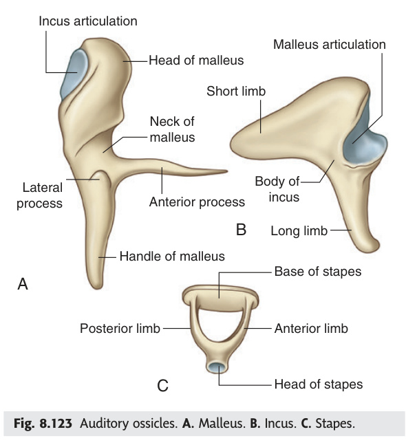
The ossicular chain — malleus, incus, stapes — forms a mechanical lever bridging the air-filled middle ear to the fluid-filled inner ear, amplifying sound pressure roughly 22-fold to overcome the impedance mismatch between air and perilymph. The malleus and incus derive from the first pharyngeal arch (Meckel's cartilage), while the stapes comes from the second arch (Reichert's cartilage); the stapes footplate plugging the oval window is the smallest bone in the body. Two intratympanic muscles protect the chain from loud sounds: tensor tympani (innervated by CN V3) dampens malleus movement, and stapedius (innervated by CN VII) stiffens the stapes — loss of stapedius in Bell's palsy unmasks hyperacusis because loud sounds can no longer be attenuated. Otosclerosis illustrates ossicular pathology at its most teachable: abnormal bone deposition fuses the stapes footplate to the oval window, producing progressive conductive hearing loss with a Carhart's notch at 2 kHz on audiometry, corrected surgically by stapedectomy.
| Ossicle | Derivation | Articulation / Function |
|---|---|---|
| Malleus (hammer) | 1st pharyngeal arch (Meckel's cartilage) | Handle attached to TM; head in epitympanum; incudomalleolar joint with incus |
| Incus (anvil) | 1st pharyngeal arch (Meckel's) | Between malleus + stapes; lenticular process articulates with stapes head |
| Stapes (stirrup) — smallest bone in body | 2nd pharyngeal arch (Reichert's cartilage) | Footplate in oval window; transmits sound vibrations to perilymph of inner ear |
| Muscle | Nerve | Action / Reflex |
|---|---|---|
| Tensor tympani | CN V3 (medial pterygoid nerve) | Pulls malleus medially → tenses TM → reduces amplitude of vibration. Acoustic reflex (bilateral, in response to loud sound) |
| Stapedius — smallest skeletal muscle in body | CN VII (facial nerve, via branch from mastoid segment) | Pulls stapes posteriorly → stiffens ossicular chain → protects inner ear from loud sounds. Paralysis (Bell's palsy) → hyperacusis (painfully loud sounds) |
Otosclerosis: autosomal dominant (incomplete penetrance); abnormal bone remodelling at the oval window → bony fixation of stapes footplate → progressive conductive hearing loss (young adult, bilateral, tinnitus, paracusis Willisii = hear better in noisy environments). Audiogram: conductive loss + Carhart's notch (dip at 2 kHz). Tympanogram: type As (reduced compliance with normal pressure). Treatment: hearing aid or stapedectomy (remove stapes + replace with Teflon/platinum prosthesis on incus).
- Which pharyngeal arch gives rise to the malleus and incus, and which to the stapes? Malleus and incus: first arch (Meckel's cartilage); stapes: second arch (Reichert's cartilage).
- What is the smallest bone in the body? The stapes.
- What happens to hearing when stapedius is paralysed in Bell's palsy? Hyperacusis — loud sounds can no longer be attenuated because the ossicular chain is not stiffened.
- Give the nerve supply of tensor tympani and stapedius. Tensor tympani: CN V3 (medial pterygoid nerve); stapedius: CN VII (branch from the mastoid segment).
- What is otosclerosis and how is it surgically corrected? AD disorder causing abnormal bone deposition that fuses the stapes footplate to the oval window → progressive conductive HL; corrected by stapedectomy (remove stapes + Teflon/platinum prosthesis).
Auditory (Eustachian/Pharyngotympanic) Tube
The auditory tube is the middle ear's pressure-equalisation and drainage channel — 35 mm long, running downward, forward, and medially from the middle ear to the nasopharynx, with its lateral third bony and its medial two-thirds fibrocartilaginous and normally collapsed at rest. It opens transiently during swallowing or yawning via tensor veli palatini, levator veli palatini, and salpingopharyngeus, equalising middle ear pressure — which is why chewing relieves the pressure change of aircraft descent. In children, the tube is shorter, more horizontal, and relatively wider, making bacterial ascent from the nasopharynx far easier and explaining why acute otitis media (AOM) is overwhelmingly a paediatric disease. Chronic Eustachian tube dysfunction creates sustained negative middle ear pressure, drawing the pars flaccida inward to form the retraction pocket that becomes an acquired cholesteatoma.
Connects the middle ear to the nasopharynx (~35 mm long). Lateral 1/3 = bony (in temporal bone). Medial 2/3 = fibrocartilaginous (collapsed at rest — opens on swallowing/yawning via tensor veli palatini, levator veli palatini, salpingopharyngeus). Orientation: runs downward, forward, medially from middle ear to nasopharynx (hence why children get more OM — tube more horizontal + shorter).
Acute otitis media (AOM): infection (Streptococcus pneumoniae, H. influenzae, Moraxella catarrhalis) ascends via auditory tube → middle ear → fever + severe otalgia + conductive deafness. TM: bulging, red, loss of light reflex. Spontaneous perforation → purulent discharge + pain relief. Treat: analgesia ± amoxicillin (if <2 yrs, severe, bilateral, or discharge). Otitis media with effusion (glue ear): chronic non-infected fluid → conductive deafness in children, delayed speech. Tympanogram type B (flat). Treat: watchful waiting → grommets (ventilation tubes in anteroinferior quadrant of TM).
- What are the proportions of the Eustachian tube? Lateral 1/3 bony (in temporal bone); medial 2/3 fibrocartilaginous, normally collapsed at rest.
- Which muscles open the auditory tube? Tensor veli palatini (primary opener), levator veli palatini, and salpingopharyngeus — active during swallowing or yawning.
- Why do children get more acute otitis media than adults? The tube is shorter, more horizontal, and relatively wider, allowing easier bacterial ascent from the nasopharynx.
- What tympanogram type is seen in glue ear (otitis media with effusion)? Type B (flat) — no compliance variation, indicating fluid in the middle ear cavity.
- How does Eustachian tube dysfunction lead to acquired cholesteatoma? Dysfunction → chronic negative middle ear pressure → pars flaccida retracts inward → retraction pocket accumulates keratin → cholesteatoma.
Inner Ear — Bony Labyrinth
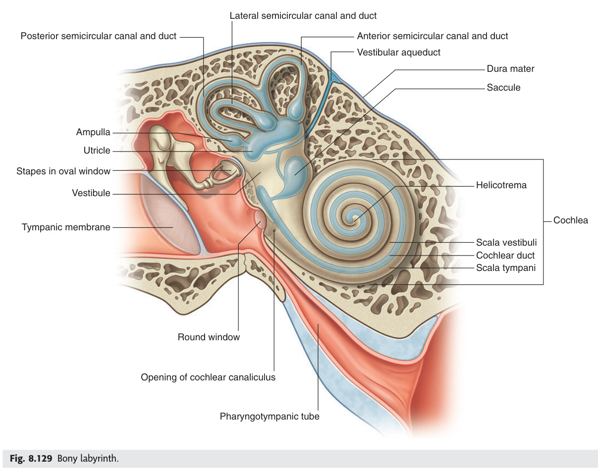
The bony labyrinth is a set of fluid-filled cavities carved into the petrous temporal bone, housing the entire apparatus for hearing and balance. The cochlea spirals 2¾ turns around its central modiolus, divided by Reissner's membrane and the basilar membrane into three channels: the perilymph-filled scala vestibuli and scala tympani (communicating at the helicotrema at the apex) flank the endolymph-filled scala media where hair cell transduction occurs. The vestibule contains the utricle and saccule for linear acceleration, while three semicircular canals set at right angles to each other detect angular acceleration in all planes. Critically, the inner ear has only one blood supply — the labyrinthine artery from AICA — with no collateral circulation, so even brief ischaemia irreversibly kills hair cells; this is why sudden sensorineural hearing loss is an audiological emergency, and why AICA infarcts (hearing loss + vertigo) are distinguished from PICA infarcts (Wallenberg syndrome, intact hearing).
| Part | Structure | Function |
|---|---|---|
| Cochlea | 2¾ turns spiralling around the modiolus (central bony pillar). Divided by vestibular (Reissner's) membrane + basilar membrane into: scala vestibuli (perilymph, top) + scala media/cochlear duct (endolymph, middle) + scala tympani (perilymph, bottom). Helicotrema connects scalae at apex | Hearing (sound transduction) |
| Vestibule | Central chamber; contains oval window (anterolateral, stapes footplate) + round window (anteroinferior). Houses utricle + saccule of membranous labyrinth | Static equilibrium + linear acceleration |
| Semicircular canals (3) | Anterior (superior), posterior, lateral (horizontal). Each has an ampulla (dilated end containing crista ampullaris). At right angles to each other to detect rotation in all 3 planes | Angular acceleration (rotation) |
Perilymph: similar to CSF (high Na⁺, low K⁺); in bony labyrinth surrounding membranous labyrinth. Endolymph: unique composition (high K⁺, low Na⁺ — like intracellular fluid); produced by stria vascularis; inside membranous labyrinth. The K⁺ gradient is essential for hair cell depolarisation.
The inner ear is supplied exclusively by the labyrinthine artery (internal auditory artery) — a branch of the anterior inferior cerebellar artery (AICA), which arises from the basilar artery. There are no collateral vessels and no anastomotic network within the cochlea or labyrinth. Even brief ischaemia causes irreversible hair cell death.
AICA territory infarct (lateral inferior pontine syndrome): ipsilateral facial numbness (CN V nucleus) + ipsilateral LMN facial palsy (CN VII nucleus) + sudden unilateral SNHL + vertigo (labyrinthine artery involvement — CN VIII and inner ear) + ipsilateral Horner's + ipsilateral limb ataxia + contralateral pain/temperature loss (spinothalamic). Hearing loss distinguishes AICA from PICA.
PICA territory infarct (Wallenberg / lateral medullary syndrome): does NOT cause hearing loss — PICA does not supply the inner ear. Features: ipsilateral face + contralateral body pain/temp loss + ipsilateral Horner's + dysphagia + ataxia — but normal hearing.
Sudden SNHL: audiological emergency — unilateral SNHL ≥30 dB over ≥3 frequencies within 72 hours. Must exclude posterior circulation stroke (MRI DWI). If idiopathic: oral prednisolone 1 mg/kg/day ± intratympanic steroid. Spontaneous recovery in 50%; poor prognostic features: profound loss, low-frequency loss, old age, delayed treatment.
- Name the three scalae of the cochlea and their fluid contents. Scala vestibuli (perilymph, top); scala media/cochlear duct (endolymph, middle); scala tympani (perilymph, bottom). Scalae vestibuli and tympani communicate at the helicotrema at the apex.
- Compare the ionic composition of endolymph and perilymph. Endolymph: high K⁺, low Na⁺ (intracellular-like); perilymph: high Na⁺, low K⁺ (CSF-like).
- What is the sole blood supply of the inner ear? The labyrinthine artery, a branch of AICA (anterior inferior cerebellar artery, from the basilar artery) — no collateral network exists.
- Why does an AICA infarct cause hearing loss but a PICA infarct does not? AICA gives the labyrinthine artery supplying the inner ear; PICA (Wallenberg syndrome) does not supply the inner ear — hearing is preserved.
- What is the treatment for sudden sensorineural hearing loss? Audiological emergency — exclude stroke (MRI DWI); if idiopathic: oral prednisolone 1 mg/kg/day ± intratympanic steroids within 72 hours.
Membranous Labyrinth & Hair Cells
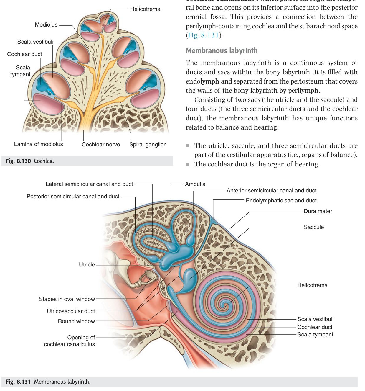
The membranous labyrinth sits within the bony labyrinth, bathed in endolymph (high K⁺, low Na⁺ — uniquely intracellular-like, produced by the stria vascularis), and contains all the sensory receptors for hearing and balance. In the cochlea, the organ of Corti rests on the basilar membrane: inner hair cells transmit the signal to CN VIII, while outer hair cells amplify it; the basilar membrane is tonotopically organised — narrow and stiff at the base encodes high frequencies, wide and floppy at the apex encodes low frequencies, which is why aminoglycoside ototoxicity and noise-induced damage cause high-frequency loss first. The otolith organs detect linear acceleration using otoconia (calcium carbonate crystals) that weigh on hair cell stereocilia; when otoconia dislodge from the utricular macula and enter the posterior semicircular canal, they cause BPPV — the most common vestibular disorder, producing brief intense positional vertigo that is both diagnosed and treated by sequential head manoeuvres (Dix-Hallpike for diagnosis; Epley for repositioning the otoconia back into the vestibule).
| Structure | Location | Stimulus / Receptor |
|---|---|---|
| Organ of Corti | On basilar membrane within cochlear duct (scala media). Inner hair cells (3,500) + outer hair cells (12,000). Tectorial membrane overlies | Sound → basilar membrane vibration → stereocilia deflection → K⁺ influx → depolarisation → CN VIII. High frequency (high pitch): base of cochlea. Low frequency: apex. Tonotopic organisation |
| Utricle | In vestibule; horizontal macula (otolith organ) | Linear horizontal acceleration + head tilt |
| Saccule | In vestibule; vertical macula (otolith organ) | Linear vertical acceleration (gravity) |
| Crista ampullaris | In ampulla of each semicircular canal; cupula (gelatinous mass) overlies hair cells | Angular rotation → endolymph flows → cupula deflects → hair cell stimulation |
Most common cause of vertigo. Otoliths (calcium carbonate crystals = otoconia) become dislodged from utricular macula → enter posterior semicircular canal → canalolithiasis. Trigger: head movement (rolling over in bed, looking up). Symptoms: brief (seconds), intense rotational vertigo + nystagmus with latency + fatigability on repeated testing (Dix-Hallpike manoeuvre positive). Treatment: Epley manoeuvre (particle repositioning — guides otoconia out of posterior canal into vestibule by sequential head positions). Resolves in weeks; recurrence common.
- What is the tonotopic organisation of the basilar membrane? Base: narrow and stiff → high frequencies; apex: wide and flexible → low frequencies. Explains why high frequencies are lost first in noise, aging, and aminoglycoside ototoxicity.
- What are otoconia and what happens when they are dislodged? Calcium carbonate crystals on the utricular macula; if dislodged into the posterior semicircular canal → canalolithiasis → BPPV.
- Which otolith organ detects horizontal linear acceleration and head tilt? Utricle (horizontal macula); saccule detects vertical linear acceleration and gravity.
- Describe the Epley manoeuvre and its purpose. A sequence of timed head repositioning steps that guide dislodged otoconia from the posterior semicircular canal back into the vestibule — treating BPPV.
- What structure produces endolymph and what is the clinical consequence of its failure? Stria vascularis; if it fails, the K⁺ gradient collapses → hair cells cannot depolarise → SNHL (e.g., endolymphatic hydrops in Ménière's).
CN VIII & Hearing Tests
The vestibulocochlear nerve (CN VIII) exits the inner ear through the internal acoustic meatus alongside CN VII and the labyrinthine artery, traverses the posterior fossa, and enters the brainstem at the cerebellopontine (CP) angle — a compact anatomical space where tumours, vessels, and cranial nerves converge. Two tuning-fork tests at the bedside localise hearing loss: Rinne compares air conduction (AC, fork near EAM) with bone conduction (BC, fork on mastoid) — normally AC > BC; a negative Rinne (BC > AC) means the middle ear is blocked and bone bypasses it to reach the cochlea directly. Weber (fork on the midline) lateralises to the deaf ear in conductive loss and to the better ear in sensorineural loss. The CP angle is the classic site of vestibular schwannoma — a slow-growing Schwann cell tumour of CN VIII's vestibular division presenting with unilateral high-frequency SNHL, tinnitus, and imbalance, confirmed by gadolinium MRI; bilateral tumours signal neurofibromatosis type 2 (NF2, chromosome 22, merlin gene).
CN VIII (vestibulocochlear nerve) = cochlear division (hearing) + vestibular division (balance). Exits inner ear via internal acoustic meatus (IAM) — in petrous temporal bone — alongside CN VII. Passes into posterior cranial fossa → enters brainstem at pontomedullary junction at the cerebellopontine (CP) angle. The CP angle also contains the anterior inferior cerebellar artery (AICA).
| Test | Method | Interpretation |
|---|---|---|
| Rinne test | 512 Hz tuning fork. AC (air conduction): fork held near EAM. BC (bone conduction): fork base on mastoid process | Normal/sensorineural deafness: AC > BC (Rinne positive). Conductive deafness: BC > AC (Rinne negative — bone bypasses blocked middle ear) |
| Weber test | 512 Hz fork on midline vertex (or forehead or teeth) | Normal: heard equally in both ears. Conductive deafness: lateralises to AFFECTED (deaf) ear (sound conducts directly to cochlea without middle ear loss). Sensorineural deafness: lateralises to NORMAL ear (better cochlea hears it) |
Benign Schwann cell tumour of vestibular division of CN VIII within the IAM / CP angle. Presents: unilateral sensorineural hearing loss (high frequency first) + tinnitus + imbalance. Large tumours compress: CN VII (facial weakness), CN V (loss of corneal reflex), cerebellum (ataxia), brainstem. MRI with gadolinium = gold standard (enhancing mass at IAM, widened IAM on CT). Treatment: observation (slow-growing) vs stereotactic radiosurgery (Gamma Knife) vs microsurgical resection. Bilateral acoustic neuromas = neurofibromatosis type 2 (NF2, chromosome 22, merlin gene mutation).
- Where does CN VIII enter the brainstem? At the pontomedullary junction at the cerebellopontine (CP) angle, alongside CN VII and the AICA.
- What does a negative Rinne test (BC > AC) indicate? Conductive deafness — bone conduction bypasses the obstructed middle ear and reaches the cochlea directly.
- In Weber test, where does sound lateralise in sensorineural deafness? To the better (normal) ear — the healthier cochlea detects the vibration more strongly.
- What is the typical presentation of vestibular schwannoma? Unilateral high-frequency sensorineural hearing loss + tinnitus + imbalance; diagnosed by gadolinium-enhanced MRI showing enhancing mass at the internal acoustic meatus.
- Bilateral acoustic neuromas are pathognomonic of which condition? Neurofibromatosis type 2 (NF2) — chromosome 22, merlin (NF2) gene mutation.
Vestibular System & Nystagmus
Vestibular disorders produce nystagmus — involuntary rhythmic eye movements with a slow vestibular-driven phase and a fast corrective phase — and the character of the nystagmus separates peripheral from central causes: peripheral nystagmus has a latency before onset, is suppressed by fixation, and fatigues on repeated testing; central nystagmus is immediate, not suppressed by fixation, and may be purely vertical or direction-changing. Ménière's disease demonstrates endolymphatic hydrops clinically as a characteristic tetrad: episodic vertigo (minutes to hours), fluctuating low-frequency sensorineural hearing loss, tinnitus, and aural fullness — the low-frequency involvement is the key that separates it from noise- or age-related loss. Vestibular neuritis causes sudden severe prolonged vertigo without hearing loss (differentiating it from labyrinthitis, which does cause SNHL), with a positive head impulse test (HIT) indicating ipsilateral peripheral vestibular hypofunction. The caloric test (COWS: Cold Opposite, Warm Same — the fast phase direction) directly tests each horizontal semicircular canal's excitability; canal paresis >25% asymmetry is the most sensitive caloric indicator of vestibular schwannoma.
| Condition | Key Features | Distinguish from central |
|---|---|---|
| BPPV | Brief positional vertigo (seconds); Dix-Hallpike positive; fatigable nystagmus (geotropic); no hearing loss | Peripheral: latency before nystagmus; fatigable; direction fixed relative to head. Central: no latency; non-fatigable; purely vertical/direction-changing = central until proven otherwise |
| Vestibular neuritis | Sudden severe prolonged vertigo (days) + nausea/vomiting; NO hearing loss; head impulse test (HIT) positive (catch-up saccade); follows viral infection | Horizontal nystagmus toward normal ear; resolves in weeks with vestibular compensation |
| Ménière's disease | Endolymphatic hydrops. Triad: episodic vertigo (minutes–hours) + fluctuating sensorineural hearing loss + tinnitus + aural fullness (4 symptoms). Attacks recur; low-frequency hearing lost first | Audiogram shows low-frequency sensorineural loss. Treat: low-salt diet + betahistine + diuretics; intratympanic gentamicin/steroid; endolymphatic sac decompression; destructive (labyrinthectomy) for severe cases |
| Labyrinthitis | Viral/bacterial. Vertigo + sensorineural hearing loss + tinnitus. Bacterial (otitis media spreading) = more severe + urgent | Differs from vestibular neuritis by presence of hearing loss |
Cold water (30°C) then warm water (44°C) instilled into the external auditory canal; temperature gradient sets up convection currents in the endolymph of the horizontal semicircular canal, simulating rotation. Stimulus generates nystagmus — assess direction of fast phase, duration, and symmetry between ears.
COWS mnemonic: Cold Opposite, Warm Same = direction of the fast phase of nystagmus. Cold water in right ear → fast phase to LEFT (away from stimulated side). Warm water in right ear → fast phase to RIGHT (toward stimulated side).
| Finding | Meaning | Typical Cause |
|---|---|---|
| Canal paresis (reduced response one side) | Reduced nystagmus from one ear compared to the other (>25% asymmetry — Jongkees formula). Indicates ipsilateral vestibular hypofunction | Acoustic neuroma (vestibular schwannoma), vestibular neuritis, labyrinthine infarct |
| Directional preponderance | Nystagmus responses stronger in one direction regardless of which ear is irrigated. Suggests vestibular imbalance | Central vestibular lesion (brainstem, cerebellum) |
| Bilateral absent caloric responses | No nystagmus from either ear — profound bilateral vestibular failure | Gentamicin ototoxicity (bilateral vestibulotoxicity), bilateral acoustic neuromas (NF2), meningitis |
| Normal caloric + spontaneous nystagmus | Peripheral compensation with persistent imbalance; or central pathology | Benign paroxysmal positional vertigo (BPPV) — caloric normal, Dix–Hallpike positive |
Cervical VEMP (cVEMP): recorded from sternocleidomastoid (SCM) during head elevation. Tests saccule (vertical linear acceleration, gravity) → inferior division of CN VIII. Absent cVEMP = saccule or inferior CN VIII pathology (e.g., superior semicircular canal dehiscence — paradoxically hyperactive cVEMP; acoustic neuroma affecting inferior nerve).
Ocular VEMP (oVEMP): recorded from contralateral inferior oblique muscle. Tests utricle (horizontal linear acceleration, head tilt) → superior division of CN VIII. Absent oVEMP = utricular or superior CN VIII pathology. Together cVEMP + oVEMP + caloric testing gives a comprehensive map of vestibular function.
Test Unit 14 knowledge
Ear anatomy, hearing tests, vertigo differentials and CP angle tumour MCQs.
- Name four features that distinguish peripheral from central nystagmus. Peripheral: latency before onset, fatigable, suppressed by fixation, unidirectional; Central: no latency, non-fatigable, not suppressed by fixation, may be vertical or direction-changing.
- Give the four symptoms of Ménière's disease. Episodic vertigo (minutes–hours); fluctuating low-frequency SNHL; tinnitus; aural fullness — caused by endolymphatic hydrops.
- What does the COWS mnemonic mean? Cold Opposite, Warm Same — describes the direction of the fast phase of nystagmus relative to the ear being irrigated in the caloric test.
- What is the clinical significance of canal paresis on caloric testing? Ipsilateral vestibular hypofunction (>25% asymmetry by Jongkees formula); most important diagnosis to exclude is vestibular schwannoma — confirm with gadolinium MRI.
- How does vestibular neuritis differ from labyrinthitis? Vestibular neuritis: sudden severe prolonged vertigo with no hearing loss; labyrinthitis: same vertigo but with sensorineural hearing loss, indicating cochlear involvement.
CN VII — Course Through Temporal Bone ★★★
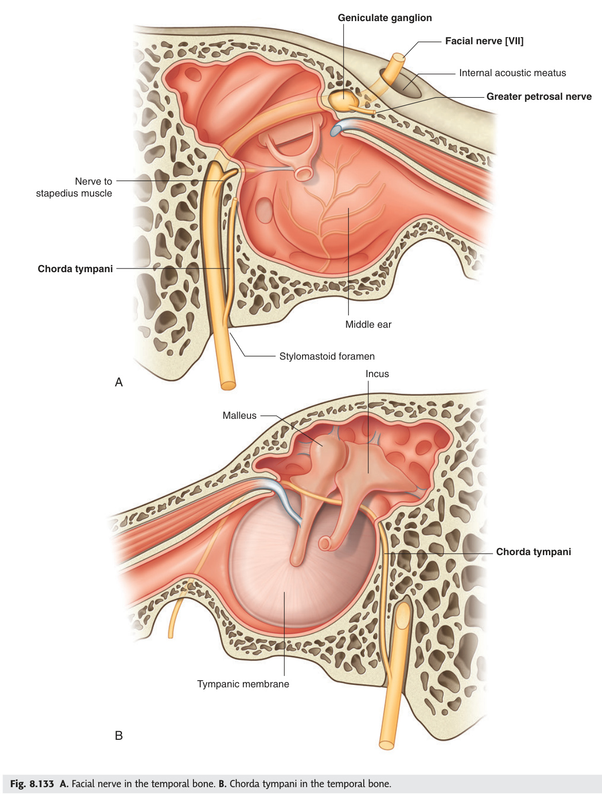
The facial nerve (CN VII) travels a longer intratemporal course than any other cranial nerve, passing through four named segments — meatal, labyrinthine, tympanic (horizontal), mastoid (vertical) — before exiting at the stylomastoid foramen. The labyrinthine segment, only 2–4 mm long and 0.68 mm wide, is the narrowest point and most vulnerable to ischaemic compression in Bell's palsy, which represents HSV-1 reactivation in the geniculate ganglion. Each segment gives off key branches that allow topographic localisation of any lesion: the greater superficial petrosal nerve (tear production via the lacrimal gland) at the geniculate ganglion, the nerve to stapedius at the tympanic segment, and the chorda tympani (taste + submandibular/sublingual parasympathetics) 6 mm above the stylomastoid foramen. The clinical rule: loss of lacrimation → lesion proximal to the geniculate; hyperacusis without taste loss → tympanic segment; isolated taste loss → between chorda tympani takeoff and stylomastoid foramen.
| Segment | Course | Key Features |
|---|---|---|
| Meatal | Brainstem → internal acoustic meatus (with CN VIII + labyrinthine artery) | No branches; runs in the IAM superior to CN VIII. Meningioma/acoustic neuroma can compress here |
| Labyrinthine | IAM fundus → geniculate ganglion (genu = bend). Between cochlea + anterior semicircular canal | Shortest (2–4 mm) + narrowest (0.68 mm) segment → most vulnerable to ischaemic compression in Bell's palsy. Geniculate ganglion (cell bodies of taste fibres) gives off: greater superficial petrosal nerve (GSP N — parasympathetics to lacrimal gland via pterygopalatine ganglion; runs under temporal lobe dura; damaged in petrous ridge fractures → dry eye) |
| Tympanic (horizontal) | Geniculate → 2nd genu. Runs horizontally above oval window + below lateral semicircular canal; forms medial wall of middle ear (visible as bulge = facial nerve prominence) | The dehiscence (congenital absence of bony covering) in 57% of cases → vulnerable to otitis media, cholesteatoma. Gives nerve to stapedius just before 2nd genu |
| Mastoid (vertical) | 2nd genu → stylomastoid foramen (exits skull). Runs vertically through mastoid bone | Gives chorda tympani (6 mm above stylomastoid foramen) → crosses middle ear (taste anterior 2/3 tongue + parasympathetics to submandibular + sublingual glands). At stylomastoid foramen gives posterior auricular nerve + nerve to digastric posterior belly + nerve to stylohyoid. Then enters parotid |
Greater superficial petrosal nerve (geniculate ganglion) → Stapedius nerve (tympanic segment, just before 2nd genu) → Chorda tympani (mastoid segment, 6 mm above stylomastoid foramen) → at stylomastoid foramen: posterior auricular nerve + digastric (post) + stylohyoid. Then in parotid → Temporal, Zygomatic, Buccal, Marginal mandibular, Cervical (To Zanzibar By Motor Car).
LMN (Bell's, Ramsay-Hunt, parotid tumour, temporal bone fracture): affects ALL branches including frontalis → CANNOT raise eyebrow, cannot close eye + Bell's phenomenon (eye rolls up on attempted closure — protective reflex). Absent corneal reflex (orbicularis efferent limb lost).
UMN (stroke, space-occupying lesion): forehead SPARED → can still raise eyebrow (frontalis has bilateral cortical representation; lower face is contralateral cortical only). Cannot move lower face on contralateral side. Always look for other UMN signs (hemiplegia, dysphasia).
Bell's palsy vs Ramsay-Hunt:
| Bell's Palsy | Ramsay-Hunt Syndrome | |
|---|---|---|
| Aetiology | Idiopathic; HSV-1 reactivation in geniculate ganglion; inflammation → compression in labyrinthine segment | VZV (Herpes zoster) reactivation in geniculate ganglion |
| Vesicles | Absent | Herpetic vesicles in EAC, pinna (concha), soft palate (zoster oticus) |
| Hearing | Normal (or mild muffling if stapedius involved) | Sensorineural hearing loss ± tinnitus ± vertigo (VZV also affects CN VIII) |
| Pain | Mild pre-auricular ache | Severe ear pain (otalgia) — often precedes rash |
| Prognosis | 80–85% full recovery; complete palsy = worse prognosis | Worse (50–70% recovery); SNHL may be permanent |
| Treatment | Prednisolone 50 mg/day × 10 days within 72h; artificial tears + eye tape at night (corneal protection) | Prednisolone + acyclovir/valaciclovir × 7–10 days; corneal protection |
The level of CN VII injury can be localised by which branches are affected:
• Above geniculate ganglion: all symptoms + dry eye (GSP N lost → no lacrimal secretion) + hyperacusis + taste loss + dry mouth.
• Between geniculate and stapedius branch: all symptoms except dry eye; still hyperacusis + taste loss + dry mouth.
• Between stapedius + chorda tympani: taste loss + dry mouth; no hyperacusis (stapedius intact).
• Between chorda tympani + stylomastoid foramen: pure motor palsy only (no taste/autonomic loss).
• Below stylomastoid foramen / parotid: selective branch palsy (depending on which branch compressed — parotid pleomorphic adenoma or malignancy).
- Name the four intratemporal segments of CN VII in order. Meatal → labyrinthine (narrowest) → tympanic/horizontal → mastoid/vertical → exits at stylomastoid foramen.
- Which segment is most vulnerable in Bell's palsy and why? Labyrinthine (2–4 mm, 0.68 mm wide) — HSV-1 reactivation at the geniculate ganglion causes oedema in this narrowest bony canal, compressing the nerve.
- What branch leaves the geniculate ganglion and what does it supply? Greater superficial petrosal nerve (GSPN) — parasympathetics to the lacrimal gland via the pterygopalatine ganglion; damaged in petrous ridge fractures → dry eye.
- How do you clinically distinguish UMN from LMN facial palsy? LMN: all branches affected including frontalis (cannot raise eyebrow, cannot close eye); UMN: forehead spared (bilateral cortical representation of frontalis).
- What distinguishes Ramsay-Hunt syndrome from Bell's palsy? Ramsay-Hunt: VZV reactivation — herpetic vesicles in EAC/pinna, SNHL, severe otalgia, worse prognosis (50–70% vs 80–85%). Treatment adds antivirals (aciclovir/valaciclovir).
Hearing Loss Classification & Audiometry ★★
All hearing loss is either conductive (failure to conduct sound to the cochlea through the external or middle ear) or sensorineural (failure of the cochlea, CN VIII, or central pathways). The audiogram distinguishes them cleanly: conductive loss shows an air-bone gap (bone conduction normal, air conduction raised), while sensorineural loss raises both equally. The same information comes from bedside tests — a negative Rinne (BC > AC) and Weber lateralising to the deaf ear both signal conductive pathology, while a positive Rinne with Weber to the better ear signals sensorineural. Tympanometry adds middle ear mechanics: type B (flat) means fluid or perforation, type As (shallow) means a stiffened ossicular chain (otosclerosis), and type Ad (deep/hypercompliant) means ossicular discontinuity. Ototoxic drugs complete the picture — aminoglycosides damage the cochlear base first (high-frequency SNHL), their pattern shifting apically with continued exposure; cisplatin causes irreversible SNHL requiring pre- and intra-treatment monitoring audiometry.
| Type | Pathology | Audiogram | Causes |
|---|---|---|---|
| Conductive | EAC, TM, ossicles, middle ear — impaired sound conduction to cochlea | Air-bone gap >10 dB (AC raised, BC normal). Rinne negative. Weber lateralises to affected ear | Wax, AOM, glue ear, TM perforation, otosclerosis, cholesteatoma, ossicular discontinuity |
| Sensorineural (SNHL) | Cochlea (most common) or CN VIII or central pathway | AC + BC both raised equally (no air-bone gap). Rinne positive (both reduced). Weber lateralises to better ear | Presbycusis (age — high frequency first), noise-induced (4 kHz notch), Ménière's (low frequency), acoustic neuroma, ototoxic drugs (aminoglycosides, cisplatin, furosemide), meningitis, congenital |
| Mixed | Both conductive + sensorineural components | Air-bone gap present + both raised. Chronic otitis media with cochlear damage (cholesteatoma) | Chronic otitis media, otosclerosis with cochlear involvement, head trauma |
Presbycusis (age-related SNHL): most common cause of hearing loss in elderly. High-frequency loss first (4–8 kHz) → bilateral, symmetrical, gradual. Outer hair cell loss at cochlear base. 4 kHz notch = noise-induced hearing loss (NIHL, occupational or recreational; temporary threshold shift → permanent with prolonged exposure). Notch at 4 kHz sparing 8 kHz is characteristic.
Cochlear implants: for severe-profound bilateral SNHL when hearing aids provide inadequate benefit. Electrode array inserted into scala tympani → directly stimulates spiral ganglion cells (bypasses damaged hair cells) → CN VIII → cortex. Best outcomes: early implantation in pre-lingual children (critical period of auditory cortex development), post-lingual adults. Requires residual spiral ganglion neurons. Meningitis is a rare complication (pneumococcal vaccine mandatory pre-implant). Bilateral implantation improves spatial hearing.
| Drug Class | Mechanism & Pattern | Key Facts |
|---|---|---|
| Aminoglycosides (gentamicin, tobramycin, amikacin, streptomycin) |
Accumulate in endolymph → ROS generation → outer hair cell (OHC) apoptosis. Damage begins at the base of the cochlea (high-frequency first) → progresses apically (low frequency) with continued exposure. Cochlear damage often irreversible; monitoring audiometry mandatory |
Gentamicin: primarily vestibulotoxic > cochleotoxic. Used therapeutically as intratympanic gentamicin in refractory Ménière's disease — deliberately ablates vestibular function to stop vertigo attacks. Tobramycin / amikacin: primarily cochleotoxic → SNHL. Streptomycin: vestibulotoxic (historical TB treatment — used in NF2 for bilateral ablation). Mitochondrial 12S rRNA A1555G mutation: renders individuals exquisitely sensitive to aminoglycoside ototoxicity even at normal doses. Maternally inherited. Genetic testing before aminoglycoside use in at-risk families. |
| Cisplatin (and carboplatin) | Platinum → ROS + direct OHC damage. Irreversible. High-frequency SNHL first. Incidence ~60% with cisplatin courses. Synergistic with noise exposure and aminoglycosides | Mandatory audiometry before and during chemotherapy. No established protective agent for routine use (sodium thiosulfate and amifostine under study). Carboplatin is less ototoxic than cisplatin |
| Loop diuretics (furosemide, ethacrynic acid) |
Inhibit Na⁺/K⁺/2Cl⁻ cotransporter (NKCC2) in stria vascularis → collapse of endocochlear potential → temporary threshold shift. Usually reversible on stopping. Ethacrynic acid is more ototoxic than furosemide | Synergistic ototoxicity with aminoglycosides — avoid co-administration; if unavoidable, separate doses maximally and monitor hearing. High-dose rapid IV furosemide most risky (e.g., acute pulmonary oedema dosing) |
| Aspirin / Salicylates (high dose) | Inhibit prestin (motor protein of OHC outer membrane) → electromotility loss → tinnitus + mild SNHL. Dose-dependent and fully reversible on stopping | Tinnitus is the sentinel symptom — appears at plasma salicylate >20 mg/dL. Hearing loss ~20–30 dB at high doses (anti-inflammatory/anti-rheumatic dosing). Used historically as a test of OHC function |
| Quinine / Chloroquine | Tinnitus + SNHL — dose-dependent, largely reversible. Mechanism: vasoconstriction of cochlear vasculature + OHC damage | Relevant in malaria treatment; high-dose IV quinine for severe malaria most risky. Chloroquine retinopathy occurs at same high doses |
LAG FC: Loop diuretics · Aminoglycosides · Gentamicin (vestibulo) · Furosemide (reversible) · Cisplatin (irreversible) — all cause SNHL. Or simply: "AMINE Loop Cisplatin Aspirin Quinine"
- How does conductive loss differ from SNHL on an audiogram? Conductive: air-bone gap (AC raised, BC normal); SNHL: both AC and BC raised equally with no air-bone gap.
- What tympanogram type is seen in otosclerosis and what is the characteristic audiogram notch? Type As (stiff/shallow) — reduced compliance; Carhart's notch (dip at 2 kHz) on audiogram.
- At what frequency is the audiogram notch in noise-induced hearing loss? 4 kHz — outer hair cell loss at the cochlear base from excessive sound exposure.
- Which aminoglycoside is primarily vestibulotoxic and how is this used therapeutically? Gentamicin — used as intratympanic gentamicin to deliberately ablate vestibular function in refractory Ménière's disease.
- Why does aminoglycoside ototoxicity cause high-frequency loss first? The drug accumulates at the cochlear base (high-frequency region), damaging outer hair cells there first; damage progresses apically (to lower frequencies) with continued exposure.
Mastoiditis & Its Complications ★★
Mastoiditis develops when acute otitis media spreads beyond the tympanic cavity into the mastoid antrum and air cells, typically within 2–3 weeks of inadequately treated AOM; coalescent mastoiditis — breakdown of bony septa into a single pus-filled cavity — requires CT to confirm and cortical mastoidectomy to treat. The pathognomonic clinical picture is a pinna displaced anteroinferiorly by post-auricular swelling and tenderness, but the real danger lies in which direction the infection spreads. Lateral cortex erosion creates a subperiosteal abscess (most common extracranial complication); medial mastoid tip erosion tracks pus under the sternocleidomastoid as a Bezold's abscess, which can descend to the mediastinum. Intracranially, meningitis is most common, but sigmoid sinus thrombosis (picket-fence fever + papilloedema), brain abscess, and Gradenigo's syndrome (petrositis — CN VI palsy + deep facial pain + otorrhoea, from petrous apex involvement) are the high-stakes complications that distinguish a thorough examination answer from a superficial one.
Spread of acute otitis media (AOM) → mastoid antrum → mastoid air cells. Develops within 2–3 weeks of inadequately treated AOM. Coalescent mastoiditis: breakdown of bony septa between air cells → single pus-filled cavity (CT temporal bone diagnosis). Classic triad: post-auricular swelling + tenderness over mastoid + pinna displaced anteroinferiorly (pathognomonic). Treatment: IV antibiotics + myringotomy; coalescent mastoiditis → cortical mastoidectomy.
| Complications of Mastoiditis | ||
|---|---|---|
| Complication | Mechanism / Features | Management |
| EXTRACRANIAL (Extra-temporal) | ||
| Subperiosteal abscess (most common) | Pus erodes through lateral cortex of mastoid → accumulates beneath periosteum → post-auricular fluctuant swelling; pinna displaced anteroinferiorly | Surgical drainage + cortical mastoidectomy |
| Bezold's abscess | Pus erodes through mastoid tip inner cortex → tracks deep to sternocleidomastoid (SCM) into deep neck → neck swelling + torticollis; can spread to mediastinum (Mouret's abscess) | Drainage via neck incision + mastoidectomy; mediastinoscopy if spread downward |
| Luc's abscess (zygomatic) | Erosion of zygomatic root of temporal bone → pre-auricular swelling anterior to ear | Drainage + mastoidectomy |
| Citelli's abscess | Erosion of medial mastoid tip → digastric triangle (posterior triangle deep to SCM) | Drainage + mastoidectomy |
| INTRACRANIAL | ||
| Facial nerve palsy | CN VII involvement at tympanic + mastoid segments (dehiscent bony canal in 57%) | Mastoidectomy; nerve decompression if no recovery |
| Labyrinthitis | Serous (reversible) → suppurative (toxic, irreversible SNHL + vertigo) → labyrinthine fistula (usually lateral semicircular canal) | IV antibiotics; fistula → conservative management of canal wall |
| Petrositis / Gradenigo's syndrome | Infection to petrous apex → triad: ipsilateral CN VI palsy (abducens) + deep facial/retro-orbital pain (CN V trigeminal) + otorrhoea | IV antibiotics; surgical drainage of petrous apex if no resolution |
| Meningitis (most common intracranial complication) | Direct spread through tegmen tympani or via venous routes → fever + nuchal rigidity + CSF changes | IV antibiotics; LP; mastoidectomy |
| Extradural / subdural abscess | Pus between dura + temporal bone (extradural) or beneath dura (subdural); subdural spreads rapidly | Neurosurgical drainage + mastoidectomy |
| Brain abscess | Temporal lobe (most common) or cerebellar abscess; insidious onset; headache + focal neurology | CT-guided aspiration/drainage + neurosurgery + IV antibiotics 6–8 weeks |
| Lateral (sigmoid) sinus thrombosis | Erosion of sinus plate → septic phlebitis → thrombosis → picket fence fever (high swinging pyrexia + rigors) + headache + papilloedema. Septic emboli → lung abscesses | IV antibiotics + anticoagulation; surgical thrombectomy if deteriorating; MRI/CT venography diagnosis |
Bezold's abscess: pus erodes the mastoid tip's inner/medial cortex → tracks deep to the SCM into the deep neck spaces → presents as a neck mass + torticollis (pain on neck movement). Danger: can track inferiorly into the mediastinum (Mouret's abscess). Requires neck incision for drainage in addition to mastoidectomy.
- What is the classic clinical triad of acute mastoiditis? Post-auricular swelling + mastoid tenderness + pinna displaced anteroinferiorly.
- How does a Bezold's abscess differ from a subperiosteal abscess? Subperiosteal: pus erodes lateral mastoid cortex → post-auricular swelling. Bezold's: pus erodes medial mastoid tip → tracks under SCM into deep neck (can descend to mediastinum = Mouret's abscess).
- What is Gradenigo's syndrome and what causes it? Triad of ipsilateral CN VI palsy + deep facial/periorbital pain (CN V) + otorrhoea — caused by petrositis with infection reaching the petrous apex.
- What is the most common intracranial complication of mastoiditis? Meningitis.
- What clinical sign suggests sigmoid sinus thrombosis in mastoiditis? Picket-fence fever (high swinging pyrexia with rigors) + headache + papilloedema; diagnosed by MRI/CT venography.
Malignant (Necrotising) Otitis Externa ★★
Malignant otitis externa is not a cancer — the name reflects its aggressive behaviour, a Pseudomonas aeruginosa osteomyelitis of the temporal bone and skull base spreading from the external auditory canal via fissures of Santorini (congenital fibrous gaps between EAC cartilage plates) through the parotid to the stylomastoid foramen and beyond. It occurs almost exclusively in elderly diabetics or the immunocompromised — glucose-rich EAC secretions nourish Pseudomonas, and impaired neutrophil function permits unchecked spread. The pathognomonic sign is granulation tissue at the bony-cartilaginous junction of the EAC floor, combined with disproportionately severe nocturnal otalgia; cranial nerve involvement worsens the prognosis stage by stage (CN VII at the stylomastoid foramen → IX/X/XI at the jugular foramen → XII → VI with intracranial extension). Treatment is ciprofloxacin for 6–8 weeks, with duration guided by Gallium-67 scintigraphy — not CT, which remains abnormal long after cure because bone remodels slowly — alongside strict glycaemic control.
Rapidly progressive, potentially life-threatening osteomyelitis of the temporal bone and skull base, spreading from the external auditory canal. Despite the name, it is NOT a malignant (cancerous) condition — the term reflects its aggressive, destructive behaviour. Organism: Pseudomonas aeruginosa (>95% of cases).
| Feature | Details |
|---|---|
| Risk factors | Elderly diabetics (most common — glucose-rich EAC secretions create ideal Pseudomonas medium + impaired leukocyte function); immunocompromised (HIV/AIDS, chemotherapy, prolonged steroids) |
| Spread route | EAC floor → fissures of Santorini (congenital fibrous bands between EAC cartilage plates) → parotid → stylomastoid foramen → temporal bone → skull base → intracranial extension |
| Key clinical sign | Granulation tissue at the bony-cartilaginous junction of the EAC floor (pathognomonic) + severe unrelenting otalgia (disproportionate to otoscopic findings, worse at night) |
| Cranial nerve involvement (in order) | CN VII (facial palsy — first and most common; at stylomastoid foramen) → CN IX/X/XI (jugular foramen = Vernet's syndrome) → CN XII (hypoglossal — tongue deviation) → CN VI (abducens — intracranial spread). Each successive CN involvement = worsening prognosis |
| Investigations | EAC swab (Pseudomonas); CT temporal bone (cortical + medullary bone erosion); MRI (soft tissue, meningeal enhancement, intracranial extent); Technetium-99m bone scan (sensitive for osteomyelitis, establishes diagnosis); Gallium-67 scan (monitors response to treatment — normalises with cure; gold standard for treatment monitoring). ESR/CRP elevated |
| Treatment | Anti-pseudomonal antibiotics: ciprofloxacin (fluoroquinolone, oral — high bioavailability = oral equivalent to IV; preferred) ± piperacillin-tazobactam/ceftazidime IV for severe cases. Duration: 6–8 weeks minimum, guided by Gallium scan normalisation. Strict glycaemic control (essential — hyperglycaemia drives infection). Surgical debridement of necrotic EAC tissue. Hyperbaric oxygen: adjunct in refractory cases (kills Pseudomonas by ↑O₂ tension in ischaemic tissue) |
| Prognosis | Mortality up to 50% with intracranial extension. CN VII palsy = poor prognostic sign (infection has reached stylomastoid foramen). Recurrence common in poorly controlled diabetics |
Management: (1) EAC swab + culture; (2) CT temporal bone + MRI to stage disease; (3) Technetium bone scan to confirm osteomyelitis; (4) Ciprofloxacin 750 mg BD orally × 6–8 weeks (or IV piperacillin-tazobactam if severe); (5) Strict glycaemic control (HbA1c optimisation); (6) Surgical debridement of necrotic tissue; (7) Gallium-67 scan at 6–8 weeks to assess treatment response.
Test Unit 14 knowledge
Ear anatomy, hearing tests, vertigo differentials and complications MCQs.
- What is the causative organism in >95% of cases? Pseudomonas aeruginosa.
- What is the pathognomonic clinical sign? Granulation tissue at the bony-cartilaginous junction of the EAC floor + disproportionately severe nocturnal otalgia.
- Which cranial nerve is affected first and why? CN VII — infection spreads via fissures of Santorini → parotid → stylomastoid foramen, where CN VII exits. Each successive nerve involved (IX/X/XI at jugular foramen, XII, VI) signals worsening prognosis.
- Why is Gallium-67 scan preferred over CT for monitoring response? Gallium-67 tracks active inflammation and normalises with cure; CT shows bone architecture and remains abnormal for months even after successful treatment.
- What is first-line antibiotic treatment and duration? Ciprofloxacin (oral, high bioavailability equivalent to IV) for 6–8 weeks minimum, guided by Gallium-67 scan normalisation; strict glycaemic control is essential.