The Eye & Orbit
Eyeball — Horizontal Cross-Section
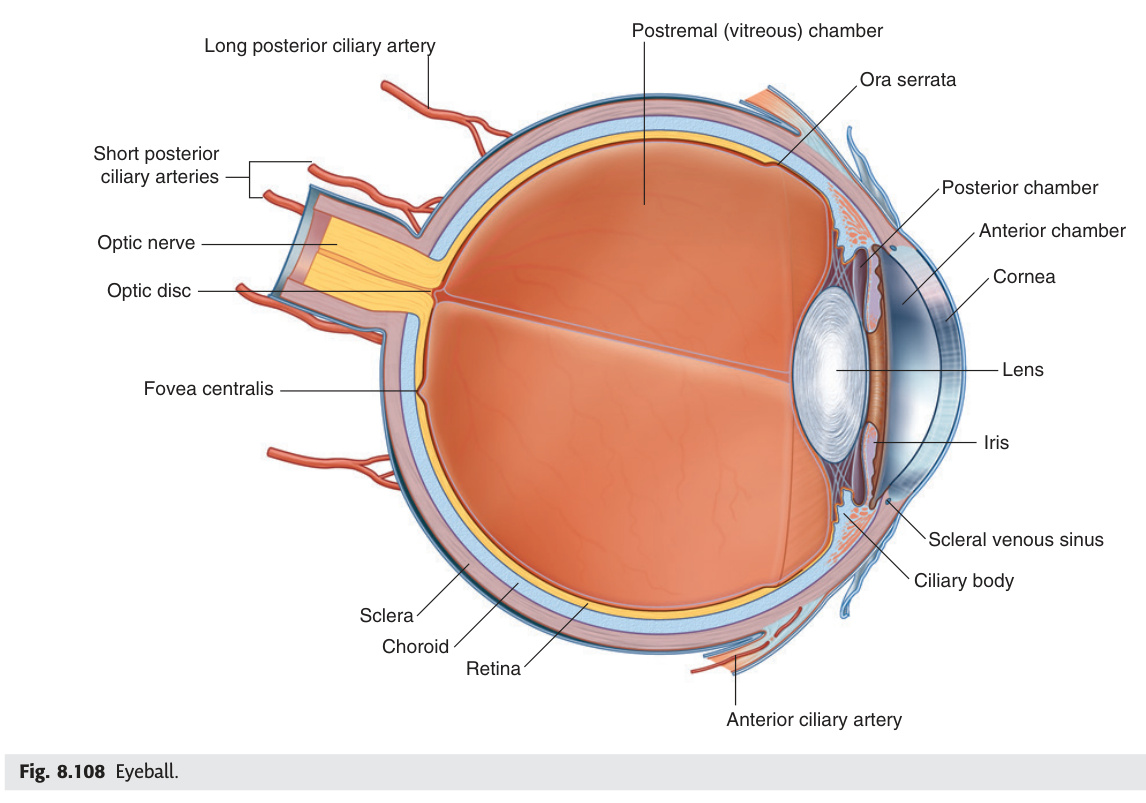
Three tunics: fibrous (sclera+cornea) · vascular (choroid+ciliary body+iris) · neural (retina) · Lens = only avascular structure in body
The Orbit
The orbit is a four-walled bony pyramid whose walls vary dramatically in strength, and knowing which wall is thinnest turns anatomy directly into clinical diagnosis. The orbital floor — made largely of the maxilla — is the weakest, which is why a blow to the eye (a fist, a squash ball) blows it downward into the maxillary sinus, trapping the inferior rectus and preventing the patient from looking upward. The medial wall, the paper-thin lamina papyracea of the ethmoid, is the reason sinusitis can become an orbital emergency — pathogens track through it silently. The optic canal and superior orbital fissure are the cable conduits of the orbit: the optic canal carries the optic nerve and ophthalmic artery, while the superior orbital fissure carries every other nerve that moves the eye (CN III, IV, VI) and the sensory branch supplying the orbit (CN V1); a lesion at the fissure paralyses all of them simultaneously. The cavernous sinus sits just behind, receiving blood from the face via connections that run without valves — which is why dental or upper-lip infections can propagate intracranially as cavernous sinus thrombosis, a neurological emergency.
| Wall | Bones | Clinical note |
|---|---|---|
| Roof | Frontal bone (orbital plate) + lesser wing of sphenoid | Meningioma of sphenoid wing → proptosis + visual loss |
| Floor | Maxilla (orbital surface) + zygomatic + palatine bones — THINNEST WALL | Blowout fracture: direct orbital trauma → floor fractures into maxillary sinus → inferior rectus entrapment → diplopia on upward gaze + enophthalmos. CT orbit confirms |
| Medial wall | Ethmoid (orbital plate = lamina papyracea) + lacrimal + frontal + sphenoid — also thin. Connects to ethmoid air cells | Ethmoid sinusitis → subperiosteal orbital abscess via lamina papyracea; orbital cellulitis → vision-threatening emergency |
| Lateral wall | Zygomatic + greater wing of sphenoid — STRONGEST WALL | Relatively resistant to fracture |
Superior orbital fissure (between greater + lesser wings of sphenoid; within/outside tendinous ring of Zinn): CN III (oculomotor), CN IV (trochlear), CN V1 branches (lacrimal + frontal + nasociliary), CN VI (abducens), superior ophthalmic vein, sympathetic fibres. Mnemonic: SO4 LR6 (Superior Oblique = CN IV; Lateral Rectus = CN VI) rest = CN III.
Inferior orbital fissure: CN V2 (infraorbital nerve), zygomatic nerve, inferior ophthalmic vein, infraorbital vessels.
Superior orbital fissure syndrome: CN III + IV + V1 + VI all affected → complete ophthalmoplegia + ptosis + mydriasis + loss of corneal reflex + forehead numbness.
Contents (lateral wall, superior → inferior): CN III (oculomotor) → CN IV (trochlear) → CN V1 (ophthalmic) → CN V2 (maxillary); within the sinus: CN VI (abducens — most medial, directly adjacent to ICA, therefore first affected by ICA aneurysm/thrombosis) + internal carotid artery (with postganglionic sympathetic plexus).
Cavernous sinus syndrome: CN III + IV + V1/V2 + VI palsy → complete ophthalmoplegia + ptosis + mydriasis + forehead/cheek numbness + loss of corneal reflex. If sympathetic plexus on ICA affected → Horner's (miosis + partial ptosis — partially masks CN III ptosis). Bilateral in cavernous sinus thrombosis.
Causes: (1) Cavernous sinus thrombosis (septic) — danger triangle of face: infection from upper lip/nose drains via facial vein → ophthalmic veins → cavernous sinus; paranasal sinusitis (sphenoid most dangerous). Signs: fever + proptosis + chemosis + bilateral ophthalmoplegia + papilloedema. Treat: IV antibiotics + anticoagulation. (2) Pituitary apoplexy (haemorrhage/infarction of pituitary → sudden severe headache + visual loss + ophthalmoplegia → neurosurgical emergency). (3) Carotid-cavernous fistula (CC fistula): ICA rupture into sinus → pulsatile exophthalmos + bruit + chemosis + raised IOP. (4) Meningioma/metastasis.
- Orbital blowout fracture: wall involved, mechanism, and 3 clinical findings? Floor (maxilla = thinnest wall). Direct blow → pressure wave → floor fractures into maxillary sinus → inferior rectus entrapment. Findings: (1) diplopia on upward gaze; (2) enophthalmos; (3) infraorbital nerve paraesthesia (cheek/upper lip numbness). Confirm: CT orbit
- Superior orbital fissure syndrome: which nerves, and what is lost clinically? CN III + CN IV + CN V1 + CN VI all pass through. Loss = complete ophthalmoplegia + ptosis + fixed dilated pupil + loss of corneal reflex + forehead numbness. Orbital apex syndrome adds CN II loss (visual loss)
- Cavernous sinus: CN VI vs other cranial nerves — why is CN VI first affected by ICA pathology? CN VI runs WITHIN the sinus directly adjacent to the ICA. Other CNs (III, IV, V1, V2) run in the lateral wall. ICA aneurysm or carotid-cavernous fistula affects CN VI first → isolated abduction failure before other palsies
- Cavernous sinus thrombosis: danger triangle, spread route, clinical features, treatment? Danger triangle = upper lip to nose. Valveless facial vein → ophthalmic vein → cavernous sinus. Features: fever + bilateral proptosis + chemosis + ophthalmoplegia + papilloedema. Treatment: IV antibiotics + anticoagulation
- Optic canal: 2 contents and clinical implication of fracture? CN II (optic nerve) + ophthalmic artery (from ICA). Fracture → traumatic optic neuropathy → sudden visual loss + RAPD. Emergency decompression if vision deteriorating
Extraocular Muscles (EOMs)
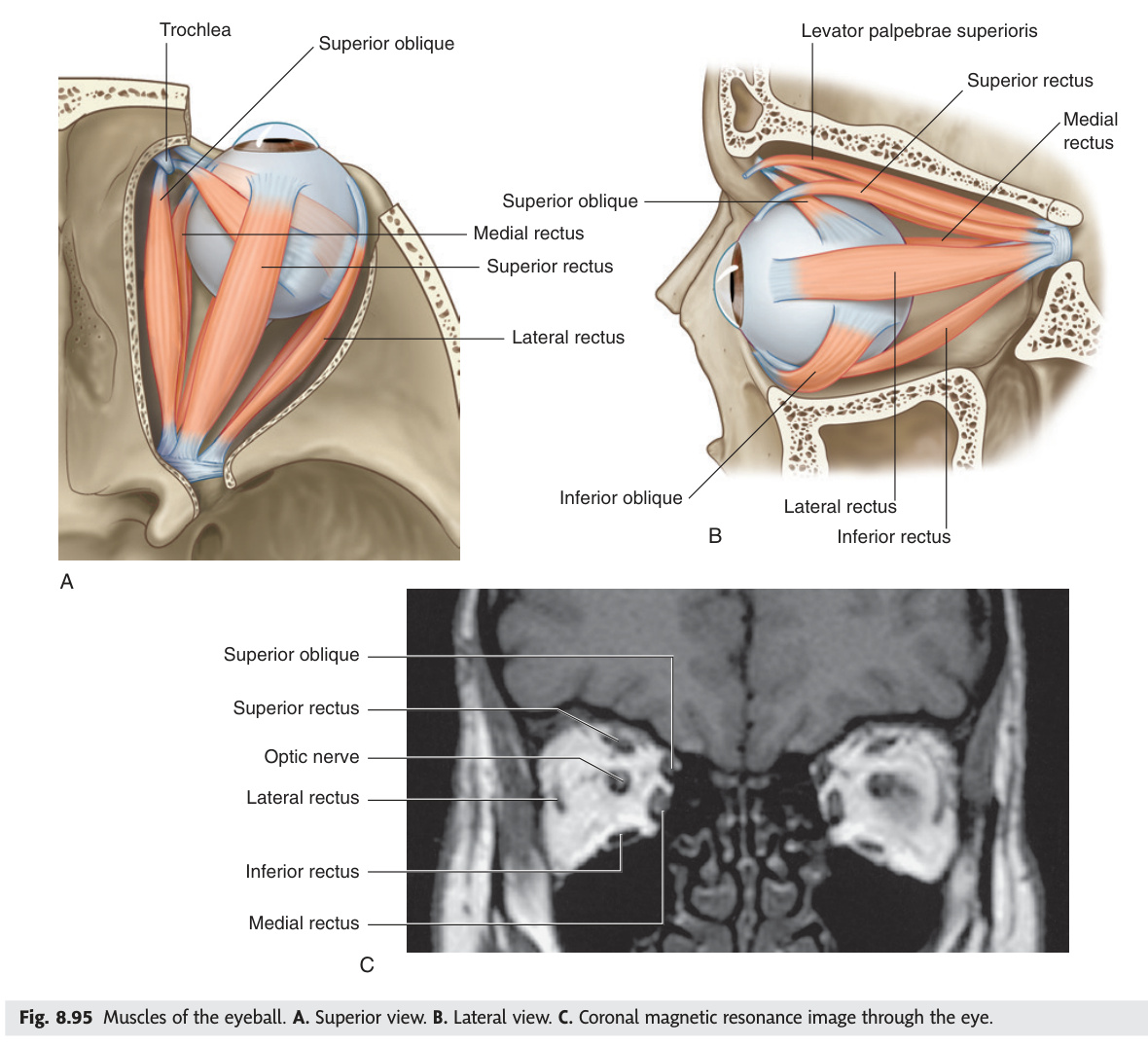
Six muscles move the eyeball, but only two nerves get their own single-muscle assignment — CN IV (trochlear) supplies only the superior oblique, and CN VI (abducens) supplies only the lateral rectus; everything else belongs to CN III. This is clinically indispensable: if a patient cannot abduct one eye at all, the lesion is CN VI until proven otherwise; if the eye drifts down and out with a drooping lid and dilated pupil, CN III is out. The superior oblique is the anatomically peculiar one — it runs forward, hooks around the trochlea (a pulley on the medial orbital wall), then turns backward to insert on the back of the eyeball, so its primary action in clinical testing is depression of the adducted eye (looking down and in toward the nose). CN IV palsy manifests as vertical diplopia worst going downstairs, and the patient unconsciously tilts their head to the opposite side to reduce double vision — the Bielschowsky head-tilt test exploits this to confirm the diagnosis.
| Muscle | Nerve | Primary action | Testing position |
|---|---|---|---|
| Superior rectus (SR) | CN III (superior division) | Elevation (in abducted position) | Look up and out |
| Inferior rectus (IR) | CN III (inferior division) | Depression (in abducted position) | Look down and out |
| Medial rectus (MR) | CN III (inferior division) | Adduction | Look medially |
| Lateral rectus (LR) | CN VI (abducens) | Abduction | Look laterally |
| Superior oblique (SO) | CN IV (trochlear) | Intorsion + depression (in adducted position) | Look down and in (toward nose) |
| Inferior oblique (IO) | CN III (inferior division) | Extorsion + elevation (in adducted position) | Look up and in |
| Levator palpebrae superioris | CN III (superior division) + sympathetic (Müller's muscle) | Elevates upper eyelid | Open eye |
Lateral Rectus = CN VI (abducens). Superior Oblique = CN IV (trochlear). All other muscles (SR, IR, MR, IO, LPS) = CN III (oculomotor). Note: CN IV has the longest intracranial course → most vulnerable to compression by raised ICP.
Most common cause of isolated vertical diplopia. Superior oblique paralysis → unopposed inferior oblique action → affected eye drifts up + extorted. Patient compensates by tilting head to the OPPOSITE side (away from lesion) → reduces diplopia. Test: Bielschowsky head tilt test — tilting to affected side worsens diplopia (confirmatory). Most common cause: closed head injury (CN IV exits dorsally from brainstem — vulnerable to contrecoup). Cover test: the affected eye drifts vertically upward when the normal eye is covered.
- "LR6 SO4 rest III" — expand the mnemonic with nerve and primary action for each? Lateral Rectus = CN VI (abduction). Superior Oblique = CN IV (intorsion + depression in adducted position). All others (SR, IR, MR, IO, LPS) = CN III. Superior division of CN III: SR + LPS. Inferior division: IR + MR + IO
- Superior oblique: trochlea anatomy and why does CN IV palsy cause vertical diplopia worst going downstairs? SO tendon passes through the trochlea (fibrocartilaginous pulley on medial orbital roof) then reflects backward to insert superolaterally. SO primary function = depression + intorsion in adduction (reading, descending stairs). CN IV palsy → cannot depress adducted eye → vertical diplopia worst looking down and inward
- Bielschowsky head-tilt test: how to perform and interpret? Tilt head toward the AFFECTED (higher) eye → diplopia WORSENS (confirms CN IV palsy). Tilt away → improves. Mechanism: head tilt toward affected side increases incyclotorsion demand on the weak superior oblique → maximal separation of images
- CN IV: anatomical feature making it uniquely vulnerable to closed head injury? Only cranial nerve to exit the DORSAL surface of the brainstem (at the midbrain-pons junction). Long, slender intracranial course around brainstem → vulnerable to contrecoup injury from frontal/occipital trauma. Most common cause of isolated vertical diplopia in young adults
- Levator palpebrae superioris: dual innervation and differential ptosis? Main motor = CN III (complete ptosis if palsy). Müller's muscle (superior tarsal muscle) = sympathetic (T1 origin). Horner's syndrome = sympathetic loss → only 2 mm partial ptosis. CN III palsy = complete ptosis (LPS fully paralysed)
CN III, IV & VI Palsies
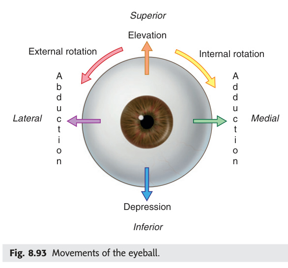
The three nerves that move the eye — CN III, IV, and VI — are among the most clinically tested in neurology, because each produces a distinctive and immediately recognisable pattern of failure. CN III is the workhorse: it runs almost every muscle in the orbit, including the lid elevator and the pupil constrictor. When it fails completely, the eye looks down and out (lateral rectus and superior oblique still working) with a drooping lid and a large fixed pupil — a presentation that demands urgent CT angiography to exclude a posterior communicating artery aneurysm compressing the nerve from outside. The key distinction between surgical and diabetic CN III palsy hinges on the pupil: compressive causes squeeze the outer parasympathetic fibres first, giving pupil involvement as the earliest sign, while ischaemic diabetic palsy spares the pupil because the peripheral fibres have better blood supply. The medial longitudinal fasciculus (MLF) connects abducens and oculomotor nuclei for conjugate gaze; a plaque of MS in the MLF at age 25 produces the classic internuclear ophthalmoplegia — failure of adduction on the side of the lesion with nystagmus in the abducting eye.
| Nerve | Palsy findings | Key causes |
|---|---|---|
| CN III (complete) | Ptosis (LPS paralysed) + eye looks "down and out" (LR + SO intact) + fixed dilated pupil (mydriasis — parasympathetic fibres run on outside of CN III, compressed early) | Posterior communicating artery (PComm) aneurysm (PAINFUL — 1st diagnosis to exclude); uncal herniation (↑ICP → coning); cavernous sinus pathology; diabetic mononeuropathy (spares pupil — ischaemic damage to nerve core spares outer parasympathetic fibres) |
| CN IV | Vertical diplopia (worse going downstairs, reading); head tilt to opposite side; Bielschowsky test positive | Closed head injury (most common); raised ICP; cavernous sinus; congenital |
| CN VI | Failure of abduction (convergent squint); diplopia worse at distance + looking to affected side; eye cannot go past midline laterally | Raised ICP (false localising sign — CN VI has long intracranial course, stretched over petrous ridge); Wernicke's encephalopathy; cavernous sinus; pontine lesion; diabetes |
Diabetic (medical) CN III palsy: ischaemic infarction of nerve core → affects central motor fibres (ptosis + ophthalmoplegia) but SPARES the pupil (parasympathetic fibres on the outer surface have better blood supply from pial vessels). Surgical (compressive) CN III palsy: PComm aneurysm or uncal herniation compresses the outer surface first → parasympathetics affected FIRST → fixed dilated pupil is the EARLIEST sign. Rule: pupil-involving CN III palsy = neurosurgical emergency until PComm aneurysm excluded by CT angiogram / digital subtraction angiography.
INO results from a lesion of the medial longitudinal fasciculus (MLF) — the white matter tract connecting the abducens nucleus (CN VI, pons) to the contralateral oculomotor medial-rectus sub-nucleus (CN III, midbrain), coordinating conjugate horizontal gaze.
Right MLF lesion: on attempted LEFT gaze → right eye fails to adduct past midline (medial rectus not activated) + left (abducting) eye has nystagmus = dissociated nystagmus. Convergence often preserved (midbrain convergence pathway bypasses MLF).
Bilateral INO = MS until proven otherwise — demyelinating plaque in MLF is a classic MS site in young adults. Unilateral INO in older patients → brainstem infarction.
WEBINO (Wall-Eyed Bilateral INO): bilateral MLF lesions → both eyes fail to adduct → exotropia at rest + nystagmus of abducting eyes.
One-and-a-half syndrome: ipsilateral PPRF lesion (horizontal gaze palsy) + ipsilateral MLF lesion → ipsilateral complete horizontal gaze palsy PLUS contralateral adduction failure → only contralateral eye can abduct. Caused by pontine infarct/MS.
- Complete CN III palsy: 3 clinical signs and most important diagnosis to exclude first? (1) Ptosis (LPS paralysed); (2) Eye "down and out" (LR + SO intact); (3) Fixed dilated pupil (parasympathetic fibres on outer surface compressed). Must exclude posterior communicating artery (PComm) aneurysm by CTA/DSA — pupil-involving CN III = neurosurgical emergency
- Diabetic CN III palsy vs surgical CN III palsy: single distinguishing feature? Pupil status. Diabetic (ischaemic) = PUPIL-SPARING (ischaemia damages central core fibres, peripheral parasympathetics spared). Surgical (compressive) = PUPIL-INVOLVED (compression hits outer parasympathetics first). Rule: any CN III palsy with pupil involvement = compressive cause until proven otherwise
- CN VI palsy: characteristic finding and why is it a false localising sign in raised ICP? Failure of abduction + convergent squint + diplopia worse at distance. CN VI has the longest intracranial course (over petrous ridge) → stretched by any process raising ICP, regardless of the actual lesion location → "false localising sign"
- INO: MLF function, findings on horizontal gaze, and age-related aetiology difference? MLF connects ipsilateral abducens nucleus (CN VI pons) to contralateral CN III medial rectus subnucleus. MLF lesion → ipsilateral adduction failure on contralateral gaze + contralateral abducting nystagmus + preserved convergence. Bilateral INO in young adult = MS. Unilateral INO in older patient = brainstem infarct
- One-and-a-half syndrome: lesion components and what eye movement remains? Ipsilateral PPRF lesion (horizontal gaze palsy to ipsilateral side) + ipsilateral MLF lesion (adduction failure). Result: ipsilateral eye completely immobile horizontally; only contralateral eye can abduct. Cause: pontine infarct or MS
Eyeball Layers
The eyeball is built in three concentric layers, each with a distinct job and a distinct set of clinical catastrophes when it fails. The outer fibrous layer — sclera and cornea — is the structural shell; the cornea's remarkable transparency depends on its avascularity and precise lamellar arrangement of collagen, which is why even a minor surface scar dramatically impairs vision and why corneal grafts succeed (no vessels means no immune rejection). The middle uveal (vascular) layer feeds the outer retina, controls the pupil via the iris, shapes the lens via the ciliary muscle, and produces aqueous humour — making it the target of both glaucoma physiology and anti-inflammatory treatment. The inner neural layer, the retina, separates into two parts at its ora serrata: a photoreceptive posterior portion and a non-visual anterior lining of the ciliary body and iris. When the neurosensory retina detaches from the retinal pigment epithelium, photoreceptors are deprived of their blood supply within hours — if the macula detaches, permanent central visual loss follows even after surgical reattachment.
| Layer (outer → inner) | Components | Notes |
|---|---|---|
| Outer fibrous layer | Sclera (posterior 5/6, opaque, white) + Cornea (anterior 1/6, transparent) | Sclera: attachment for EOMs + tendons. Cornea: avascular + no lymphatics (why corneal grafts succeed — immune privileged). Corneal reflex: CN V1 (afferent, nasociliary → ophthalmic) + CN VII (efferent, orbicularis oculi) |
| Middle vascular layer (uveal tract) | Choroid (posterior) + ciliary body + iris (anterior) | Choroid: highly vascular, nourishes outer retina. Iris: pigmented diaphragm with pupil opening. Ciliary body: contains ciliary muscle (parasympathetic → contracts → relaxes zonule fibres → lens rounds up = accommodation) + produces aqueous humour |
| Inner neural layer | Retina | Optic part (photoreceptors: rods [peripheral, low light] + cones [macula, colour, high acuity]) + non-visual part (lines ciliary body + iris). Macula lutea (yellow spot) + fovea centralis (highest cone density = point of maximum visual acuity) |
Separation of the neurosensory retina (photoreceptors) from the retinal pigment epithelium (RPE). Symptoms: sudden flashes (photopsia) + floaters (vitreous gel detaches = posterior vitreous detachment) → then "curtain/shadow" descending across vision as retina detaches. Risk factors: myopia, trauma, diabetic retinopathy, previous cataract surgery. Direct ophthalmoscopy: cannot see detached area; indirect ophthalmoscopy sees grey, billowing retinal folds. Emergency treatment: scleral buckle or vitrectomy. If macula detaches → permanent central visual loss even if reattached.
- Why is the cornea avascular, and what clinical advantage does this provide? Cornea is avascular (receives nutrients from aqueous humour + oxygen from atmosphere + limbal vessels at periphery only). Avascularity = no blood group antigens to trigger immune rejection → corneal grafts (keratoplasty) are the most successful transplants in medicine
- Uveal tract: 3 components and their functions? (1) Choroid: vascular layer nourishing outer retina. (2) Ciliary body: produces aqueous humour + ciliary muscle controls accommodation. (3) Iris: pigmented diaphragm controlling pupil size (sphincter pupillae = parasympathetic; dilator pupillae = sympathetic)
- Retinal detachment: symptom sequence, macula significance, and treatment urgency? Posterior vitreous detachment (floaters + photopsia) → "curtain/shadow" descending. If macula ON = surgical emergency (hours), visual prognosis good. If macula OFF = irreversible central visual loss even after successful reattachment. Surgery: scleral buckle or pars plana vitrectomy
- Ciliary muscle contraction: what happens to zonules and lens shape? What nerve drives it? Ciliary muscle contracts (parasympathetic, CN III) → zonule fibres RELAX → lens becomes more spherical (higher power) → accommodation for near vision. Opposite: ciliary relaxes → zonules taut → lens flattens → distance vision
- Macula lutea vs fovea centralis: structural and functional distinction? Macula = area lateral to optic disc; yellow pigment (lutein/zeaxanthin). Fovea = central pit of macula; highest cone density + no rods + no blood vessels + no nerve fibres overlying cones → point of maximum visual acuity. Foveal sparing in cortical lesions reflects dual blood supply
Chambers of the Eye & Aqueous Humour
Aqueous humour is a crystal-clear fluid produced by the ciliary body, flowing from the posterior chamber through the pupil into the anterior chamber, then draining at the iridocorneal angle into the trabecular meshwork and the canal of Schlemm. This circulation is the pressure regulator of the eye, and when it fails — either because the drain is slowly clogged (open-angle glaucoma) or because the iris suddenly blocks it (acute angle-closure) — intraocular pressure rises and silently destroys the optic nerve. Open-angle glaucoma is the quiet one: raised IOP erodes peripheral visual fields over years, often unnoticed until late, and is treated with drops that either reduce production (beta-blockers like timolol) or increase drainage (prostaglandins like latanoprost). Acute angle-closure is the dramatic one: sudden pain, a rock-hard red eye, corneal haze, nausea, and a mid-dilated non-reactive pupil — a genuine emergency treated with IV acetazolamide and pilocarpine to force the pupil constricted and restore drainage, followed by laser peripheral iridotomy to prevent recurrence.
| Compartment | Contents / Boundaries | Clinical |
|---|---|---|
| Anterior chamber | Between cornea + iris. Contains aqueous humour. Anterior chamber angle = iridocorneal angle = site of aqueous drainage (trabecular meshwork → canal of Schlemm → episcleral veins) | Primary open-angle glaucoma (POAG): most common; trabecular meshwork resistance ↑ → ↑ IOP → optic nerve damage. Gradual peripheral visual field loss. Treat: timolol (β-blocker, ↓ production), latanoprost (prostaglandin ↑ drainage), acetazolamide (↓ production) |
| Posterior chamber | Between iris + lens. Aqueous produced here by ciliary body → flows through pupil → anterior chamber → drains at iridocorneal angle | Angle-closure glaucoma: acute attack — iris root blocks angle. Painful red eye + corneal haziness + dilated non-reactive pupil + N+V. EMERGENCY: IV acetazolamide + pilocarpine (constricts pupil, opens angle) → laser iridotomy |
| Vitreous chamber | Behind lens; filled with vitreous humour (gel, 99% water + collagen + hyaluronic acid). Does NOT regenerate | Vitreous haemorrhage: diabetic retinopathy (neovascularisation), trauma → sudden painless visual loss + floaters |
| Lens | Biconvex, transparent, avascular. Held by zonule fibres from ciliary body. Accommodation: ciliary muscle contracts → zonule relaxes → lens rounds up | Cataract: lens opacity. Most common cause of preventable blindness globally. Risk: age, UV, diabetes, steroids, trauma. Treatment: phacoemulsification + intraocular lens (IOL) implant |
| POAG (Chronic open-angle) | PACG (Acute angle-closure) | |
|---|---|---|
| Mechanism | Trabecular meshwork resistance ↑ → impaired outflow → slowly ↑IOP despite open angle | Iris root physically blocks iridocorneal angle → sudden total aqueous outflow obstruction |
| Onset | Insidious, painless, bilateral (asymmetric) | Acute attack, PAINFUL, unilateral; precipitated by mydriasis (dim light, anticholinergics, stress) |
| Symptoms | Gradual peripheral visual field loss (arcuate scotoma, tunnel vision); often asymptomatic until late | Severe eye pain + headache + N+V + blurred vision + halos around lights; may mimic acute abdomen/migraine |
| Examination | Optic disc cupping (cup:disc ratio >0.6, notching, disc haemorrhage); IOP >21 mmHg; open angle on gonioscopy | Red eye + corneal haziness (oedema) + dilated non-reactive oval pupil + shallow anterior chamber + very high IOP (>50 mmHg) |
| Risk factors | Age, FH (50× risk), Afro-Caribbean, myopia, diabetes, corticosteroids | Hypermetropia (short axial length), shallow AC, age, female, pupil dilation |
| Treatment | Topical: prostaglandin analogues (latanoprost 1st line, ↑uveoscleral outflow); β-blockers (timolol, ↓ production); carbonic anhydrase inhibitors (dorzolamide); α-agonists (brimonidine). Surgery: trabeculectomy (filtration bleb) | EMERGENCY: IV acetazolamide (↓ production) + pilocarpine 4% (miosis → opens angle) + topical β-blocker + analgesia/antiemetics. Definitive: bilateral laser peripheral iridotomy (PI) — creates hole in iris to bypass pupil block |
- Aqueous humour flow: production site → route → drainage site? Produced by ciliary body (posterior chamber) → flows through pupil → anterior chamber → iridocorneal angle → trabecular meshwork → canal of Schlemm → episcleral veins. Blockage at any point → raised IOP
- POAG vs PACG: single most important difference in presentation? POAG = painless, insidious, bilateral peripheral field loss (arcuate scotoma → tunnel vision). PACG = painful, acute, unilateral; precipitated by pupil dilation (dark, anticholinergics). Both raise IOP but by completely different mechanisms (resistance vs physical blockage)
- Acute angle-closure glaucoma: 5 clinical signs? (1) Painful red eye; (2) Corneal haziness (oedema from high IOP); (3) Dilated non-reactive oval pupil; (4) Shallow anterior chamber; (5) Very high IOP (>50 mmHg). N+V common — may mimic acute abdomen/migraine
- Acute angle-closure treatment: immediate pharmacological steps and definitive procedure? IV acetazolamide (↓ production) + pilocarpine 4% (miosis → mechanically opens angle) + topical β-blocker + analgesia/antiemetics. Definitive: bilateral laser peripheral iridotomy (creates iris bypass hole to prevent pupil block)
- Accommodation mechanism: ciliary muscle, zonules, lens shape — complete sequence? Near object → parasympathetic (CN III) → ciliary muscle CONTRACTS → zonule fibres RELAX → lens rounds up (increases dioptric power) → near focus. Cycloplegic drops (atropine) → ciliary paralysis → zonules taut → lens flat → cannot accommodate (used in refraction of children)
Visual Pathway & Field Defects
The visual pathway is one of the most rigorously tested anatomical pathways in clinical medicine because every relay station from the retina to the occipital cortex produces a uniquely localising pattern of visual field loss. The key anatomical fact is the partial decussation at the optic chiasm: nasal fibres from each retina cross to the opposite optic tract, while temporal fibres stay ipsilateral. This means a lesion at the chiasm (the pituitary tumour pressing upward from below) destroys only the crossing nasal fibres, producing bitemporal hemianopia — each eye loses its outer (temporal) visual field. Anterior to the chiasm, only one eye is affected; posterior to it, both eyes lose the same half of the visual field (homonymous hemianopia). Quadrantanopias localise even more precisely: the inferior fibres of the optic radiation loop forward into the temporal lobe (Meyer's loop) before heading posteriorly, so a temporal lobe lesion gives a superior quadrantanopia — "pie in the sky." Macular sparing with occipital lesions occurs because the macular cortex at the occipital pole has dual blood supply from both PCA and MCA collaterals.
- Optic chiasm lesion: which fibres, which field, and most common cause? Decussating NASAL fibres (carrying temporal visual field information) from both eyes are destroyed. Result: bitemporal hemianopia (both eyes lose their outer temporal field). Most common cause: pituitary macroadenoma pressing upward from below
- Temporal lobe vs parietal lobe optic radiation lesion — field defect difference? Temporal lobe (Meyer's loop = inferior radiation fibres carrying superior field) → contralateral superior quadrantanopia ("pie in the sky"). Parietal lobe (superior radiation fibres carrying inferior field) → contralateral inferior quadrantanopia ("pie on the floor")
- Macular sparing in occipital lobe infarction: mechanism? Occipital pole (macular representation) has dual blood supply: PCA (primary) + MCA collaterals. When PCA occludes, MCA collaterals maintain macular cortex → central vision preserved despite peripheral homonymous hemianopia
- Right optic tract lesion: which eye loses which field? Right optic tract carries fibres from right temporal retina (right eye) + left nasal retina (left eye), both representing the LEFT visual field. Lesion → left homonymous hemianopia (both eyes lose left field). Optic tract lesions typically show incongruous hemianopia (asymmetric between eyes)
- Monocular visual loss vs homonymous hemianopia: what does laterality tell you? Monocular = lesion anterior to chiasm (retina or optic nerve — ipsilateral only). Homonymous (same field in both eyes) = lesion posterior to chiasm (optic tract, radiation, or cortex). Bitemporal = lesion AT the chiasm
Pupillary Reflexes
The pupil is a two-nerve system: the parasympathetic pathway constricts it (the afferent arm runs in CN II to the pretectal nucleus, then the efferent arm runs in CN III to the ciliary ganglion), and the sympathetic pathway dilates it (a three-neuron chain descending from the hypothalamus through the ciliospinal centre at C8–T2, then up to the superior cervical ganglion, then along the ICA into the orbit). Examining the pupil in the dark and in light, testing the swinging flashlight sign, and looking for ptosis and anhidrosis together gives you localising information that no scan can replace. The RAPD (relative afferent pupillary defect) is the most sensitive bedside sign of unilateral optic nerve disease — when you shine the torch in the affected eye, both pupils dilate because the damaged optic nerve sends a weaker signal than the normal side. Argyll Robertson pupils (small, irregular, bilateral, accommodating but not reacting to light) are the hallmark of neurosyphilis and represent selective destruction of the pretectal light-reflex relay while the accommodation pathway through the superior colliculus is spared.
| Reflex | Pathway | Clinical test |
|---|---|---|
| Light reflex (direct) | Light → retina → CN II → pretectal nucleus (midbrain) → bilateral Edinger-Westphal nuclei → CN III → ciliary ganglion → short ciliary nerves → constrictor pupillae (sphincter). Bilateral → explains consensual reflex | Shine light in one eye → ipsilateral pupil constricts (direct) + contralateral pupil constricts (consensual/indirect). Test each eye separately |
| Accommodation reflex | Visual cortex → midbrain → CN III → bilateral: ciliary muscle (lens rounds) + medial rectus (convergence) + constrictor pupillae (miosis) | Ask patient to look at near object: convergence + miosis + accommodation (3 components) |
| Swinging flashlight test (RAPD) | Alternately shine light in each eye; compare pupillary response | Relative afferent pupillary defect (RAPD/Marcus Gunn pupil): when light shines in AFFECTED eye → BOTH pupils DILATE (afferent limb defect → less light signal → less constriction). Cause: optic neuritis, optic nerve damage, severe retinal disease |
Interruption of sympathetic supply to the eye. Signs: partial ptosis (Müller's muscle, upper lid) + miosis (dilator pupillae paralysed) + anhidrosis (ipsilateral face — if 1st/2nd order lesion) + enophthalmos (apparent). Causes: 1st order neuron (hypothalamus → ciliospinal centre C8–T2): Pancoast tumour, syringomyelia, lateral medullary syndrome. 2nd order (C8–T2 → superior cervical ganglion): thyroid carcinoma, cervical rib, apical lung carcinoma, subclavian artery aneurysm. 3rd order (superior cervical ganglion → orbit): carotid artery dissection (PAINFUL Horner's = emergency), cavernous sinus. Pharmacological testing: cocaine (won't dilate in Horner's); hydroxyamphetamine (dilates in 1st/2nd order but not 3rd order lesion).
| Feature | Argyll Robertson | Adie's Tonic Pupil | CN III Palsy (complete) |
|---|---|---|---|
| Size | Small (miotic), irregular, bilateral | Large (mydriatic), slightly irregular, unilateral (80%) | Large, fixed dilated |
| Light reaction | Absent | Absent or very slow | Fixed — no reaction |
| Accommodation | Intact — "accommodates but does not react" (ABCR: Accommodates Brisk, Constriction to light Reduced) | Slow tonic constriction to near; slow redilation (tonic response) | Impaired (ciliary muscle paralysed) |
| Ptosis/EOM | Absent | Absent; DTRs reduced (Holmes-Adie syndrome) | Complete ptosis + down-and-out eye |
| Pharmacological test | 0.1% pilocarpine — no hypersensitivity | 0.1% pilocarpine → brisk constriction (denervation hypersensitivity of ciliary ganglion) | 1% pilocarpine constricts normally |
| Cause / Lesion | Neurosyphilis — selective pretectal nucleus destruction → light-near dissociation | Postganglionic parasympathetic denervation of ciliary ganglion (viral/autoimmune). Benign | PComm aneurysm, uncal herniation |
Pupil accommodates (near) but does NOT react to light. Causes: (1) Argyll Robertson — neurosyphilis, pretectal lesion; (2) Adie's tonic pupil — ciliary ganglion (slow not truly preserved); (3) severe optic neuropathy (no afferent limb); (4) Parinaud's dorsal midbrain syndrome (pineal tumour/hydrocephalus compressing dorsal midbrain) → bilateral large pupils + light-near dissociation + upgaze palsy + convergence-retraction nystagmus + eyelid retraction (Collier's sign).
- RAPD: what causes both pupils to DILATE when light is shone in the affected eye? The affected optic nerve (CN II) sends a weaker afferent signal → pretectal nucleus receives less input → less Edinger-Westphal activation → less pupillary constriction bilaterally → relative dilation when torch moves to affected eye. RAPD = unilateral optic nerve or severe retinal disease; NOT present in bilateral symmetric optic neuropathy
- Horner's syndrome: 4 signs and the 3-neuron pathway? (1) Partial ptosis (Müller's muscle); (2) Miosis; (3) Anhidrosis (ipsilateral face — 1st/2nd order only); (4) Apparent enophthalmos. Pathway: 1st order = hypothalamus → ciliospinal centre (C8–T2). 2nd order = C8–T2 → superior cervical ganglion. 3rd order = SCG → orbit via ICA. Painful 3rd order Horner's = carotid artery dissection (emergency)
- Argyll Robertson: size, light reaction, accommodation, cause? Small miotic irregular pupils (bilateral). Light = ABSENT. Accommodation = INTACT (ABCR: Accommodates Brisk, Constriction to light Reduced). Cause: neurosyphilis — selective pretectal nucleus destruction. Distinguish from Adie's: Adie's = large, unilateral, 0.1% pilocarpine → brisk constriction (denervation hypersensitivity)
- Adie's tonic pupil: size, pharmacological test, associated systemic finding? Large unilateral pupil. Slow tonic constriction to near, slow redilation. 0.1% pilocarpine → brisk constriction (dilated ciliary ganglion denervated → supersensitive). Holmes-Adie syndrome = Adie's pupil + reduced/absent deep tendon reflexes. Benign, idiopathic
- Light-near dissociation: 4 causes to know? (1) Argyll Robertson (neurosyphilis). (2) Adie's tonic pupil (ciliary ganglion). (3) Severe optic neuropathy (no afferent limb). (4) Parinaud's dorsal midbrain syndrome (pineal tumour/hydrocephalus) → bilateral large pupils + upgaze palsy + convergence-retraction nystagmus + Collier's sign (lid retraction)
Eyelids & Lacrimal Apparatus
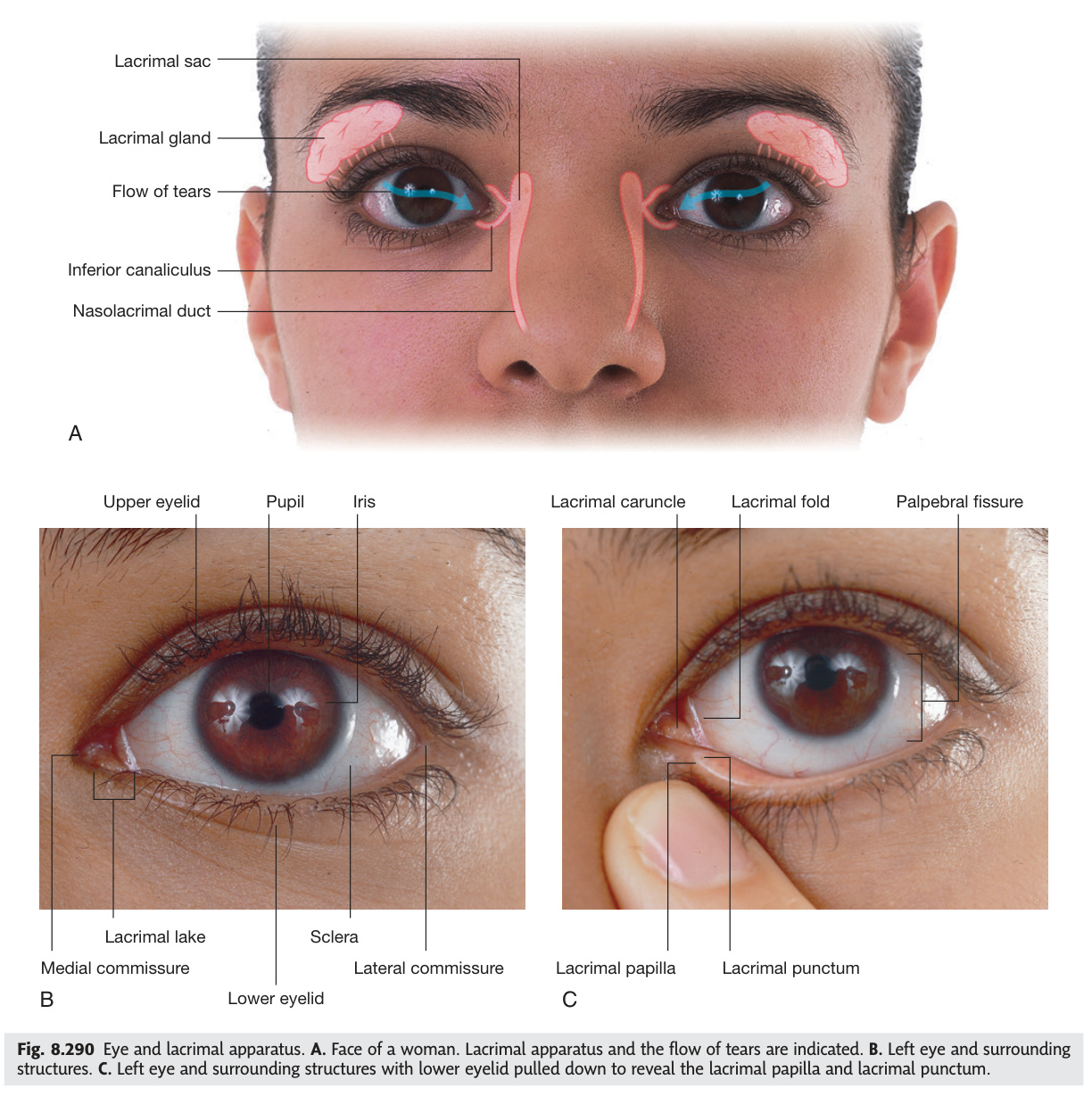
The eyelid is a precisely engineered structure that opens, closes, blinks, and produces tears in perfect co-ordination — and when any one component fails, the clinical consequences are immediate. Two muscles govern lid position: orbicularis oculi (CN VII) closes the eye; levator palpebrae superioris (CN III) and Müller's muscle (sympathetic) open it. Bell's palsy knocks out orbicularis, leaving the eye unable to close and the cornea exposed to drying and ulceration — lagophthalmos requiring lubricating drops and nocturnal taping. The Meibomian glands in the tarsal plates produce the oily outer layer of the tear film; when their ducts clog, a lipogranuloma (chalazion) forms; when they become acutely infected (hordeolum/stye), the lid becomes hot and tender. Tears drain medially through the lacrimal puncta into the lacrimal sac and down the nasolacrimal duct to the inferior meatus — when the nasolacrimal duct is blocked in a newborn, epiphora (overflow tearing) follows and can be treated by massage or syringing.
| Structure | Details |
|---|---|
| Orbicularis oculi | Closes eyelid. Orbital part (forceful closure) + palpebral part (blinking). Nerve: CN VII (facial). Paralysed in Bell's palsy → lagophthalmos + exposure keratitis |
| Levator palpebrae superioris (LPS) | Opens upper eyelid. Nerve: CN III (main) + sympathetic (Müller's muscle, upper 2 mm). CN III palsy → complete ptosis; sympathetic loss (Horner's) → partial ptosis (2 mm) |
| Tarsal plates | Fibrocartilaginous supports of upper + lower lids. Contain Meibomian glands (sebaceous) → secrete oily layer of tear film. Blockage → chalazion (painless granuloma); infection → hordeolum (stye, painful) |
| Lacrimal gland | Superolateral orbit. Secretes tears → lacrimal puncta (medial lid margins) → canaliculi → lacrimal sac (in lacrimal fossa, medial orbital wall) → nasolacrimal duct → inferior meatus of nasal cavity. Nerve: CN VII (secretomotor via pterygopalatine ganglion) |
Test Unit 13 knowledge
Orbital anatomy, visual pathway defects, and CN III/IV/VI palsy MCQs.
- Bell's palsy: which muscle is paralysed, mechanism of corneal damage, and management? Orbicularis oculi (CN VII) paralysed → lagophthalmos (lid cannot close fully) → corneal exposure → drying + ulceration + exposure keratitis. Management: hourly lubricating drops (daytime) + viscous gel at night + tape/moisture chamber closure of lid. If no recovery → gold weight implant in upper lid
- Chalazion vs hordeolum (stye): pathology, gland, symptoms? Chalazion = blocked Meibomian gland duct → lipogranuloma → painless lid lump, no erythema. Hordeolum (stye) = acute infection of Meibomian gland (or Zeis/Moll glands) → painful, red, hot, pointed swelling at lid margin. Chalazion: warm compress ± intralesional steroid ± incision + curettage. Hordeolum: warm compress, topical antibiotics
- Lacrimal drainage: complete route from punctum to nose? Lacrimal puncta (medial upper + lower lids) → upper/lower canaliculi → common canaliculus → lacrimal sac (medial orbital wall, lacrimal fossa) → nasolacrimal duct → inferior meatus of nasal cavity. Nasolacrimal duct obstruction in newborn = congenital dacryostenosis → epiphora + discharge; treat: massage + syringing
- Secretomotor nerve to lacrimal gland: complete pathway? CN VII → greater petrosal nerve → pterygopalatine ganglion (synapse) → zygomatic nerve → lacrimal nerve (CN V1) → lacrimal gland. Disruption (e.g., lesion at geniculate ganglion) → reduced lacrimation + dry eye. Crocodile tears = aberrant CN VII regeneration → lacrimal gland stimulated by eating
- Ptosis: complete CN III palsy vs Horner's — how to distinguish at bedside? CN III = COMPLETE ptosis (LPS fully paralysed; lid at pupil level); pupil fixed dilated; eye down and out. Horner's = PARTIAL ptosis (~2 mm; Müller's muscle only); pupil SMALL (miosis); anhidrosis; no EOM involvement; look for other Horner features
Orbital Pathology & Proptosis ★★
Proptosis — forward displacement of the eyeball beyond the orbital rim — is the orbital symptom that unifies a surprisingly wide range of pathologies, from autoimmune thyroid disease to carotid-cavernous fistula to orbital cellulitis. The single most important first question is bilateral versus unilateral: bilateral proptosis almost always means thyroid eye disease (Graves' ophthalmopathy), while unilateral proptosis demands a systematic differential including tumour, inflammation, vascular malformation, and infection. Thyroid eye disease operates through TSH-receptor antibody cross-reactivity with orbital fibroblasts, causing glycosaminoglycan accumulation and muscle belly swelling — inferior rectus is first and most often affected, so the patient's first complaint is diplopia looking upward. The most feared complication is dysthyroid optic neuropathy from crowding at the orbital apex: emergency decompression surgery or high-dose steroids are required to preserve vision. Orbital cellulitis and preseptal cellulitis look superficially similar (swollen, red, tender periorbital tissue) but are separated by the orbital septum — orbital cellulitis causes proptosis and ophthalmoplegia with vision threat, while preseptal cellulitis does not.
Causes — RITZ mnemonic:
(R) Retrobulbar tumour: orbital haemangioma (most common benign), dermoid cyst, lacrimal gland tumour, metastases (from breast, lung, prostate — unilateral, scirrhous metastasis → enophthalmos not proptosis), lymphoma, optic nerve glioma, meningioma.
(I) Inflammation/Infection: orbital cellulitis, orbital pseudotumour (idiopathic orbital inflammation, painful, responds to steroids), dacryoadenitis.
(T) Thyroid: thyroid eye disease (Graves' — most common cause of bilateral proptosis in adults, see below).
(Z) Vascular: carotid-cavernous fistula (pulsatile proptosis + bruit), orbital varix (positional).
Investigation: CT orbit (bone lesions, blow-out fracture, sinus disease), MRI orbit (soft tissue — muscle belly enlargement in thyroid eye disease vs tendon sparing differentiates from myositis), USS (cystic vs solid), TFTs/TRAb (thyroid eye disease).
Pathophysiology: TSH receptor antibodies (TRAb) cross-react with orbital fibroblasts → glycosaminoglycan deposition → extraocular muscle swelling + orbital fat expansion → proptosis. Active in 10% of Graves', but can occur with hypothyroidism/euthyroid state. Smoking major risk factor.
NOSPECS classification: No signs → Only signs (lid retraction, lid lag) → Soft tissue (chemosis, periorbital oedema) → Proptosis → EOM restriction (inferior rectus most affected → diplopia looking up; then medial rectus → diplopia looking laterally) → Corneal involvement (exposure keratitis from lagophthalmos) → Sight loss (dysthyroid optic neuropathy — compressive at orbital apex — EMERGENCY).
Lid retraction (Dalrymple's sign): sympathetic overactivation of superior tarsal muscle (Müller's) + LPS contracture → sclera visible above limbus. Lid lag on downgaze (von Graefe's sign). Infrequent blinking (Stellwag's sign).
Treatment: Selenium (active mild disease); IV methylprednisolone (active moderate-severe); orbital decompression (remove medial/inferior wall into ethmoidal/maxillary sinus — for optic neuropathy or severe proptosis); squint surgery; lid surgery (in that order).
| Feature | Preseptal (Periorbital) | Orbital (Postseptal) |
|---|---|---|
| Location | Anterior to orbital septum — eyelid only | Posterior to orbital septum — within orbit proper |
| Cause | Skin trauma, insect bite, hordeolum, dacryocystitis, spread from sinusitis | Ethmoid sinusitis (most common, especially in children), trauma, dental infection, endophthalmitis |
| Proptosis | Absent | Present |
| Ophthalmoplegia | Absent | Present (pain on eye movement) |
| Vision | Normal | May be reduced → EMERGENCY (optic nerve at risk) |
| Treatment | Oral antibiotics (amoxicillin-clavulanate), outpatient if mild | IV antibiotics + ENT consult for drainage; CT orbit to identify subperiosteal abscess (→ surgical drainage) |
- Bilateral vs unilateral proptosis: what does bilaterality suggest, and most common cause? Bilateral = systemic cause, overwhelmingly thyroid eye disease (Graves' ophthalmopathy). Unilateral = local orbital pathology (tumour, cellulitis, vascular). Measure with Hertel exophthalmometer (>2 mm beyond orbital rim = proptosis)
- Thyroid eye disease: pathophysiology, most affected muscle, and sight-threatening complication? TRAb cross-react with orbital fibroblast TSH receptors → glycosaminoglycan deposition → muscle belly swelling + orbital fat expansion. Inferior rectus most affected → diplopia on upward gaze. Sight-threatening: dysthyroid optic neuropathy (orbital apex compression of CN II) → emergency IV methylprednisolone ± surgical decompression
- NOSPECS: which stages require urgent ophthalmology referral? Stage S (Sight loss) = emergency — compressive optic neuropathy → IV steroids ± orbital decompression. Stage C (Corneal involvement, exposure keratitis) = urgent. Stage E (EOM restriction, diplopia) = moderate. Stages No/O/P (early) = monitor
- Orbital cellulitis vs preseptal cellulitis: 3 key distinguishing features? Orbital: (1) proptosis present; (2) ophthalmoplegia + pain on eye movement; (3) vision may be reduced. Preseptal: all three absent (normal vision, no proptosis, no EOM restriction). Both may have periorbital swelling/erythema. CT orbit separates them
- Carotid-cavernous fistula: triad of clinical features and investigation of choice? (1) Pulsatile (pulsating) exophthalmos; (2) Bruit over the eye (auscultate with stethoscope); (3) Chemosis (conjunctival oedema) + raised IOP. Investigation: CT/MRI angiography or digital subtraction angiography. Treatment: endovascular coil embolisation
Retinal Vascular Disease ★★★
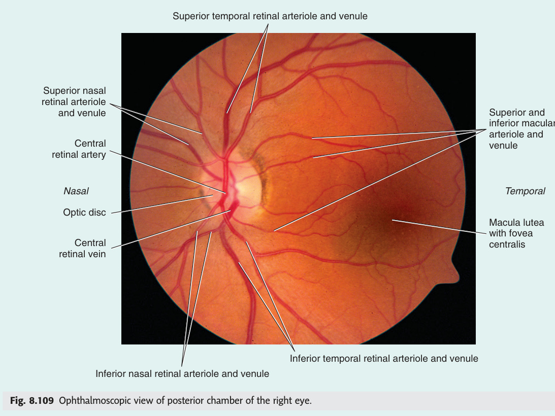
The retina is one of the most metabolically active tissues in the body, and its blood supply — the central retinal artery branching from the ophthalmic artery — leaves absolutely no collateral reserve. Central retinal artery occlusion produces ischaemic whitening of the entire retina within minutes, with the fovea remaining pink as an isolated cherry-red spot because it is fed by the choroid rather than the retinal circulation. This is an ophthalmic emergency comparable to a stroke — the treatment window is brief and outcome is rarely complete recovery. Central retinal vein occlusion produces the opposite appearance: an explosion of haemorrhages in all four quadrants (the "blood and thunder" fundus) from backed-up venous pressure. Diabetic retinopathy follows a predictable staging progression driven by pericyte loss, microaneurysm formation, and ultimately neovascularisation under the drive of VEGF — and the entire disease process is modifiable through glycaemic and blood pressure control long before intravitreal injections are needed. AMD is now the leading cause of irreversible visual loss in the developed world, and the shift from dry to wet disease — driven by choroidal neovascularisation — is the moment when treatment with anti-VEGF injections must begin within days to preserve central vision.
| Condition | Fundoscopy | Causes / Notes |
|---|---|---|
| CRAO (Central Retinal Artery Occlusion) | Pale/white oedematous retina (ischaemic swelling of nerve fibre layer) + cherry-red spot at fovea (fovea avascular zone — underlying choroid visible through thin foveal retina) + "box-carring" of vessels (segmented blood column) | Embolic (from ICA atheroma, cardiac — AF, valve disease, paradoxical from DVT) or thrombotic. Ophthalmic artery occlusion = more severe (no cherry-red spot, all pale). Amaurosis fugax = transient CRAO (ophthalmic artery TIA — "curtain coming down"). Emergency: ocular massage + AC paracentesis + IV thrombolysis (if within 4.5h in some centres) + embolic workup (echocardiogram, carotid USS, ECG for AF) |
| CRVO (Central Retinal Vein Occlusion) | "Blood and thunder": flame haemorrhages in ALL 4 quadrants + disc swelling + dilated tortuous veins + cotton-wool spots. "Stormy sunset" appearance | Hypertension (most important), diabetes, hyperviscosity (polycythaemia, myeloma), glaucoma (raised IOP compresses CRV at lamina cribrosa). Non-ischaemic (good prognosis) vs ischaemic (neovascular glaucoma risk → "100-day glaucoma"). Branch RVO: sector of haemorrhage at AV crossing. Anti-VEGF injections for macular oedema |
Background DR (mild-moderate NPDR): Microaneurysms (earliest sign — dot haemorrhages) + dot-blot haemorrhages (deep, intraretinal) + hard exudates (lipid deposits at edge of leaking microaneurysms) + retinal oedema. Vision affected only if macula involved (clinically significant macular oedema, CSMO).
Preproliferative DR (severe NPDR): Cotton-wool spots (nerve fibre layer infarcts = soft exudates; due to arteriolar occlusion) + venous beading/looping + intraretinal microvascular abnormalities (IRMA). "4-2-1" rule: haemorrhages in 4 quadrants OR venous beading in 2 quadrants OR IRMA in 1 quadrant = severe NPDR.
Proliferative DR (PDR): Neovascularisation at disc (NVD) or elsewhere (NVE) → vitreous haemorrhage (sudden painless visual loss + floaters) → fibrovascular proliferation → tractional retinal detachment (TRD).
Diabetic maculopathy: Most common cause of visual loss in diabetics. CSMO = hard exudates ± oedema within 500 μm of fovea centre OR retinal thickening within 500 μm of fovea. OCT = best assessment.
Treatment: Systemic: optimize HbA1c (each 1% reduction → 35% risk reduction), BP control (<130/80), lipids. Ocular: laser photocoagulation (panretinal photocoagulation PRP for PDR — destroys ischaemic retina → reduces VEGF); focal/grid laser for maculopathy; intravitreal anti-VEGF (ranibizumab, aflibercept, bevacizumab — gold standard for CSMO); vitrectomy for TRD/vitreous haemorrhage.
| Dry AMD (Geographic Atrophy) | Wet AMD (Neovascular) | |
|---|---|---|
| Prevalence | 85–90% of AMD | 10–15% of AMD, causes 90% of AMD blindness |
| Pathology | Drusen (lipid/protein deposits between RPE and Bruch's membrane) → RPE atrophy → photoreceptor loss. Slow progression over years | Choroidal neovascularisation (CNV) breaks through Bruch's membrane → subretinal fluid + haemorrhage + fibrosis → rapid (days–weeks) central visual loss |
| Symptoms | Slow loss of central vision, metamorphopsia (distortion — use Amsler grid) | Sudden distortion + scotoma; straight lines appear wavy (Amsler grid) |
| Investigation | OCT (drusen, RPE changes); fluorescein angiography (FA) | OCT (subretinal fluid, pigment epithelial detachment); FA + OCT-angiography (CNV mapping) |
| Treatment | AREDS2 supplements (zinc + antioxidants — vitamin C, E, β-carotene, lutein/zeaxanthin) slow progression in intermediate-advanced dry AMD | Intravitreal anti-VEGF injections (ranibizumab, aflibercept, bevacizumab) — monthly initially then PRN (treat-and-extend); visual outcome depends on speed of treatment |
- CRAO fundoscopy: 2 key findings and their pathophysiological basis? (1) Pale/white retina — ischaemic swelling of nerve fibre layer from arterial occlusion. (2) Cherry-red spot at fovea — fovea supplied by choroid (not retinal artery), choroidal red colour visible through avascular foveal retina against white surrounding. Amaurosis fugax = transient CRAO (TIA of ophthalmic artery)
- CRVO fundoscopy appearance and highest-risk complication? "Blood and thunder" = flame haemorrhages in ALL 4 quadrants + disc swelling + dilated tortuous veins + cotton-wool spots. Highest-risk: ischaemic CRVO → VEGF-driven iris neovascularisation → neovascular glaucoma ("100-day glaucoma")
- Diabetic retinopathy earliest sign and the 4-2-1 rule for severe NPDR? Earliest sign = microaneurysms (dot haemorrhages). Severe NPDR (4-2-1 rule): haemorrhages in ALL 4 quadrants OR venous beading in ≥2 quadrants OR IRMA in ≥1 quadrant → high risk of progression to proliferative DR
- Proliferative DR: mechanism of neovascularisation and treatment? Retinal ischaemia → VEGF production → new vessels at disc (NVD) or elsewhere (NVE) → vitreous haemorrhage (sudden painless visual loss) + tractional retinal detachment. Treatment: panretinal photocoagulation (PRP) — destroys ischaemic peripheral retina → reduces VEGF drive
- Wet AMD vs dry AMD: prevalence, pathology, symptom onset, treatment? Dry = 85–90%; drusen → slow RPE atrophy; gradual central loss + metamorphopsia; AREDS2 supplements slow progression. Wet = 10–15% cases, 90% of AMD blindness; choroidal neovascularisation → rapid (days–weeks) central loss; emergency intravitreal anti-VEGF (ranibizumab/aflibercept)
Cataract Classification & Surgery ★
A cataract is any opacity of the crystalline lens, and despite being the world's most common cause of treatable blindness, the underlying biology is straightforward — protein denaturation, cross-linking, or disruption of the normally transparent lens fibres. The type of cataract tells you the aetiology: posterior subcapsular cataracts in a young patient on steroids, nuclear sclerosis in an elderly patient complaining of worsening distance vision, or a dense central opacity in a child born to a mother with rubella. Posterior subcapsular cataract is the most visually disabling type because it sits at the visual axis and is maximally exposed when the pupil constricts for near tasks and bright light. Congenital cataract is a paediatric emergency: the visual cortex must receive a clear image during the critical period (birth to ~7 years) or it will permanently suppress that eye — amblyopia that no operation can later reverse. Phacoemulsification is now the world's most performed elective operation, and posterior capsule opacification — its commonest late complication — is treated definitively with a single outpatient Nd:YAG laser session.
| Type | Slit-lamp appearance | Visual impact / Causes |
|---|---|---|
| Nuclear sclerosis | Brunescent yellowing/browning of nucleus; graded I–IV by density | Gradual distance blur; early "myopic shift" (↑ refractive index → reading improves transiently = "second sight"). Age most common cause |
| Cortical cataract | Spoke-like (cuneiform) grey-white opacities radiating from periphery of cortex toward centre; retroillumination shows "spokes" | Glare, dazzle, monocular diplopia. Age, UV-B radiation, diabetes |
| Posterior subcapsular (PSC) | Fine granular opacity immediately anterior to posterior capsule; seen best on retroillumination as a central plaque | Most visually disabling: affects near vision + causes severe glare (pupil constricts for near → opacification at centre of pupil). Causes: corticosteroids (systemic or topical), diabetes, trauma, irradiation, uveitis. Youngest age group affected |
| Anterior subcapsular | Opacity just beneath anterior capsule; may be associated with fibrous metaplasia of lens epithelium | Trauma (blunt or perforating), uveitis, atopic dermatitis |
| Congenital cataract | Variable — lamellar/zonular most common (opacification of specific fetal nucleus zone); posterior lenticonus, polar opacities | Causes: Rubella (most common infectious cause — dense central opacity); TORCH infections; galactosaemia (oil-droplet cataract); Down's syndrome (sutural + flocculi); Lowe syndrome (oculocerebrorenal); Marfan (ectopia lentis > cataract). Urgency: must treat within weeks to prevent amblyopia (visual deprivation during critical period) |
Most common elective surgery performed worldwide. Technique: 2.5–3 mm clear corneal incision → continuous curvilinear capsulorhexis (CCC, opening in anterior capsule) → hydrodissection → ultrasonic phacoemulsification of nucleus → cortex aspiration → foldable intraocular lens (IOL) implant in posterior capsule bag. Biometry pre-op (IOLMaster) calculates IOL power for target refraction.
Intraoperative complications: Posterior capsule rupture (PCR, ~1–3%) → vitreous loss → nucleus drop into vitreous → pars plana vitrectomy needed. Iris trauma, suprachoroidal haemorrhage (rare, catastrophic).
Post-operative complications:
• Posterior capsule opacification (PCO): most common late complication (up to 50% within 5 years). Residual lens epithelial cells on posterior capsule proliferate → Elschnig pearls or fibrous membrane → blurred vision + glare. Treatment: Nd:YAG laser posterior capsulotomy (outpatient, definitive).
• Cystoid macular oedema (CMO/Irvine-Gass syndrome): inflammation-mediated leakage at fovea → reduced acuity 4–12 weeks post-op. OCT diagnostic. Topical NSAIDs/steroids.
• Endophthalmitis: acute (<6 weeks, Staphylococcus epidermidis commonest) or delayed/chronic (Propionibacterium acnes — white plaque on posterior capsule). Presents: pain + visual loss + hypopyon. Emergency: vitreous tap + intravitreal antibiotics (vancomycin + ceftazidime).
• Retinal detachment: risk ↑ in myopes + after PCR.
- Posterior subcapsular cataract: why most visually disabling, and 3 causes? Sits at visual axis + maximally exposed when pupil CONSTRICTS (near tasks, bright light) → severe near-vision loss + glare. Causes: (1) corticosteroids (systemic or topical — commonest drug cause); (2) diabetes; (3) irradiation/uveitis. Youngest age group. Slit-lamp: granular opacity anterior to posterior capsule
- Nuclear sclerosis: visual complaint and the "second sight" phenomenon? Gradual distance blur (decreased far vision). Nucleus hardens → refractive index increases → myopic shift → patient can read without glasses temporarily ("second sight of the aged") before opacity progresses. Brunescent (amber→brown) nucleus on slit-lamp
- Posterior capsule opacification: timing, mechanism, and treatment? Most common LATE complication of phacoemulsification (up to 50% within 5 years). Residual lens epithelial cells proliferate on posterior capsule → Elschnig pearls/fibrous membrane → blurred vision + glare. Treatment: Nd:YAG laser posterior capsulotomy (outpatient, rapid, definitive)
- Congenital cataract — 3 infectious causes and urgency of treatment? Rubella (most common infectious cause — dense central nuclear opacity); other TORCH (CMV, toxoplasmosis, herpes); also galactosaemia (oil-droplet cataract). Surgery within weeks (before 8 weeks ideally for dense bilateral cataract) to prevent irreversible amblyopia; followed by optical correction + occlusion therapy
- Endophthalmitis post-cataract: presentation, organisms, and emergency treatment? Acute (<6 weeks): pain + visual loss + hypopyon (pus in anterior chamber). Organism: Staphylococcus epidermidis (most common). Treatment: vitreous tap (for culture) + intravitreal antibiotics (vancomycin + ceftazidime) — emergency. Chronic: Propionibacterium acnes → white plaque on posterior capsule
Optic Disc Pathology — Papilloedema vs Papillitis ★★
The optic disc is the only place in the body where you can directly observe a nerve and its vasculature without surgery — making fundoscopy one of the most information-dense clinical examinations available. Papilloedema and papillitis look similar on fundoscopy (both cause disc swelling), but they are completely different diseases: papilloedema is bilateral disc swelling driven by raised intracranial pressure, vision is preserved until late, and there is no pain with eye movement; papillitis (optic neuritis at the disc) is typically unilateral, vision falls severely and acutely, colour vision is early affected, and the patient experiences pain on eye movement. The swinging flashlight test separates them clinically — papillitis gives a RAPD, bilateral papilloedema does not. Giant cell arteritis deserves special emphasis because it is one of the few true ophthalmic emergencies: any patient over 50 with new headache, jaw claudication, and raised ESR has GCA until proven otherwise, and the fellow eye can go blind within 24 hours without immediate high-dose corticosteroids — biopsy must never delay treatment.
Papilloedema: bilateral optic disc swelling caused specifically by raised intracranial pressure. Axoplasmic flow in optic nerve blocked by raised ICP → accumulation of axoplasm at disc → swelling. The term papilloedema is ONLY used for raised-ICP aetiology — disc swelling from other causes (optic neuritis, malignant hypertension, CRVO) should be called "disc swelling" or "disc oedema."
Papillitis: optic disc swelling due to optic neuritis at the disc. Retrobulbar optic neuritis (retrobulbar neuritis): optic nerve inflammation posterior to disc → disc appears normal initially ("doctor sees nothing, patient sees nothing").
| Feature | Papilloedema (↑ICP) | Papillitis / Optic Neuritis |
|---|---|---|
| Laterality | Bilateral (both discs swell equally from ICP) | Usually unilateral (bilateral in NMO, sarcoid) |
| Visual acuity | Preserved early (may be normal for weeks/months); reduced only in advanced/chronic papilloedema or if macular threatened | Severely reduced acutely (often 6/36 to hand movements) |
| Visual field | Enlarged blind spot (earliest sign, due to disc swelling); later peripheral constriction; central vision last | Central scotoma (loss of central vision); altitudinal defect in ischaemic optic neuropathy |
| Colour vision | Normal early | Reduced — red desaturation (red appears "washed out") is characteristic and often present before acuity drops |
| RAPD | Absent (bilateral optic nerve involvement; neither afferent limb has asymmetric deficit) | Present (unilateral optic nerve lesion → asymmetric afferent deficit → Marcus Gunn pupil) |
| Pain | Headache (raised ICP); no eye pain; positional (worse lying flat, stooping, Valsalva) | Pain on eye movement (retrobulbar pain on upgaze, medial gaze); characteristic of optic neuritis |
| Fundoscopy | Disc margin blurring (nasal first); disc elevation; peripapillary haemorrhages; cotton-wool spots; absent venous pulsations (reliable early sign — absent in raised ICP because pulsation needs ICP < venous pressure) | Disc swelling ± peripapillary haemorrhages (papillitis) OR normal disc (retrobulbar neuritis). No haemorrhages typically |
| Causes | Space-occupying lesion (tumour, abscess, SDH, EDH), venous sinus thrombosis, IIH (idiopathic intracranial hypertension), hydrocephalus, hypertensive encephalopathy, meningitis, CO₂ retention, vitamin A toxicity | MS (50% of optic neuritis cases → 50% risk of MS), neuromyelitis optica (NMO, AQP4-IgG antibody), sarcoidosis, viral (post-infectious), Leber's hereditary optic neuropathy (LHON — bilateral, young males, mitochondrial) |
| Treatment | Treat underlying cause. IIH: weight loss (most important — 5–10% reduction) + acetazolamide (↓CSF production); topiramate. Threatening vision: LP shunting (lumboperitoneal/ventriculoperitoneal) or optic nerve sheath fenestration (ONSF — emergency for visual loss from papilloedema) | IV methylprednisolone 1 g/day × 3 days (speeds recovery but doesn't improve final acuity); oral prednisolone alone CONTRAINDICATED (↑ relapse risk). Investigate + treat underlying cause (MRI brain/spine for MS plaques; VEPs delayed in demyelination) |
Grade 1: C-shaped (circumferential) blurring of nasal disc margin only; subtle elevation; venous pulsations may be lost (earliest sign).
Grade 2: All disc margins obscured (360°); disc elevation; venous pulsations absent.
Grade 3: Disc elevation ≥1 dioptre; all margins obscured; peripapillary halo (grey zone); obscuration of one or more vessels leaving disc.
Grade 4: Total obscuration of a segment of major vessels on the disc.
Grade 5: Total obscuration of all vessels on disc; dome-shaped protrusion; may have subretinal fluid.
Grades 3–5 = severe papilloedema → high risk of visual loss → urgent treatment.
Modified Dandy criteria: (1) signs/symptoms of raised ICP; (2) awake, alert patient; (3) elevated CSF opening pressure >25 cmCSF on LP; (4) normal CSF composition; (5) no causative lesion on neuroimaging (MRI: small ventricles, empty sella, posterior globe flattening, transverse sinus stenosis). Bilateral papilloedema required for diagnosis (unilateral papilloedema = atypical).
Treatment: Weight loss (10% body weight may normalise ICP); acetazolamide (↓CSF production); topiramate (also causes weight loss). Refractory/progressive: LP shunting or ONSF.
Pseudo-Foster Kennedy syndrome: sequential (non-simultaneous) bilateral optic neuritis — one nerve already atrophic from a prior episode cannot swell; the second nerve swells with new attack → mimics Foster Kennedy but no tumour. Distinguish with MRI and VEPs.
| Feature | Arteritic AION (GCA) | Non-Arteritic AION (NAION) |
|---|---|---|
| Mechanism | Giant cell arteritis → inflammation + occlusion of posterior ciliary arteries → optic nerve head ischaemia | Small vessel ischaemia at "disc at risk" (small C:D ratio, crowded disc with no physiological cup) |
| Demographics | >50 yrs; F>M; Caucasian; ESR >50 mm/h (often >100) | 50–70 yrs; hypertension, diabetes, hyperlipidaemia, sleep apnoea |
| Vision loss | Profound, sudden, painless; fellow eye at 25% risk within 24 hours if untreated | Sudden painless; often inferior altitudinal field defect; less severe than arteritic |
| Fundoscopy | Pale chalky-white disc swelling; peripapillary haemorrhages; RAPD present | Hyperaemic or segmental pale disc oedema; RAPD present |
| Systemic features | New headache, jaw claudication (PATHOGNOMONIC), scalp tenderness, temporal artery tenderness/absent pulse, PMR (shoulder/hip girdle stiffness), fever, weight loss | None; vascular risk factors only |
| Investigations | ESR + CRP (↑↑); temporal artery biopsy (≥2 cm; skip lesions; do NOT delay treatment) | ESR/CRP normal; BP, HbA1c, lipids, sleep study |
| Treatment | IV methylprednisolone immediately if visual loss present → oral prednisolone 1 mg/kg/day, taper over 1–2 years. Aspirin. Never delay for biopsy (biopsy positive for 2 wks post-steroids) | No proven treatment to restore vision; modify vascular risk factors; aspirin |
GCA (temporal arteritis) = most common systemic vasculitis in adults >50. Jaw claudication (pain on chewing — external maxillary artery involvement) is the most specific symptom. Once visual loss occurs it is irreversible; the fellow eye is at 25% risk within 24 hours without treatment.
Rule: Any patient >50 with new headache + visual symptoms + raised ESR/CRP = GCA until proven otherwise. Start 60 mg oral prednisolone immediately (IV if visual loss already present). Never delay for biopsy.
Immediate management: (1) IV methylprednisolone 500–1000 mg (visual loss present → IV route); (2) Aspirin 75 mg; (3) Do NOT delay for biopsy — start steroids NOW; (4) Temporal artery biopsy within 1 week (skip lesions → take ≥2 cm); (5) Oral prednisolone 1 mg/kg/day long-term. Fellow eye at 25% risk within 24 hours without treatment → bilateral blindness is preventable.
- Papilloedema vs papillitis: 4 clinical differences? (1) Laterality: papilloedema = bilateral; papillitis = usually unilateral. (2) Visual acuity: papilloedema = preserved early; papillitis = severely reduced acutely. (3) RAPD: papilloedema = absent; papillitis = present. (4) Pain on eye movement: papilloedema = none (headache); papillitis = yes (retrobulbar pain)
- Frisen Grade 1 vs Grade 2 papilloedema: key difference? Grade 1 = C-shaped blurring of NASAL margin only (earliest change; nasal fibres most crowded at disc). Grade 2 = ALL margins obscured 360° + absent venous pulsations. Absent venous pulsations = reliable early sign of raised ICP (venous pulsations require ICP < venous pressure)
- IIH: typical patient, CSF pressure threshold, and first-line treatment? Young obese female. CSF opening pressure >25 cmCSF on LP with normal composition + no structural lesion. First-line: weight loss (5–10% body weight can normalise ICP) + acetazolamide. Refractory/visual loss: LP shunting or optic nerve sheath fenestration
- Foster Kennedy syndrome: lesion, 2 signs, and pseudo-Foster Kennedy? Frontal lobe / olfactory groove meningioma → ipsilateral optic atrophy (direct compression) + contralateral papilloedema (raised ICP). Also: anosmia. Pseudo-Foster Kennedy = sequential bilateral optic neuritis (one nerve already atrophic, second swells with new attack) — no tumour; distinguish with MRI + VEPs
- GCA arteritic AION: most pathognomonic symptom, treatment timing rule, and fellow-eye risk? Jaw claudication (external maxillary artery involvement) = most specific symptom. Rule: NEVER delay steroids for biopsy (biopsy remains positive for 2 weeks post-steroids). Start IV methylprednisolone IMMEDIATELY if visual loss present. Fellow eye: 25% risk of blindness within 24 hours without treatment