Veins & Lymphatics
Portal Venous System & Portosystemic Anastomoses
Superior Vena Cava (SVC)
The superior vena cava collects all venous blood from above the diaphragm — both upper limbs, the head and neck, and the thoracic wall — and returns it to the right atrium. Its formation at the first costal cartilage marks the sternal angle level, which is simultaneously the aortic arch, the carina, and the azygos vein's entry point. The left brachiocephalic vein is anatomically important in surgery and central line placement because it crosses anterior to all three aortic arch branches — the right brachiocephalic vein simply drops almost vertically. SVC obstruction is a clinical syndrome, not a diagnosis: the key exam feature is non-pulsatile JVP elevation with facial and arm swelling, caused most commonly by a right upper lobe bronchogenic carcinoma pressing on the thin-walled SVC.
Formed by union of left + right brachiocephalic veins behind the right 1st costal cartilage. Descends to enter right atrium at T3. Lower half is intrapericardial. Azygos vein arches over the right lung root and drains into the posterior SVC (at T4).
| Brachiocephalic vein | Formation | Tributaries |
|---|---|---|
| Right (shorter, ~2.5 cm) | Union of right subclavian + right internal jugular behind right sternoclavicular joint | Right internal thoracic + right inferior thyroid + right superior intercostal veins |
| Left (longer, ~6 cm) | Union of left subclavian + left internal jugular; crosses midline anterior to arch branches | Left superior intercostal + left internal thoracic + left inferior thyroid + thymic veins |
SVC obstruction: most common cause = bronchogenic carcinoma (right upper lobe compresses SVC) or mediastinal lymphoma. Features: non-pulsatile JVP elevation + facial/arm oedema + venous distension of neck + chest wall collaterals (superficial veins on anterior chest wall carry blood to IVC via abdominal wall veins). Pemberton's sign: raising arms above head → worsens obstruction → facial flushing + increased JVP + stridor (thoracic inlet narrowing). Treatment: corticosteroids + radiotherapy/chemo + SVC stenting for malignant obstruction.
- SVC formation, level, and termination? Forms at right 1st costal cartilage (union of brachiocephalic veins) → terminates at right 3rd costal cartilage / right atrium. Lower half is intrapericardial. Receives azygos vein at T4
- Left vs right brachiocephalic vein — key anatomical difference? Left (~6 cm) crosses midline anterior to all three aortic arch branches. Right (~2.5 cm) descends almost vertically. Left is longer and surgically more vulnerable
- SVC obstruction (SVCO): most common cause and Pemberton's sign? Right upper lobe bronchogenic carcinoma (or mediastinal lymphoma). Pemberton's sign: raising arms above head → facial flushing + ↑JVP + stridor (thoracic inlet narrowing)
- Features distinguishing SVCO from right heart failure? SVCO: JVP is non-pulsatile (no waveform), facial oedema prominent, chest wall collaterals visible. Right heart failure: JVP pulsatile, hepatomegaly, bilateral leg oedema
- Why are collateral veins visible on the anterior chest wall in SVCO? Blood bypasses the obstructed SVC via internal thoracic → epigastric → femoral veins, distending the superficial chest/abdominal wall veins as collateral channels
Inferior Vena Cava (IVC)
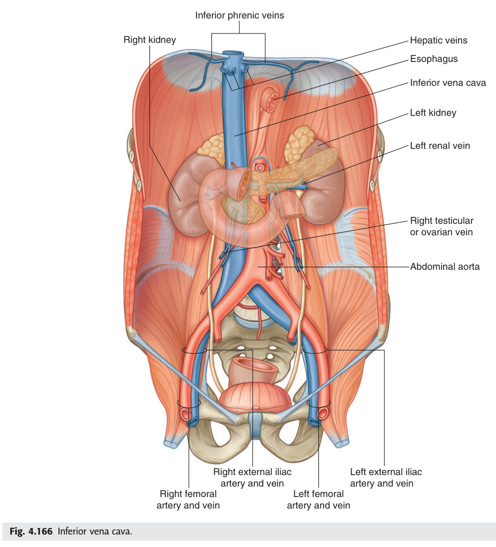
The IVC is a right-sided structure — it runs to the right of the aorta throughout the abdomen, which has surgical consequences: during right adrenalectomy the short right suprarenal vein empties directly into the IVC and tears easily; during left adrenalectomy the longer left suprarenal vein drains into the left renal vein and is less immediately dangerous. The gonadal vein asymmetry is one of the highest-yield venous anatomy facts: the left gonadal vein drains into the left renal vein at a right angle under higher hydrostatic pressure, explaining why varicoceles are almost always left-sided (90%). A left-sided varicocele that does not decompress when the patient lies down is a red flag for left renal vein obstruction — most commonly renal cell carcinoma extending tumour thrombus into the renal vein or IVC.
Formed at L5 (just below aortic bifurcation at L4) by union of right and left common iliac veins. Ascends right of aorta → passes through caval opening in diaphragm at T8 → right atrium. Right-sided structure throughout (aorta is left of midline, IVC is right).
| Tributaries (inferior → superior) | Level | Note |
|---|---|---|
| Common iliac veins | L5 | Formed from external + internal iliac veins |
| Lumbar veins (4 pairs) | L1–L4 | Drain posterior abdominal wall; connected by ascending lumbar veins (collateral if IVC obstructed) |
| Right gonadal vein | L2 | Drains directly into IVC (left gonadal → left renal vein) |
| Renal veins | L1–L2 | Left renal vein longer; receives left gonadal + left suprarenal |
| Right suprarenal vein | T12–L1 | Very short; drains directly into IVC (at risk in right adrenalectomy) |
| Hepatic veins (3) | T8 | Right, middle, left hepatic veins drain into IVC just below diaphragm |
IVC filter (e.g., Greenfield): percutaneous insertion via femoral or jugular vein; placed below renal veins. Indication: DVT/PE with contraindication to anticoagulation, or recurrent PE despite anticoagulation. Traps thrombi from lower limb/pelvic veins before they reach pulmonary circulation. Note: does NOT prevent new clot formation; anticoagulate once safe to do so. IVC thrombosis: renal cell carcinoma (RCC) commonly extends tumour thrombus up IVC → Budd-Chiari syndrome if it reaches hepatic veins.
Anatomical asymmetry (from the IVC table above):
• Right gonadal vein → drains directly into IVC at an oblique angle → low back-pressure
• Left gonadal vein → drains into the left renal vein at a right angle → higher hydrostatic pressure from the longer venous column + right-angle junction → venous incompetence and retrograde flow
Consequence: Varicocele (dilated pampiniform plexus in spermatic cord) is left-sided in 90% of cases for this anatomical reason. "Bag of worms" felt in the scrotum above and around the testis; disappears on lying down (decompression).
Grading: Grade I = palpable only on Valsalva; Grade II = palpable at rest; Grade III = visible through scrotal skin.
Consequences: ↑ scrotal temperature → impaired spermatogenesis → oligospermia + reduced sperm motility → subfertility (present in 15% of infertile men, 40% of men attending infertility clinics). The testis may atrophy over time.
Secondary varicocele — important red flag: A sudden-onset left-sided varicocele that does NOT decompress on lying down suggests left renal vein obstruction (most commonly left renal cell carcinoma with tumour thrombus in the left renal vein). A right-sided varicocele or bilateral varicocele suggests retroperitoneal mass or IVC obstruction — always investigate. Treatment: Grade II–III or infertility → percutaneous embolisation (gold standard) or surgical high ligation (Palomo procedure). [Gray's 4e Ch5 p338; surgical anatomy of spermatic cord]
- IVC formation level and which side of the aorta? Forms at L5 (union of common iliac veins, just below aortic bifurcation at L4). Right-sided structure throughout abdomen
- Right vs left gonadal vein drainage asymmetry? Right gonadal → IVC directly (oblique angle, low pressure). Left gonadal → left renal vein at right angle (higher hydrostatic pressure → prone to incompetence → left varicocele in 90%)
- Left renal vein tributaries (three)? Left gonadal vein + left suprarenal vein + left inferior phrenic vein. All drain into the left renal vein before it crosses the aorta to reach the IVC
- Red flag varicocele — what does it suggest? Left varicocele not decompressing on lying down = left renal vein obstruction (renal cell carcinoma tumour thrombus). Right-sided or bilateral varicocele = retroperitoneal mass / IVC obstruction
- Budd-Chiari syndrome: definition, caudate lobe finding, and commonest cause? Hepatic venous outflow obstruction (hepatic vein thrombosis). Caudate lobe hypertrophy (drains directly to IVC via short veins, spared from congestion). Commonest cause: polycythaemia vera (JAK2 V617F)
Portal Venous System
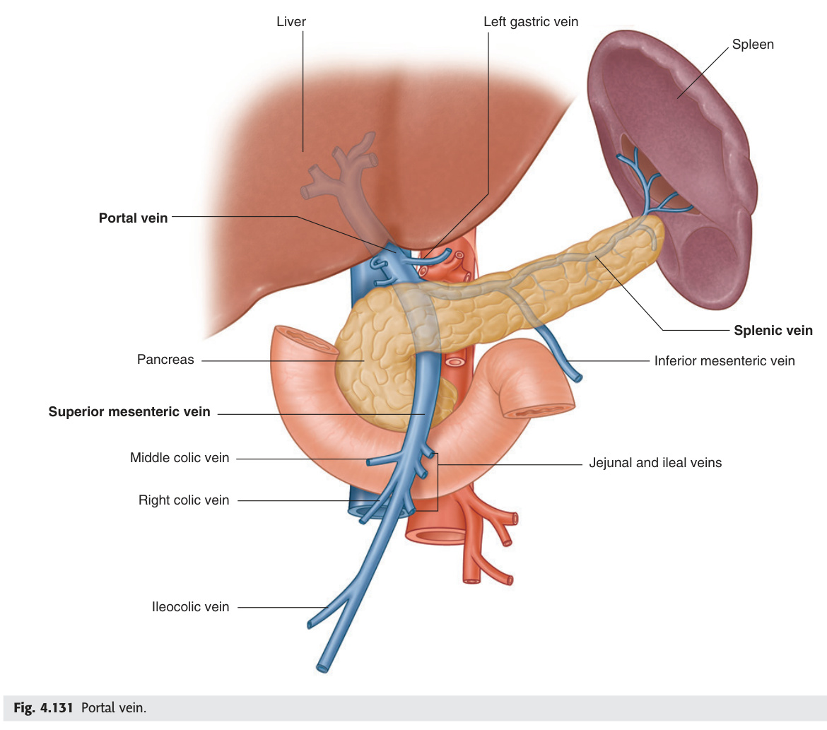
The portal venous system is the liver's first-pass filtration highway: all blood drained from the GI tract, spleen, and pancreas must pass through the liver sinusoids before reaching the systemic circulation. This is why oral drugs are absorbed in the gut but metabolised (and sometimes inactivated) before reaching the systemic circulation — the first-pass effect. The portal vein forms behind the neck of the pancreas at L2, the SMV and splenic vein meeting there after having collected blood from the entire bowel and spleen. When portal pressure rises above 12 mmHg — most commonly from cirrhosis — blood backs up and finds collateral routes to the systemic circulation wherever the two venous systems are in anatomical contact: oesophagus (varices that bleed fatally), rectum (rectal varices, not haemorrhoids), anterior abdominal wall (caput medusae), and retroperitoneum. Budd-Chiari syndrome, by contrast, blocks the hepatic venous outflow, and its imaging hallmark — caudate lobe hypertrophy — is the anatomical consequence of that lobe's unique direct drainage into the IVC.
Portal pressure >12 mmHg (normal 5–10 mmHg). Causes: pre-hepatic (portal vein thrombosis), intrahepatic (cirrhosis = most common — fibrosis increases resistance), post-hepatic (Budd-Chiari, right heart failure). Consequences of portosystemic shunting: oesophageal varices (haematemesis = #1 cause of variceal bleeding), rectal varices (not haemorrhoids), caput medusae (paraumbilical veins dilated around umbilicus), splenomegaly (hypersplenism → pancytopenia). Hepatic encephalopathy: ammonia bypasses liver via portosystemic shunts → CNS toxicity. Treat: non-selective β-blockers (propranolol) → reduce portal pressure; banding of varices; TIPSS (transjugular intrahepatic portosystemic shunt) in refractory cases.
Definition: Hepatic venous outflow obstruction — from hepatic vein thrombosis (most common) or IVC occlusion at the level of the hepatic veins.
Causes (thrombotic): Myeloproliferative disorders — especially polycythaemia vera (JAK2 V617F mutation), essential thrombocythaemia; PNH (paroxysmal nocturnal haemoglobinuria); thrombophilia (Factor V Leiden, antiphospholipid syndrome, antithrombin III deficiency); OCP/pregnancy; Behçet's disease; abdominal malignancy compressing hepatic veins.
Clinical triad: RUQ pain + hepatomegaly + ascites (onset over days–weeks in acute; insidious in chronic). Jaundice is late. Splenomegaly + portal hypertension as hepatic congestion progresses.
Anatomical key — caudate lobe sparing: The caudate lobe drains via multiple small, direct veins directly into the IVC — bypassing the main hepatic veins. In Budd-Chiari, these short veins remain patent → caudate lobe is not congested → it hypertrophies (enlarges) while the rest of the liver is congested and atrophied. Caudate lobe hypertrophy is the imaging hallmark of Budd-Chiari syndrome.
Investigation: Doppler USS liver (absent/reversed hepatic vein flow, congested liver parenchyma); CT/MRI (heterogeneous liver with peripheral atrophy + central caudate hypertrophy; comma-shaped caudate lobe); hepatic venography + IVC pressure measurement (gold standard for planning).
Treatment: Anticoagulation (LMWH → warfarin, or DOAC); treat underlying cause (hydroxyurea for PV, eculizumab for PNH); TIPSS (decompress portal system via hepatic parenchymal shunt — bypasses occluded hepatic veins); liver transplantation for fulminant or end-stage disease. [Gray's 4e Ch4 p290; clinical anatomy of hepatic veins]
- Portal vein formation: vessels, level, and landmark structure? SMV + splenic vein unite behind the neck of the pancreas at L2. IMV usually drains into splenic vein
- Portal vein course in the hepatoduodenal ligament — position relative to bile duct and hepatic artery? Portal vein = posterior; hepatic artery = medial/left; bile duct = lateral/right. "Portal vein Posterior, Artery Anterior-left, Bile duct rightmost" (mnemonic: PAB from back to front)
- Portal hypertension: threshold pressure and commonest cause? >12 mmHg (normal 5–10). Commonest cause: cirrhosis (fibrosis → ↑ resistance). Pre-hepatic: portal vein thrombosis. Post-hepatic: Budd-Chiari, right heart failure
- Four consequences of portosystemic shunting in portal hypertension? Oesophageal varices (haematemesis); rectal varices; caput medusae (paraumbilical veins); splenomegaly + hypersplenism (pancytopenia)
- Budd-Chiari imaging hallmark and its anatomical basis? Caudate lobe hypertrophy — caudate drains via small direct veins into IVC (bypassing hepatic veins), so it remains unperfused-congestion-free and hypertrophies while the rest of the liver is congested
Portosystemic Anastomoses
Portosystemic anastomoses are the anatomical sites where the portal venous system is in direct communication with systemic veins. Normally these connections carry little flow — but in portal hypertension they dilate into varices that can rupture and bleed catastrophically. The four sites are mnemonised as OARU: Oesophagus (left gastric vein meets azygos, via oesophageal veins), Anal canal (superior rectal meets middle/inferior rectal), Retroperitoneum (Veins of Retzius), and Umbilicus (paraumbilical veins radiating as caput medusae). The oesophageal site is the most dangerous: the submucosal oesophageal veins are thin-walled and unsupported, so rupture produces haematemesis that kills in minutes. Understanding which portal tributary connects to which systemic vein at each site explains exactly which vessel to look for on endoscopy, which direction to embolise, and why banding works where it does.
| Site | Portal vessel | Systemic vessel | Clinical consequence |
|---|---|---|---|
| Lower oesophagus | Left gastric (coronary) vein → portal | Oesophageal veins → azygos → SVC | Oesophageal varices — most dangerous (torrential haematemesis) |
| Anal canal (above pectinate line) | Superior rectal vein → IMV → portal | Middle + inferior rectal veins → internal iliac → IVC | Rectal varices (NOT haemorrhoids — haemorrhoids are arteriovenous plexuses) |
| Umbilicus | Paraumbilical veins → left portal vein (remnant of umbilical vein in ligamentum teres) | Epigastric veins → SVC/IVC | Caput medusae — dilated veins radiating from umbilicus |
| Retroperitoneum | Veins of retroperitoneal viscera (ascending/descending colon, duodenum, pancreas) | Posterior abdominal wall veins → lumbar veins → IVC | Veins of Retzius — develop with portal hypertension; can cause significant surgical haemorrhage |
Oesophagus + Anal canal + Retroperitoneum + Umbilicus. In portal hypertension, these become dilated collateral channels. Knowing which portal vessel connects to which systemic vessel explains which vein dilates clinically.
- Four sites of portosystemic anastomosis (mnemonic)? OARU: Oesophagus, Anal canal, Retroperitoneum (Veins of Retzius), Umbilicus (paraumbilical veins)
- Oesophageal varices: portal vessel + systemic vessel? Left gastric (coronary) vein (portal) ↔ oesophageal veins → azygos → SVC. Most dangerous site — thin-walled submucosal veins rupture → fatal haematemesis
- Anal canal anastomosis: portal vessel + systemic vessel + what is NOT a varix? Superior rectal vein (portal) ↔ middle/inferior rectal veins → internal iliac → IVC. Haemorrhoids are NOT rectal varices — they are dilated arteriovenous submucosal cushions
- Caput medusae: anatomical basis? Paraumbilical veins (in ligamentum teres, remnant of umbilical vein) dilate and carry portal blood to anterior abdominal wall → epigastric veins → SVC/IVC. Veins radiate from umbilicus like Medusa's head
- Veins of Retzius: where and clinical significance? Retroperitoneal portosystemic connections between retroperitoneal visceral veins and posterior abdominal wall veins. Cause significant intra-operative haemorrhage during retroperitoneal dissection in portal hypertension
Azygos Venous System
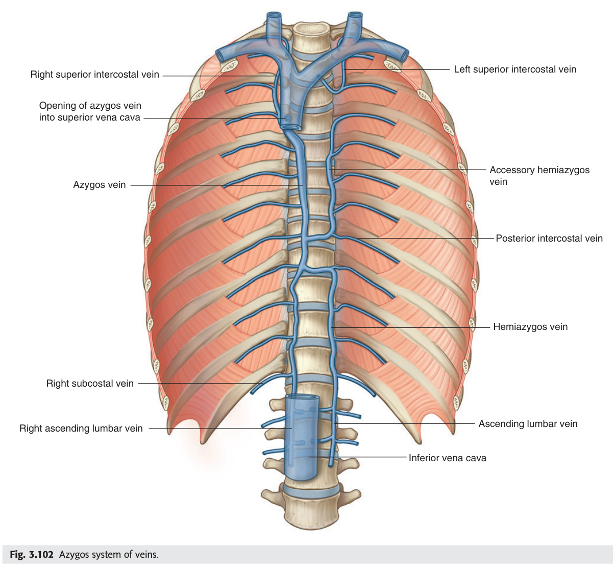
The azygos system is the body's great venous bypass highway — running alongside the vertebral column in the posterior mediastinum, it drains the thoracic wall (posterior intercostal veins), the oesophagus, and the bronchi, and crucially connects the IVC below to the SVC above. This connection is what makes IVC occlusion survivable: blood from the lower limbs and pelvis can still reach the heart by flowing up the ascending lumbar veins into the azygos/hemiazygos, then into the SVC. Batson's vertebral venous plexus is the clinically crucial extension of this system — a valveless network surrounding the entire spine that explains haematogenous vertebral metastases from pelvic tumours (prostate → lumbar spine), vertebral osteomyelitis from pelvic sepsis, and even "skip" brain metastases that bypass the lungs entirely during Valsalva-type pressure rises.
| Vein | Course | Drains |
|---|---|---|
| Azygos vein | Right of vertebral column; arises from right subcostal + ascending lumbar veins at T12. Arches over right lung root at T4 → posterior SVC | Right posterior intercostal veins (4–11) + right superior intercostal + hemiazygos + accessory hemiazygos + bronchial veins + oesophageal veins |
| Hemiazygos vein | Left side, T8–T12; crosses midline at T8–T9 → azygos | Left posterior intercostal veins (9–11) + lower oesophageal veins |
| Accessory hemiazygos | Left side, T4–T8; crosses midline at T7–T8 → azygos | Left posterior intercostal veins (4–8) |
If the infrarenal IVC is occluded (thrombosis, tumour, surgical ligation), blood can return to the heart via: ascending lumbar veins → azygos/hemiazygos → SVC. This explains why IVC thrombosis or ligation is survivable. On CXR: enlarged azygos vein (>7 mm shadow at right tracheobronchial angle) = right heart failure, SVC obstruction, IVC obstruction, or azygos continuation of IVC (congenital absence of infrarenal IVC). Azygos lobe: accessory fissure formed when azygos vein migrates through right upper lobe during development → visible as curved line + small "teardrop" shadow at right lung apex on CXR.
A network of valveless veins surrounding the vertebral column, extending from the pelvis to the cranium. Described by Oscar Batson (1940). Consists of an internal vertebral plexus (within the spinal canal, around the dura mater) and an external vertebral plexus (around the external surface of vertebral bodies and neural arches).
Connections: Basivertebral veins (drain vertebral cancellous bone) → internal plexus ↔ intervertebral veins ↔ azygos/hemiazygos · lumbar veins · intercostal veins · pelvic venous plexuses (prostatic, vesical, uterine) · IVC.
Why valveless matters: During raised intraabdominal/intrathoracic pressure (coughing, Valsalva, straining), blood is forced retrogradely from pelvic and abdominal veins into the vertebral plexus and superiorly toward the skull — bypassing the IVC entirely.
Clinical importance:
• Haematogenous vertebral metastases: Prostate cancer → lumbar spine (blastic mets); breast cancer → thoracic spine; lung cancer → any vertebral level; thyroid and RCC also common. Tumour cells enter pelvic/intercostal veins → retrograde into Batson's plexus → seed vertebral body. Mnemonic PBLTK: Prostate · Breast · Lung · Thyroid · Kidney.
• Vertebral osteomyelitis: Pelvic sepsis (UTI, prostatic abscess, pelvic inflammatory disease) can spread retrogradely via Batson's plexus → vertebral body infection → discitis → epidural abscess.
• “Skip” brain metastases from pelvic tumours without lung involvement are explained by retrograde flow bypassing pulmonary capillaries.
- Azygos vein: origin, where it arches, and where it drains? Arises at T12 from right subcostal + ascending lumbar veins → ascends right side → arches over right lung root at T4 → posterior SVC. Hemiazygos (left T8–T12) and accessory hemiazygos (left T4–T8) cross midline to drain into it
- Why is IVC occlusion survivable? Ascending lumbar veins → azygos/hemiazygos → SVC provides a collateral bypass allowing lower limb/pelvic venous blood to reach the right atrium
- Batson's plexus: what is it and clinical significance? Valveless internal + external vertebral venous plexus. During Valsalva/raised intraabdominal pressure, blood refluxes retrogradely from pelvic veins through vertebral plexus toward skull, seeding metastases and infection
- Tumours metastasising via Batson's plexus (mnemonic)? PBLTK: Prostate (→ lumbar blastic mets), Breast (→ thoracic), Lung (→ any level), Thyroid, Kidney (RCC)
- CXR enlarged azygos vein: threshold and causes? >7 mm at right tracheobronchial angle = right heart failure, SVC obstruction, IVC obstruction, congenital azygos continuation of IVC
Jugular Veins
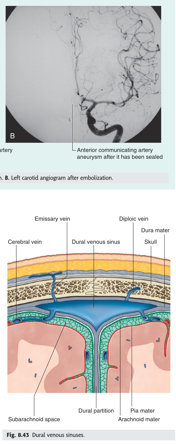
The internal jugular vein is the most direct window into right heart haemodynamics — because it communicates almost directly with the right atrium via the SVC, its filling height and waveform reflect right atrial pressure in real time. The JVP assessment uses the right IJV because the right brachiocephalic vein is short and nearly vertical; the left IJV has a longer, more angled path and is less reliable. The "danger triangle" of the face is not a surface anatomy curiosity — it is a life-threatening anatomical principle: the facial vein is valveless, so squeezing a boil between the upper lip and nose can push septic emboli retrogradely through the angular vein → superior ophthalmic vein → cavernous sinus. Cavernous sinus thrombosis presents as a septic patient with proptosis, chemosis, and painful ophthalmoplegia — CN VI palsy appears first because the abducens nerve has the least protection as it runs free through the sinus.
| Vein | Course / Formation | Clinical |
|---|---|---|
| Internal jugular vein (IJV) | Continuation of sigmoid sinus at jugular foramen. Descends in carotid sheath (lateral to ICA then CCA; anterior to vagus nerve). Joins subclavian → brachiocephalic. Dilatable in right heart failure | JVP assessment (right IJV used — more direct communication with RA). Normal JVP ≤4 cm above sternal angle. Central line access (right IJV preferred — straight course to SVC + RA). Complications: pneumothorax, carotid puncture, haematoma, air embolism |
| External jugular vein (EJV) | Forms at angle of mandible (posterior division of retromandibular v. + posterior auricular v.). Runs superficially over SCM → subclavian vein. More visible than IJV | JVP can be assessed in EJV if IJV not visible. EJV engorged in heart failure, SVC obstruction, tension pneumothorax. Cuts easily with superficial lacerations over SCM |
| Anterior jugular veins | Small veins lateral to midline; connect via jugular venous arch above suprasternal notch | Jugular venous arch: risk in low neck incisions + tracheostomy (venous bleeding if divided) |
The facial vein begins at the angular vein (medial canthus of the eye), runs obliquely downward and posteriorly along the side of the nose and mouth, crosses the lower border of the mandible, and drains into the internal jugular vein (directly, or via the retromandibular vein). KEY: The facial vein has NO venous valves — blood can flow in either direction. This creates a valveless connection between the face and the cavernous sinus.
The “danger area” (danger triangle) of the face is the region between the angles of the mouth and the bridge of the nose (upper lip, nose, nasolabial folds). Infection here (furuncle, infected acne, boil) can spread retrogradely to the cavernous sinus via two valveless routes:
Route 1: Facial v. → angular vein → superior ophthalmic vein → cavernous sinus (retrograde, no valves)
Route 2: Facial v. → deep facial vein → pterygoid venous plexus → emissary veins → cavernous sinus
Cavernous sinus thrombosis (CST): Septic thrombosis of the cavernous sinus arising from facial infection. Contents of the cavernous sinus: CN III (oculomotor), CN IV (trochlear), CN V1 (ophthalmic), CN V2 (maxillary), CN VI (abducens) in the lateral wall + ICA + sympathetic plexus traversing the sinus. Clinical features: fever + severe orbital headache + proptosis (orbital venous congestion) + chemosis (conjunctival oedema) + painful ophthalmoplegia (all extraocular movements painful/restricted; CN VI palsy appears first — abducens has least protected course through sinus). Bilateral involvement via intercavernous sinuses is characteristic. Most common organism: Staphylococcus aureus. Treatment: IV antibiotics (vancomycin + broad-spectrum cover) ± anticoagulation. Mortality ~30% untreated; visual loss in survivors common.
c wave = tricuspid valve closure (usually not visible clinically).
x descent = atrial relaxation + tricuspid valve draws downward during RV contraction; preserved in cardiac tamponade (x descent present, y absent in tamponade).
v wave = venous filling with tricuspid valve closed; Giant 'v' wave in tricuspid regurgitation (systolic venous regurgitation = pulsatile liver).
y descent = tricuspid valve opens, right atrium empties into RV; Absent/blunted in cardiac tamponade; Steep y descent in constrictive pericarditis (Friedreich's sign).
JVP elevated (>4 cm above sternal angle): right heart failure, cardiac tamponade, constrictive pericarditis, SVCO, fluid overload, tension pneumothorax.
Kussmaul's sign: JVP rises on inspiration (paradoxical — normally falls) = constrictive pericarditis / RV failure / cardiac tamponade.
- IJV course: where does it begin, what sheath, and where does it end? Begins at jugular foramen (continuation of sigmoid sinus) → descends in carotid sheath (lateral to ICA/CCA, anterior to vagus) → joins subclavian → brachiocephalic vein
- Why use the RIGHT IJV for JVP assessment? Right IJV → right brachiocephalic vein (short, vertical) → SVC → right atrium — most direct, unobstructed route. Left IJV has a longer angled path and is less reliable
- Facial vein "danger triangle": route of spread to cavernous sinus? Facial v. → angular v. → superior ophthalmic v. → cavernous sinus (valveless, retrograde). Also: facial v. → deep facial v. → pterygoid plexus → cavernous sinus
- Cavernous sinus thrombosis: which CN palsy appears first and why? CN VI (abducens) — runs free through the sinus, least protected. Presents: fever + proptosis + chemosis + painful ophthalmoplegia. Organism: Staph aureus
- JVP waveform: giant 'a' wave vs giant 'v' wave? Giant 'a' = atrial contracts against resistance (tricuspid stenosis, pulmonary hypertension). Giant 'v' = tricuspid regurgitation (systolic venous regurgitation → pulsatile liver). Absent 'a' wave = AF
Upper Limb Superficial Veins ★★★
The two great superficial veins of the upper limb — cephalic and basilic — are not symmetrical, and the asymmetry is exactly what examiners test. Both arise from the dorsal venous rete of the hand but at opposite sides: cephalic from the radial (lateral/thumb) side, basilic from the ulnar (medial/little-finger) side. They meet in the cubital fossa via the median cubital vein, which is where clinical staff cannulate most reliably because it is large, superficial, and usually visible. In the upper arm the cephalic vein runs in the lateral bicipital groove all the way to the deltopectoral groove, then pierces the clavipectoral fascia to drain into the axillary vein — it does not drain into the brachial vein. The basilic vein, by contrast, pierces the deep fascia at mid-arm to join the brachial veins and becomes the axillary vein at the lower border of teres major. Past paper MCQ Q11 (2020) specifically tests cephalic vein anatomy.
| Vein | Origin | Course | Termination & Notes |
|---|---|---|---|
| Cephalic vein | Lateral (radial) side of dorsal venous rete of hand | Winds around lateral aspect of forearm → lateral side of biceps brachii (bicipital groove) → deltopectoral groove (between deltoid + pectoralis major) → pierces clavipectoral fascia | Drains into axillary vein (or subclavian at thoracic inlet). Does NOT accompany radial artery. Does NOT drain into brachial vein. Arises from LATERAL (radial) side, not medial. Used for PICC lines + IV access. PAST PAPER MCQ Q11 |
| Basilic vein | Medial (ulnar) side of dorsal venous rete of hand | Medial forearm → medial side of arm → pierces deep fascia at mid-arm → joins brachial veins (venae comitantes) | Forms axillary vein at lower border of teres major. Preferred for PICC line insertion (straighter path to subclavian + SVC). Piercing of deep fascia at mid-arm = point of tethering (entrapment possible) |
| Median cubital vein | Connects cephalic to basilic at cubital fossa | Passes obliquely across the antecubital fossa (cephalic laterally, basilic medially). Communicates with deep veins of forearm via a perforator passing deep to the bicipital aponeurosis | Most common site for venepuncture and blood donation. Brachial artery lies deep and medial (beneath bicipital aponeurosis) — do not penetrate medially. Median nerve lies medial to the brachial artery |
| Dorsal venous rete (arch) of hand | Dorsal digital veins from all fingers converge on dorsum | Lies in subcutaneous tissue on dorsum of hand | Cephalic arises from RADIAL (lateral) side; basilic from ULNAR (medial) side. IV cannulae often placed here |
(A) arises from the medial side of dorsal venous rete of hand
(B) accompanies the radial artery
(C) drains into the brachial vein
(D) receives the superficial veins of the hand and the medial side of the forearm
(E) runs along the lateral side of the biceps brachii
A = wrong: cephalic arises from the lateral (radial) side of the dorsal venous rete, not medial (basilic arises from medial/ulnar side).
B = wrong: the cephalic vein is a superficial vein; it does NOT accompany the radial artery (which is deep).
C = wrong: the cephalic vein drains into the axillary vein (via deltopectoral groove); the basilic vein joins brachial veins to form the axillary vein.
D = wrong: that describes tributaries of the basilic/median veins (medial forearm drainage).
E = correct: the cephalic vein runs along the lateral groove of the biceps brachii (lateral bicipital groove) before ascending to the deltopectoral groove and axillary vein.
Cephalic = Crown of the arm = lateral (radial/thumb side). Basilic = Basement of the arm = medial (ulnar/little-finger side). Median cubital connects them in the cubital fossa like a bridge: C — Bridge — B. The cephalic vein runs in the deltopectoral groove (“the groove between the shoulder and chest”) — a landmark for surgical access and pacemaker lead insertion.
- Cephalic vein: origin side, upper arm course, and termination? Lateral (radial/thumb) side of dorsal venous rete → lateral bicipital groove → deltopectoral groove → pierces clavipectoral fascia → axillary vein (NOT brachial vein)
- Basilic vein: origin side, where it pierces deep fascia, and what it becomes? Medial (ulnar/little-finger) side of dorsal venous rete → medial forearm → mid-arm pierces deep fascia → joins brachial veins → axillary vein at lower border of teres major
- Median cubital vein: connects what, where, and clinical use? Connects cephalic (lateral) to basilic (medial) in the antecubital fossa. Most common site for venepuncture and blood donation. Brachial artery lies deep and medial — do NOT penetrate medially
- Past paper MCQ Q11 (2020): which statement about cephalic vein is correct? E — runs along the lateral side of biceps brachii. (NOT medial origin; NOT accompanies radial artery; NOT drains into brachial vein; NOT medial forearm drainage)
- Preferred vein for PICC line and why? Basilic vein — straighter, more direct path to subclavian vein and SVC. Cephalic vein has an acute angle at clavipectoral fascia that can obstruct catheter advancement
Thoracic Duct & Right Lymphatic Duct
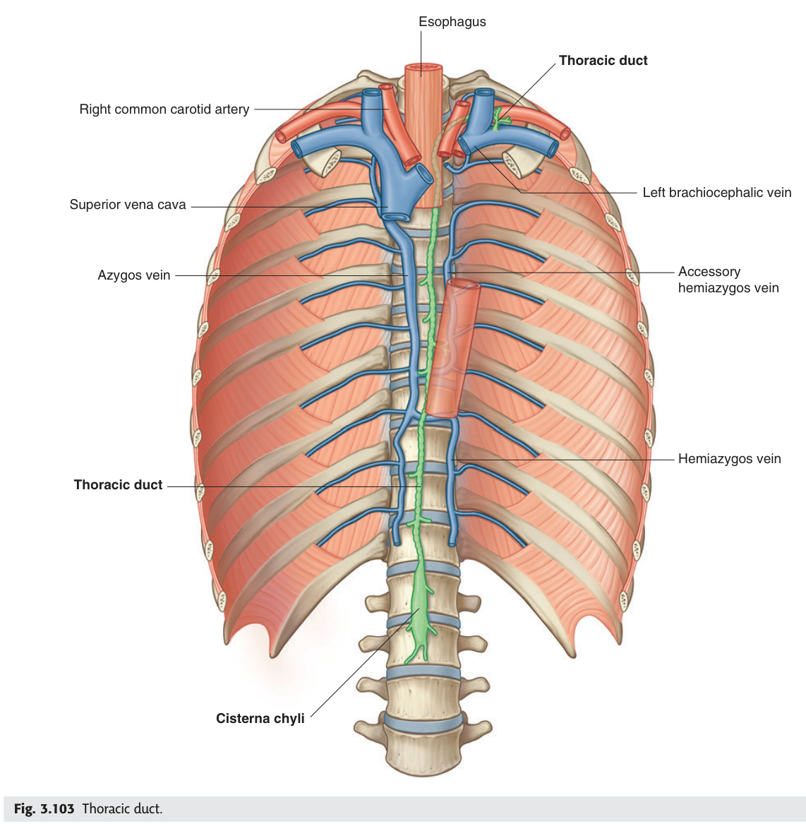
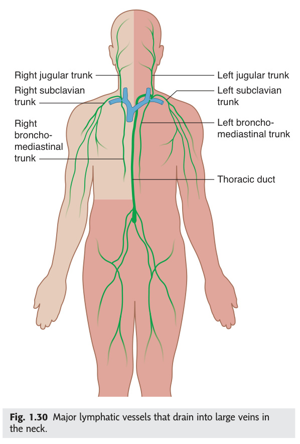
The thoracic duct is the largest lymphatic vessel in the body, draining everything except the right upper quadrant — that means roughly 75% of the body's lymph, including all intestinal chyle and lymph from both lower limbs, the pelvis, the left chest, and the left side of the head and neck. It originates from the cisterna chyli at L1/L2, enters the thorax through the aortic hiatus, climbs on the right side of the spine, then crosses left at T4/T5 and terminates at the left subclavian–internal jugular junction. This crossing is why chyle leaks from left neck dissections but rarely right ones. The right lymphatic duct drains the right upper quadrant into the right venous angle — and in 25% of people it doesn't exist as a single duct at all, just three separate trunks draining individually. Virchow's node (left supraclavicular) is the clinical sentinel: it receives the thoracic duct's drainage, so abdominal malignancies — especially gastric carcinoma — metastasise there first.
Chylothorax: thoracic duct injury (trauma, surgery, malignant obstruction) → milky pleural effusion (triglycerides >110 mg/dL). Treat: low-fat diet (medium-chain triglycerides bypass lymphatics) + pleural drainage; surgical ligation at T5 or thoracic duct embolisation if conservative fails.
Formation: The right lymphatic duct (or more commonly, three separate trunks) is formed by the convergence of:
• Right jugular trunk — drains right side of head and neck
• Right subclavian trunk — drains right arm and right chest wall
• Right bronchomediastinal trunk — drains right lung, right pleura, right heart, right mediastinum
Drainage site: Right venous angle (junction of right internal jugular + right subclavian veins). In ~25% of individuals, a true single right lymphatic duct is absent — each trunk drains separately.
Territory: Right upper quadrant of the body = right side of head/neck, right arm, right thorax including right lobe of lung, right heart, and part of the right lobe of the liver (though bare area of liver drains to thoracic duct).
Key comparison — Thoracic duct vs Right lymphatic duct:
| Thoracic Duct | Right Lymphatic Duct | |
|---|---|---|
| Drains | All of body EXCEPT right upper quadrant | Right side of head/neck + right arm + right thorax |
| Drains into | LEFT venous angle (left IJV + left subclavian junction) | RIGHT venous angle |
| Origin | Cisterna chyli (L1–L2) | Three lymphatic trunks (often no single duct) |
| Clinical injury | Left neck dissection → chylothorax / chyle fistula | Right neck dissection → right-sided chyle leak (less common) |
- Thoracic duct: origin, midline crossing, and termination? Cisterna chyli (L1–L2) → aortic hiatus (T12) → right of aorta → crosses left at T4/5 → arches over left subclavian → drains into LEFT venous angle (left IJV + left subclavian junction)
- What does the thoracic duct drain? Entire body EXCEPT right upper quadrant (right head/neck, right arm, right thorax). Approximately 75% of body lymph including all intestinal chyle
- Right lymphatic duct: territory and termination? Anatomical variant? Right head/neck + right arm + right thorax → RIGHT venous angle. In ~25% no single duct exists — three trunks drain separately
- Chylothorax: cause, fluid character, and treatment? Thoracic duct injury (surgery, trauma, malignant obstruction) → milky pleural effusion (triglycerides >110 mg/dL). Treat: low-fat diet (MCT diet bypasses lymphatics) + drainage; surgical ligation at T5 if fails
- Virchow's node: location, what drains into it, and clinical significance? Left supraclavicular node. Receives thoracic duct drainage → sentinel node for abdominal/pelvic malignancy. Troisier's sign = palpable Virchow's node → gastric carcinoma until proven otherwise
Major Lymph Node Groups
Lymph nodes are the surveillance posts of the immune system — bean-shaped filters stationed along lymphatic channels that intercept antigens, bacteria, tumour cells, and pathogens before they reach the bloodstream. Their anatomical position defines which region each node drains, making them invaluable clinical indicators: a tender, enlarged node signals local infection, while a hard, fixed, non-tender node suggests metastatic malignancy. Knowing which station drains which territory lets you locate the primary tumour from the involved node alone — testicular cancer spreads to para-aortic nodes, not inguinal ones, because the testis descends from L1 carrying its lymphatics with it. The clinical examination of lymph nodes (size, consistency, tenderness, mobility, overlying skin) distinguishes reactive from malignant lymphadenopathy before biopsy is needed.
| Group | Location | Drains / Clinical |
|---|---|---|
| Cervical nodes | Deep cervical chain (along IJV); superficial; submandibular; submental | Head and neck cancer drainage. Virchow's node = left supraclavicular node (sentinel node for abdominal malignancy via thoracic duct). Troisier's sign = palpable Virchow's node → gastric carcinoma |
| Axillary nodes | 5 groups: anterior (pectoral) + posterior (subscapular) + lateral + central + apical. Level I (below pec minor) → Level II (behind) → Level III (medial to pec minor → subclavian) | Breast cancer staging: sentinel node biopsy (first draining node, usually Level I). >4 positive nodes → poor prognosis. Axillary clearance complications: lymphoedema, intercostobrachial nerve injury (medial arm numbness) |
| Para-aortic nodes | Around abdominal aorta at L1–L2 | Drain testes/ovaries + kidneys + suprarenal + upper abdominal viscera. Testicular cancer: paraaortic nodes on CT (not inguinal). Bulky para-aortic LAP on CT → lymphoma, testicular, cervical, ovarian primary |
| Mesenteric nodes | Within mesentery | Drain GI tract. Mesenteric adenitis (children, Yersinia/viral) → mimics appendicitis |
| Inguinal nodes | Superficial (below inguinal ligament) + deep (medial to femoral vein) | Drain lower limb + perineum + lower anal canal + scrotum/labia majora. Anal canal cancer below pectinate line → inguinal nodes (NOT mesenteric). Penile cancer → inguinal nodes |
| Mediastinal nodes | Paratracheal + subcarinal + hilar + anterior | Lung cancer staging: N1 = ipsilateral hilar; N2 = ipsilateral mediastinal/subcarinal; N3 = contralateral/supraclavicular. Sarcoidosis: bilateral hilar lymphadenopathy (BHL) |
Test Unit 12 knowledge
Portal system, thoracic duct, and lymph node drainage MCQs.
- Testicular cancer: why para-aortic nodes, not inguinal? Testes descend from retroperitoneum at L1–L2 carrying their lymphatic drainage with them → para-aortic nodes. Inguinal nodes drain scrotum/perineum. Exception: scrotal involvement or prior scrotal surgery may involve inguinal nodes
- Anal canal above vs below pectinate line — lymphatic drainage? Above pectinate line → internal iliac nodes (same as rectum). Below pectinate line → superficial inguinal nodes. Carcinoma BELOW pectinate line presents as inguinal lymphadenopathy
- Axillary node levels I, II, III: anatomical landmark and breast cancer staging? Level I = lateral to pec minor; Level II = behind pec minor; Level III = medial to pec minor (apical). Sentinel node = first draining node, usually Level I. ≥4 positive axillary nodes = high-risk classification
- Mediastinal nodes: N1 vs N2 vs N3 in lung cancer staging? N1 = ipsilateral hilar/peribronchial nodes; N2 = ipsilateral mediastinal or subcarinal nodes; N3 = contralateral mediastinal/hilar or any supraclavicular nodes. N3 = inoperable (Stage IIIB)
- Sarcoidosis lymphadenopathy: characteristic CXR finding and differential? Bilateral hilar lymphadenopathy (BHL) = characteristic of sarcoidosis. Differential: lymphoma, primary TB, metastatic disease. BHL + erythema nodosum = Löfgren's syndrome (good prognosis)
Spleen — Anatomy & Clinical ★★
The spleen is simultaneously an immune organ and a blood filter — it destroys aged red cells, phagocytoses opsonised bacteria, and produces IgM antibodies, making it the first line of defence against encapsulated organisms. Anatomically it hides under the 9th–11th ribs in the left hypochondrium, which is why you only feel it when it is at least twice its normal size. Its unique clinical signature is the splenic notch — a notch on the superior border that you can palpate as it pushes below the costal margin; no other abdominal mass has this feature. The splenorenal ligament tethers the splenic vessels and tail of the pancreas directly to the hilum, which is why distal pancreatitis or surgery can injure the splenic vasculature simultaneously. After splenectomy, the patient loses polysaccharide capsule immunity permanently, making vaccination and prophylactic antibiotics non-negotiable for life.
The spleen is the largest lymphoid organ, lying in the left hypochondriac region between the stomach fundus and the diaphragm. It lies deep to the 9th–11th ribs, with its long axis corresponding to the 10th rib. Entirely covered by peritoneum (intraperitoneal). Relations: lateral/superior = diaphragm; medial = stomach (gastrosplenic ligament with short gastric arteries) + left kidney + tail of pancreas (at splenic hilum); inferior = splenic flexure of colon. Weight: 150 g (adult); size ~12 × 7 × 3 cm.
| Feature | Description |
|---|---|
| Surfaces | Diaphragmatic: smooth, convex (posterolateral, against diaphragm). Visceral: concave; bears the hilum (entry of splenic artery + exit of splenic vein, lymphatics, and nerves) |
| Borders | Superior border: has 2–3 splenic notches — palpation landmark when enlarged; pathognomonic of splenomegaly on clinical exam (vs enlarged left kidney, which has NO notch). Inferior border: rounder. Normal spleen is NOT palpable below the costal margin |
| Blood supply | Splenic artery (largest branch of celiac trunk; tortuous along upper border of pancreas → hilum). Splenic vein (travels with tail/body of pancreas → joins SMV behind neck of pancreas to form portal vein) |
| Ligaments | Gastrosplenic ligament (short gastric + left gastroepiploic arteries); Splenorenal (lienorenal) ligament (splenic vessels + tail of pancreas) |
| Functions | (1) Lymphocyte + monocyte production; (2) Phagocytosis of bacteria (especially encapsulated organisms), inert particles, old/abnormal WBCs + platelets; (3) Destruction of effete (aged) RBCs; (4) Antibody production (IgM); (5) Fetal haemopoiesis (3rd–5th month gestation); extramedullary haemopoiesis in adults with bone marrow failure |
Causes of splenomegaly (TIPICAL mnemonic — Top 6):
• Infections: EBV (infectious mononucleosis — tender spleen; monospot/heterophile antibody test), malaria (massive in chronic), typhoid, brucellosis, kala-azar/visceral leishmaniasis (can be massive)
• Haematological: CML (massive splenomegaly — characteristic; spleen may reach iliac fossa), lymphoma, leukaemia, hereditary spherocytosis, myelofibrosis
• Portal hypertension: cirrhosis → congestive splenomegaly + hypersplenism (pancytopenia from sequestration)
• Autoimmune: SLE, Felty's syndrome (RA + neutropenia + splenomegaly)
• Storage diseases: Gaucher's (glucocerebrosidase deficiency — most common lipid storage disease)
Spleen vs kidney on clinical exam (4 differences):
(1) Splenic notch palpable superomedially (kidney has no notch).
(2) Spleen moves inferomedially on inspiration (follows diaphragm); kidney moves inferiorly (vertical drop).
(3) Spleen is dull to percussion (no bowel in front); kidney is resonant (colon in front).
(4) Kidney can be ballotted bimanually (anterior + posterior hands); spleen cannot.
(5) You cannot “get above” a spleen (extends to costal margin); you may get above a kidney.
Splenic rupture: most common from blunt abdominal trauma. Left upper quadrant pain + Kehr's sign (referred left shoulder-tip pain: blood under left diaphragm irritates left phrenic nerve → referred pain C3–C5 dermatome = left shoulder tip). Haemodynamic instability. CT abdomen with contrast = investigation of choice. Treatment: emergency splenectomy or (if stable) splenic artery embolisation/conservative.
Post-splenectomy precautions: loss of splenic phagocytosis of opsonised encapsulated bacteria → lifelong risk of OPSI (Overwhelming Post-Splenectomy Infection — rapidly fatal sepsis within hours; mortality ~50%). Organisms: Streptococcus pneumoniae (most common), Haemophilus influenzae type b, Neisseria meningitidis. Management: (1) Vaccinations: Pneumococcus + Meningococcus + Hib (given 2 weeks before elective or ASAP after emergency splenectomy). (2) Lifelong penicillin V (amoxicillin) prophylaxis especially first 2 years. (3) Medical alert bracelet. (4) Annual influenza vaccine. (5) Malaria prevention when travelling.
- Spleen surface anatomy: rib level, long axis rib, and normal palpability? Deep to 9th–11th ribs; long axis = 10th rib. Normal spleen NOT palpable below the costal margin — only palpable when ≥twice normal size (>300–400 g)
- 5 clinical differences between splenomegaly and an enlarged left kidney? (1) Splenic notch palpable superomedially; kidney has none. (2) Spleen moves inferomedially on inspiration; kidney drops vertically. (3) Spleen dull to percussion (no bowel in front); kidney resonant (colon overlies it). (4) Kidney ballottable bimanually; spleen is not. (5) Cannot get above spleen; may get above kidney
- Kehr's sign: mechanism, and what causes it? Left shoulder-tip pain from blood/fluid under left diaphragm irritating the left phrenic nerve (C3–5) → referred pain to C3–5 dermatome (left shoulder tip). Classic in splenic rupture. Also: sub-phrenic abscess, ruptured ectopic pregnancy
- OPSI organisms (3) and prophylaxis strategy? Streptococcus pneumoniae (most common) + Haemophilus influenzae type b + Neisseria meningitidis. Prophylaxis: vaccinate all three (2 weeks pre-op elective / ASAP post-emergency) + lifelong penicillin V + annual influenza + medical alert bracelet
- CML splenomegaly: why is it so massive, and what sign does it produce? CML causes massive extramedullary haemopoiesis in the spleen → spleen can reach the iliac fossa. The notch is palpable even at this size. Massive splenomegaly differentials: CML, myelofibrosis, kala-azar (visceral leishmaniasis), chronic malaria
Leg Veins & Varicose Veins ★★
The leg has two venous systems — a deep system buried within the muscle compartments that carries 90% of returning blood, and a superficial system sitting in the subcutaneous fat that is visible, strippable, and clinically notorious for dilating into varicose veins. The two systems communicate through perforating veins whose valves normally allow flow only from superficial to deep; when those valves fail, high-pressure deep venous blood refluxes back into the superficial system, distending and torturing it into the ropes of varicose veins. The great saphenous vein is the longest vein in the body and the surgeon's friend — used as a bypass conduit in CABG and peripheral arterial reconstruction — so understanding its course (anterior to medial malleolus, up the medial thigh, into the femoral vein at the SFJ) matters beyond venous surgery. Varicose veins are often dismissed cosmetically, but the CEAP classification recognises a spectrum from thread veins to venous ulcers, all driven by the same underlying venous hypertension.
| Vein | Course | Clinical Importance |
|---|---|---|
| Great (long) saphenous vein (GSV) | Begins at medial dorsal venous arch → anterior to medial malleolus → medial leg and thigh → saphenofemoral junction (SFJ) → femoral vein (~4 cm below inguinal ligament) | Longest vein in body; used for CABG grafts (reversed); accessed for IV in emergencies; saphenofemoral incompetence = most common cause of varicose veins (90%). Important tributary at SFJ: superficial external pudendal, circumflex iliac, inferior epigastric veins |
| Small (short) saphenous vein (SSV) | Begins posterior to lateral malleolus → runs up posterior calf → saphenopopliteal junction (SPJ) at posterior knee (variable level — duplex USS to map) | Saphenopopliteal incompetence = posterior calf varicose veins; variable SPJ level makes surgery precise duplex marking essential |
| Perforating veins | Connect superficial to deep veins; valves direct flow inward (superficial → deep); Cockett's perforators (medial lower leg — Cockett I, II, III most important); Dodd's (mid-thigh); Boyd's (upper calf) | Incompetent Cockett's perforators → high-pressure deep venous blood refluxes into superficial system → medial lower leg skin damage + venous ulcers. SEPS (subfascial endoscopic perforator surgery) for refractory venous ulcers |
| Deep veins | Femoral (with femoral artery in adductor canal) + popliteal + posterior tibial + peroneal + anterior tibial + plantar veins; have valves directing flow superiorly | DVT most dangerous in popliteal + above (proximal DVT → PE risk). Calf DVT: lower PE risk but can propagate proximally. Deep venous insufficiency after DVT = post-thrombotic syndrome |
Dilated, tortuous, lengthened superficial veins due to valvular incompetence. 90% = saphenofemoral junction incompetence. Risk factors: female, pregnancy, prolonged standing, obesity, FH, previous DVT.
| CEAP Class | Clinical Features |
|---|---|
| C0 | No visible signs of venous disease |
| C1 | Telangiectasiae (thread veins) or reticular veins |
| C2 | Varicose veins |
| C3 | Ankle oedema |
| C4a | Venous eczema (skin changes medial lower leg) or pigmentation (haemosiderin) |
| C4b | Lipodermatosclerosis (fibrosis + atrophie blanche — "inverted champagne bottle" appearance) |
| C5 | Healed venous ulcer |
| C6 | Active venous ulcer (above medial malleolus, gaiter area; sloping edges; fibrinous base; minimal pain; treat: 4-layer compression bandaging) |
- GSV: origin, course, and termination? Why clinically important beyond varicose veins? Medial dorsal venous arch → anterior to medial malleolus → medial leg and thigh → SFJ (4 cm below inguinal ligament) into femoral vein. Used as CABG conduit (reversed, valves face away from flow). IV access in emergencies at the ankle
- Cockett's perforators: location and what happens when they fail? Medial lower leg (Cockett I, II, III). Normally direct flow from superficial → deep. When incompetent: deep venous high-pressure blood refluxes into superficial system → medial lower leg venous hypertension → haemosiderin pigmentation, lipodermatosclerosis, venous ulcers (CEAP C4–C6)
- CEAP C6 venous ulcer: characteristics, location, and treatment? Active venous ulcer. Medial lower leg (gaiter area, above medial malleolus). Sloping edges, fibrinous base, surrounding haemosiderin/eczema, minimal pain (unlike arterial ulcers). Treatment: 4-layer compression bandaging + wound care + underlying venous insufficiency treatment
- Trendelenburg test: procedure and what each result means? Leg elevated to empty veins → tourniquet at SFJ → patient stands. Varices stay empty = SFJ incompetence only (tourniquet controlled it). Varices refill despite tourniquet = perforator incompetence below tourniquet
- SSV course and SPJ level significance for surgery? Posterior to lateral malleolus → posterior calf → saphenopopliteal junction (SPJ) at posterior knee. SPJ level is variable (may be at knee crease or higher) → duplex USS MANDATORY before surgery to mark exact SPJ level and prevent sural nerve injury
Deep Vein Thrombosis & Pulmonary Embolism ★★★
DVT and pulmonary embolism are the same disease at different stages of its natural history — a clot that forms in the deep venous system of the leg can detach, travel through the right heart, and lodge in the pulmonary arterial tree, converting a local problem into a life-threatening emergency. Virchow's triad (stasis, hypercoagulability, endothelial damage) remains the conceptual framework for every risk factor you will memorise, from long-haul flights to Factor V Leiden. The Wells score turns clinical suspicion into a probability, and D-dimer — highly sensitive but notoriously non-specific — is powerful only as a rule-out tool in low-probability patients. The great challenge of PE is that its classic ECG finding (S1Q3T3) appears in only 20% of cases, so a high index of clinical suspicion followed by CTPA is the diagnostic cornerstone. Treatment has shifted dramatically toward DOACs, which have replaced warfarin in most situations without the need for INR monitoring.
(1) Stasis: immobility (long-haul flight, post-op, paralysis), heart failure, pregnancy, varicose veins.
(2) Hypercoagulability: malignancy, thrombophilia (Factor V Leiden most common, antithrombin III deficiency, protein C/S deficiency, antiphospholipid syndrome), OCP (especially oestrogen-containing), pregnancy, dehydration.
(3) Endothelial damage: surgery, trauma, central venous catheters, infection.
Wells DVT score (pre-test probability): active cancer (+1), paralysis/cast (+1), immobility >3 days (+1), tenderness along deep vein (+1), entire leg swollen (+1), calf >3 cm vs opposite (+1), pitting oedema (+1), previous DVT (+1), alternative diagnosis as likely (−2). Score ≤1 = low probability; ≥2 = high probability.
Diagnosis pathway: Low Wells + negative D-dimer = DVT excluded (D-dimer sensitive but not specific — elevated in pregnancy, cancer, infection, post-op, elderly). High Wells or elevated D-dimer → compression duplex USS (incompressibility of vein = DVT; direct visualisation of thrombus).
Treatment: Direct oral anticoagulants (DOACs) = first-line for most DVTs: rivaroxaban (15 mg BD × 3 weeks → 20 mg OD) or apixaban (10 mg BD × 7 days → 5 mg BD). Alternatively: LMWH bridging to warfarin (INR 2–3). Duration: 3 months (provoked DVT with transient risk factor); 6 months minimum (unprovoked); indefinite (recurrent, antiphospholipid syndrome, malignancy). Calf DVT: if not extending → serial USS at 1 week; if extending proximally → treat. IVC filter: if anticoagulation absolutely contraindicated or recurrent PE despite anticoagulation.
Post-thrombotic syndrome (PTS): chronic venous insufficiency after DVT → deep vein valve destruction → reflux → venous hypertension → oedema + pain + skin changes + venous ulcers. Affects 30–50% of DVT patients at 2 years. Prevention: graduated compression stockings worn for 2 years after DVT reduces incidence by 50%.
| Feature | Detail |
|---|---|
| Source | 50–80% from proximal lower limb DVT; also pelvic, renal vein, right heart thrombus |
| Symptoms | Dyspnoea (most common) + pleuritic chest pain + haemoptysis + tachycardia; massive PE → haemodynamic collapse + hypotension + syncope + right heart strain |
| ECG | Sinus tachycardia (most common); S1Q3T3 = S wave in I + Q wave and T-wave inversion in III = right heart strain pattern (classic but only in 20% of PE); new RBBB; right axis deviation; AF |
| CXR | Often normal; Hampton's hump (wedge-shaped pleural-based opacity = pulmonary infarction); Westermark sign (oligaemia/hypovascular region); elevated hemidiaphragm; pleural effusion |
| Diagnosis | CTPA (CT pulmonary angiography) = gold standard (filling defects in pulmonary arteries); V/Q scan (if contrast contraindicated or CTPA equivocal); USS lower limb (find DVT = treat as PE) |
| Risk stratification | Massive PE (haemodynamic instability) = high mortality (30%); submassive PE (RV dysfunction on echo, elevated troponin/BNP, no hypotension); low-risk PE (clinically stable, normal RV) |
Massive PE (SBP <90 mmHg or collapse): IV unfractionated heparin + systemic thrombolysis (alteplase 100 mg over 2 h) if no contraindications; surgical embolectomy or catheter-directed thrombolysis if thrombolysis contraindicated/fails.
Submassive/non-massive PE: DOACs (rivaroxaban/apixaban) or LMWH. Duration: 3–6 months minimum; reassess underlying cause (exclude occult malignancy). Echocardiography to assess RV function — if RV dilation + hypotension = consider thrombolysis.
CTEPH (chronic thromboembolic pulmonary hypertension): unresolved/recurrent PE → fixed pulmonary artery obstruction + vascular remodelling → progressive RV failure; V/Q scan + CTPA + RHC for diagnosis; pulmonary endarterectomy (PEA) = curative in operable disease; riociguat (sGC stimulator) for inoperable.
- Virchow's triad: 3 components with 2 examples each? (1) Stasis: immobility, heart failure. (2) Hypercoagulability: malignancy, Factor V Leiden / OCP. (3) Endothelial damage: surgery, central venous catheter. All three often coexist post-operatively
- Wells DVT score: which finding subtracts points, and what threshold rules out DVT? "Alternative diagnosis as likely" = −2 points. Wells ≤1 (low probability) + negative D-dimer = DVT excluded. D-dimer is sensitive but non-specific; elevated in pregnancy, cancer, infection, surgery, elderly
- PE: ECG pattern, frequency of occurrence, and what it represents physiologically? S1Q3T3 (S wave in lead I + Q wave + T inversion in lead III) = classic right heart strain pattern. Seen in only ~20% of PE. Sinus tachycardia is the most common ECG finding. Mechanism: acute RV pressure overload → RV dilation → right axis deviation + strain pattern
- Massive PE treatment: first-line agent, dose, and when to withhold? Systemic thrombolysis: alteplase 100 mg IV over 2 hours. Contraindications: recent surgery/trauma, haemorrhagic stroke, active bleeding. Alternative if contraindicated: surgical embolectomy or catheter-directed thrombolysis
- Post-thrombotic syndrome: mechanism, incidence at 2 years, and prevention? DVT damages deep vein valves → chronic reflux → venous hypertension → oedema + skin changes + venous ulcers. Affects 30–50% of DVT patients at 2 years. Prevention: graduated compression stockings worn for 2 years post-DVT reduces incidence by 50%
Lymphoedema ★★
Lymphoedema is not just "swelling" — it is a chronic, progressive disease caused by the failure of lymphatic drainage to remove the daily protein load from the interstitium. Unlike venous oedema, which is a low-protein transudate that pits easily and resolves overnight with leg elevation, lymphoedema is protein-rich, triggering a relentless inflammatory response that over months to years converts the soft, pitting swelling into leathery, non-pitting fibrosis. The clinical hallmark is Stemmer's sign: inability to pinch the dorsal skin of the base of the second toe because it is thickened and fibrotic. Globally, filariasis (Wuchereria bancrofti) is the leading cause; in clinical practice in Sri Lanka and the UK, post-cancer treatment lymphoedema — especially after breast cancer axillary clearance — dominates. The most feared long-term complication is Stewart-Treves syndrome: lymphangiosarcoma arising in chronically lymphoedematous tissue, carrying a 5-year survival of only ~10%.
Accumulation of protein-rich interstitial fluid due to impaired lymphatic drainage. Unlike venous oedema (transudate, pitting, low protein), lymphoedema is protein-rich → chronic inflammatory response → dermal fibrosis → becomes non-pitting over time.
| Primary Lymphoedema | Secondary Lymphoedema | |
|---|---|---|
| Cause | Congenital/developmental failure of lymphatics | Acquired lymphatic damage |
| Types | Milroy's disease (congenital, VEGFR-3 mutation, AD, bilateral lower limb); Lymphoedema praecox (Meige's disease, onset puberty–35 yrs, F>M); Lymphoedema tarda (>35 yrs) | Worldwide most common: filariasis (Wuchereria bancrofti, mosquito-borne, blocks inguinal lymphatics → elephantiasis). In developed world: post-cancer treatment (breast cancer axillary clearance/radiotherapy → arm lymphoedema most common) |
| Other secondary causes | — | Recurrent cellulitis → lymphatic scarring; tumour invasion; tuberculosis (tropical); post-surgical; obesity (functional lymphatic overload) |
Venous oedema: pitting, resolves overnight with elevation, medial lower leg/gaiter area, haemosiderin pigmentation, lipodermatosclerosis, venous ulcers, varicosities visible.
International Society of Lymphology (ISL) Staging:
Stage 0 = latent — lymphatic damage present, no visible oedema yet (subclinical)
Stage I = early pitting oedema, reduces on elevation overnight
Stage II = pitting or non-pitting, does NOT reduce with elevation alone; fibrosis begins
Stage III = elephantiasis — gross non-pitting oedema, severe skin changes (hyperkeratosis, papillomatosis), hugely increased limb circumference
Treatment — Complete Decongestive Therapy (CDT):
Phase 1 (intensive): Manual lymphatic drainage (MLD) by trained therapist (specific massage technique redirecting lymph to functioning nodes) + multilayer compression bandaging + skin care + exercise
Phase 2 (maintenance): Compression garments (class II–III, 20–40 mmHg), MLD self-massage, exercise, skin hygiene
Adjuncts: pneumatic compression pumps; weight management
Surgical: liposuction (for fat-replaced non-pitting stage II/III); lymphaticovenular anastomosis (LVA) — microsurgical bypass connecting lymphatic vessels to venules; vascularised lymph node transfer
Key complication — Stewart-Treves syndrome: Development of lymphangiosarcoma (malignant vascular tumour) in chronically lymphoedematous tissue. Classically occurs 10+ years after radical mastectomy + axillary clearance in breast cancer patients → purple/blue nodules or bruising on the oedematous arm. Rare but lethal (5-year survival ~10%). Diagnosis: biopsy. Treatment: wide excision ± amputation + chemotherapy (poor prognosis). [Gray's 4e Ch2 p66; lymphatic anatomy of upper limb]
Lymphoedema: Lasting (chronic), Limb dorsum affected (especially feet), Leathery (non-pitting late), Loss of toe taper, Lacunae/vesicles sometimes visible, Stemmer's sign Locked positive.
Venous oedema: pitting, gaiter area, overnight reduction with leg elevation, haemosiderin staining, varicosities.
- Stemmer's sign: how to elicit, what a positive result means, and why it occurs? Attempt to pinch/tent the skin at the dorsum of the base of the 2nd toe. Positive (inability to tent) = lymphoedema. Due to protein accumulation → dermal fibrosis → thickened, non-mobile skin. Most specific clinical sign for lymphoedema
- Primary lymphoedema types by age of onset? Milroy's disease = congenital onset (VEGFR-3 mutation, autosomal dominant, bilateral lower limbs). Lymphoedema praecox (Meige's disease) = onset 10–35 years, female predominance. Lymphoedema tarda = onset >35 years
- Worldwide vs developed-world most common cause of secondary lymphoedema? Worldwide: filariasis (Wuchereria bancrofti) — mosquito-borne, blocks inguinal lymphatics → elephantiasis. Developed world: post-cancer treatment (breast cancer axillary clearance + radiotherapy → arm lymphoedema most common)
- ISL staging: key feature distinguishing Stage I from Stage II? Stage I = early pitting oedema that REDUCES ON ELEVATION overnight (still reversible). Stage II = oedema does NOT reduce with elevation alone (fibrosis established); may be pitting or non-pitting. Stage III = elephantiasis with gross non-pitting oedema + severe skin changes
- Stewart-Treves syndrome: what is it, classic setting, and prognosis? Lymphangiosarcoma (malignant vascular tumour) arising in chronic lymphoedema. Classic: 10+ years after radical mastectomy + axillary clearance → purple/blue nodules on oedematous arm. 5-year survival ~10%. Treatment: wide excision ± amputation + chemotherapy