Systemic Arteries
Aorta & Major Branches — Schematic
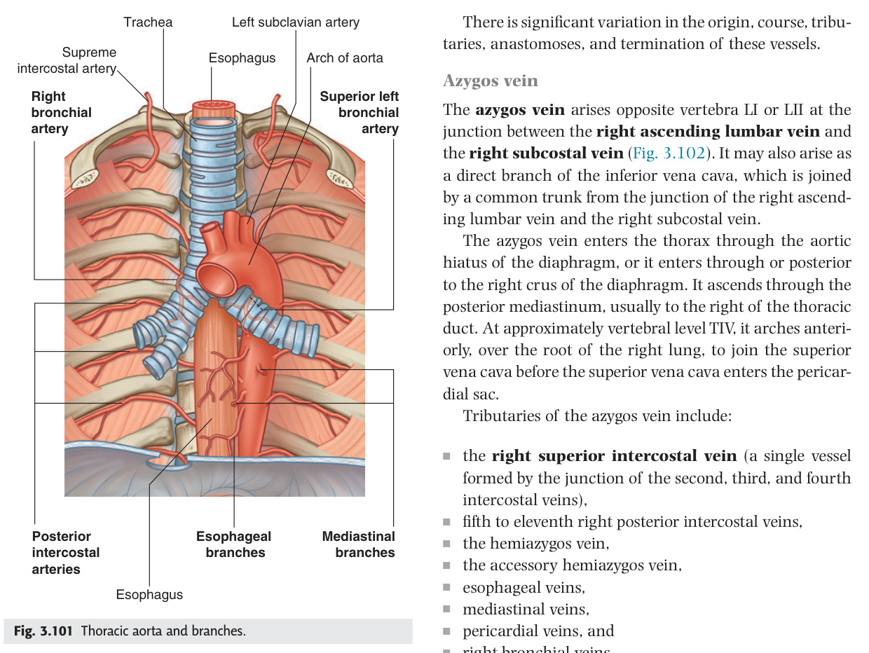
The Aorta — Overview
The aorta is a single continuous tube divided into four functional segments: ascending (coronaries only), arch (three great vessels to head and arms), thoracic descending (intercostals and bronchials), and abdominal (solid organs and bowel, bifurcating at L4). Three key openings pierce the diaphragm — T8 for the IVC (+ right phrenic nerve), T10 for the oesophagus (+ both vagal trunks), T12 for the aorta (+ thoracic duct + azygos vein) — memorised as "I Eat Apples." The aortic hiatus is a fibro-osseous gap (not muscular), so unlike the oesophageal hiatus it does not herniate, but it is the level where traumatic aortic transection occurs most commonly in deceleration injuries.
| Part | Location | Key Branches |
|---|---|---|
| Ascending aorta | Intrapericardial; from aortic valve (T4) to arch; length ~5 cm. Aortic sinuses (of Valsalva) at base | Left + right coronary arteries only |
| Arch of aorta | Posterior to manubrium; curves from right to left over left pulmonary artery + left main bronchus; T4–T5 level. Ligamentum arteriosum (ductus arteriosus remnant) from arch to left pulmonary artery | Brachiocephalic trunk + left common carotid + left subclavian (see 11.2) |
| Thoracic (descending) aorta | Begins at T4 (level of sternal angle/angle of Louis), descends left of vertebral column → through aortic hiatus at T12 | Posterior intercostals 3–11 + subcostal + bronchial + oesophageal (see 11.3) |
| Abdominal aorta | T12 to L4 (bifurcation at level of umbilicus/iliac crest). Lies left of midline on vertebral bodies. Aortic pulsation palpable in thin individuals | Coeliac, SMA, suprarenal, renal, gonadal, IMA, lumbar, median sacral (see 11.4) |
T8 = IVC (caval opening — vena cava + right phrenic nerve). T10 = Oesophagus (oesophageal hiatus — oesophagus + both vagal trunks). T12 = Aorta (aortic hiatus — aorta + thoracic duct + azygos vein). Mnemonic: I Eat Apples (IVC 8, Oesophagus 10, Aorta 12).
- Four parts of the aorta, location of each? Ascending (intrapericardial, T4); Arch (posterior to manubrium, T4–T5, 3 great branches); Thoracic descending (T4–T12, intercostals + bronchials); Abdominal (T12–L4, coeliac/SMA/renals/IMA)
- "I Eat Apples" — what diaphragm openings? T8 = IVC + right phrenic; T10 = oesophagus + both vagal trunks; T12 = aorta + thoracic duct + azygos vein
- Where does the thoracic aorta begin and at what landmark? At T4 (sternal angle / angle of Louis) — the same level as the aortic arch end, tracheal bifurcation, and 2nd costal cartilage
- Where does the abdominal aorta bifurcate? L4 — level of the iliac crest and umbilicus; lies just left of midline on vertebral bodies
- Only branches of the ascending aorta? Left and right coronary arteries (arising from aortic sinuses of Valsalva)
Arch of Aorta & Its Branches
The arch of aorta gives three branches in order from right to left: brachiocephalic trunk (first and largest, dividing behind the right sternoclavicular joint into right common carotid and right subclavian), left common carotid, and left subclavian. The subclavian artery's branches are remembered as "VIT C D" — Vertebral (→ basilar → Circle of Willis), Internal thoracic (→ anterior intercostals; harvested for CABG as the gold-standard left ITA-to-LAD graft), Thyrocervical trunk, Costocervical trunk, Dorsal scapular. Subclavian steal syndrome occurs when proximal stenosis reverses vertebral artery flow during arm exercise, causing vertebrobasilar symptoms — a BP difference >15 mmHg between arms is the bedside clue.
| Branch (left→right) | First branch | Key divisions |
|---|---|---|
| Brachiocephalic trunk (innominate) | First and largest branch; arises right of midline | Divides behind right sternoclavicular joint into: right common carotid + right subclavian arteries |
| Left common carotid artery | Second branch; longer thoracic course than right | No thoracic branches; divides at C4 (thyroid cartilage level) into left ICA + ECA |
| Left subclavian artery | Third branch; longest intrathoracic course | Thyrocervical trunk + costocervical trunk + vertebral artery + internal thoracic artery (see below) |
Stenosis of proximal subclavian artery (before vertebral origin) → exercising ipsilateral arm increases demand → blood 'stolen' from posterior circulation via retrograde flow in ipsilateral vertebral artery → vertebrobasilar ischaemia (dizziness, diplopia, drop attacks during arm exercise). BP difference >15 mmHg between arms = significant subclavian stenosis. Treat: subclavian angioplasty/stenting or carotid-subclavian bypass.
- Three branches of the aortic arch in order? (1) Brachiocephalic trunk → right common carotid + right subclavian; (2) Left common carotid; (3) Left subclavian. "VIT C D" = subclavian branches
- "VIT C D" subclavian branches? Vertebral → basilar → Circle of Willis; Internal thoracic → anterior intercostals + superior epigastric; Thyrocervical trunk; Costocervical trunk; Dorsal scapular
- Subclavian steal syndrome: mechanism and diagnosis? Proximal subclavian stenosis → exercising arm reverses vertebral artery flow → vertebrobasilar ischaemia (dizziness, diplopia). Diagnosed by BP difference >15 mmHg between arms
- Why is the internal thoracic artery used in CABG? Long-term patency rate superior to saphenous vein grafts; left ITA to LAD is the gold standard; single use limits number of grafts possible from this source
- Where does the vertebral artery enter the transverse foramina? Enters at C6 (not C7); ascends through C6–C1 foramina → curves posterior to atlas → through foramen magnum → joins opposite VA → basilar artery
Thoracic (Descending) Aorta
The thoracic aorta supplies posterior intercostal arteries to spaces 3–11 (spaces 1–2 are supplied by the costocervical trunk of the subclavian), bronchial arteries to the airway walls, and oesophageal branches to the middle oesophagus. The key clinical application is coarctation: narrowing at the ligamentum arteriosum forces blood through intercostal collaterals that enlarge and erode the inferior rib margins — rib notching on CXR — a sign that only appears after age 5–6 when collaterals have had time to hypertrophy. The lower oesophageal arteries connect the left gastric artery (portal system) with the azygos (systemic), making the lower oesophageal mucosa a site of portosystemic varices in portal hypertension — and a cause of life-threatening haematemesis.
| Branch | Distribution | Clinical |
|---|---|---|
| Posterior intercostal arteries (3rd–11th) | Supply intercostal spaces 3–11; anastomose with anterior intercostals (from ITA). 1st + 2nd = from costocervical trunk | Coarctation of aorta: enlarged intercostal arteries cause rib notching (inferior rib margins) on CXR — classic sign |
| Subcostal artery | Below 12th rib | |
| Bronchial arteries | Nourish bronchial walls + lung parenchyma (not gas exchange — that's pulmonary). Left: from aorta directly. Right: usually from 3rd right posterior intercostal artery | Bronchiectasis: bronchial arteries hypertrophied → massive haemoptysis. Embolisation of bronchial arteries = treatment |
| Oesophageal arteries (4–5) | Middle oesophagus. Upper = inferior thyroid; lower = left gastric | Oesophageal varices: lower oesophagus drained by both azygos (systemic) + left gastric (portal) → portosystemic anastomosis |
| Pericardial + mediastinal branches | Small, numerous |
Congenital narrowing of the aorta, usually just distal to the origin of the left subclavian artery at the level of the ligamentum arteriosum. Results in: hypertension in upper limbs + hypotension/weak pulses in lower limbs. Collateral circulation develops via intercostal arteries (posterior intercostals bypass the coarctation via internal thoracic anastomoses) → rib notching on CXR (3rd–8th ribs bilaterally). Also: bicuspid aortic valve (50–80% association), berry aneurysms in Circle of Willis. Repair: balloon angioplasty ± stenting or surgical resection + end-to-end anastomosis.
- Which intercostal spaces get their supply from the costocervical trunk (not aorta)? 1st and 2nd intercostal spaces — supplied by superior intercostal artery from costocervical trunk (subclavian); 3rd–11th come from thoracic aorta
- Rib notching on CXR — cause and which ribs? Coarctation → enlarged intercostal collaterals erode inferior rib margins; seen on 3rd–8th ribs bilaterally; appears after age 5–6 when collaterals hypertrophy
- Lower oesophagus portosystemic anastomosis — vessels involved? Left gastric vein (portal) anastomoses with oesophageal tributaries of azygos vein (systemic) → oesophageal varices in portal hypertension
- Bronchial arteries supply what, and what is the clinical relevance in bronchiectasis? Supply bronchial walls + lung parenchyma (not gas exchange); in bronchiectasis they hypertrophy → massive haemoptysis; treated by bronchial artery embolisation
- Where does the thoracic aorta pass through the diaphragm and with what structures? Aortic hiatus at T12; carries aorta + thoracic duct + azygos vein
Abdominal Aorta Branches
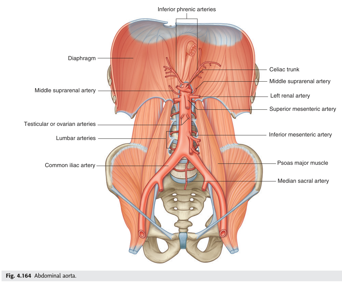
The abdominal aorta's ventral branches supply the gut in three embryological blocks: coeliac (T12/L1) for foregut, SMA (L1) for midgut up to the splenic flexure, IMA (L3) for hindgut. The critical watershed between SMA and IMA territory sits at the splenic flexure — its tenuous arc of Riolan collateral makes this the commonest site of ischaemic colitis after AAA repair or low-flow states. Renal arteries are end-artery systems with no cortical anastomosis, which is why bilateral renal artery stenosis combined with ACE inhibitor therapy — which removes the efferent arteriolar tone needed to maintain glomerular filtration pressure — precipitates acute kidney injury.
| Branch | Level | Territory / Notes |
|---|---|---|
| Coeliac trunk | T12/L1; first ventral branch | Left gastric + splenic + common hepatic. Supplies: foregut (oesophagus lower → duodenojejunal flexure). Everything from coeliac to SMA = superior mesenteric = midgut |
| Superior mesenteric artery (SMA) | L1; 1 cm below coeliac | Supplies: duodenum (2nd–4th) + jejunum + ileum + right half colon (caecum, ascending, transverse to splenic flexure). Branches: inferior pancreaticoduodenal + jejunal/ileal + ileocolic + right colic + middle colic |
| Suprarenal arteries | L1; 3 arteries each side | Superior (phrenic), middle (aorta), inferior (renal) |
| Renal arteries | L1–L2 (below SMA) | Right longer (passes posterior to IVC + right renal vein). Each: segmental branches (end arteries = no anastomosis) |
| Gonadal arteries | L2; from aorta | Testicular (right → IVC; left → left renal vein) / ovarian |
| Inferior mesenteric artery (IMA) | L3 | Supplies: hindgut — left transverse (distal 1/3) + descending + sigmoid + upper rectum. Branches: left colic + sigmoid arteries + superior rectal |
| Lumbar arteries (×4) | L1–L4 | Posterior abdominal wall + spinal cord supply (important for AAA repair) |
| Median sacral artery | L4; unpaired midline | Sacrum + coccyx; remnant of embryonic axial artery. May be large source of haemorrhage in pelvic fractures |
Two main causes: (1) Atherosclerosis (90%) — elderly patients with diffuse vascular disease; proximal renal artery (within 1 cm of aortic ostium); bilateral in ~30%; flash pulmonary oedema is a classic presentation of bilateral stenosis. (2) Fibromuscular dysplasia (FMD, 10%) — young women (15–40 yrs); medial fibromuscular hyperplasia of mid/distal renal artery; "string of beads" appearance on angiography; no atherosclerosis.
Mechanism — renovascular hypertension: ↓ renal perfusion pressure → ↑ renin (juxtaglomerular cells) → ↑ angiotensin II → vasoconstriction + ↑ aldosterone → sodium/water retention + hypokalaemia → hypertension.
Critical: Bilateral stenosis + ACE inhibitor/ARB → acute kidney injury. ACEi block Ang II-mediated efferent arteriolar constriction → ↓ filtration pressure → acute oliguric AKI. Always check renal function 1–2 weeks after starting ACEi in patients with risk factors for renovascular disease (elderly, diffuse atherosclerosis, unexplained renal impairment).
Investigation: Renal Doppler USS (first-line; peak systolic velocity >200 cm/s = significant); CT/MR angiography (gold standard). Treatment: FMD → PTA (percutaneous transluminal angioplasty) — ~80% cure rate. Atherosclerotic → medical management (ACEi/ARB if unilateral; statin; aspirin); the ASTRAL trial showed angioplasty offered no benefit over medical therapy for most atherosclerotic cases. [Gray's 4e Ch2 p78]
The splenic flexure of the colon lies at the junction between SMA territory (transverse colon via middle colic) and IMA territory (descending colon via left colic). The anastomosis here (arc of Riolan / marginal artery of Drummond) is the most tenuous part of the colonic blood supply. Ischaemic colitis most commonly affects the splenic flexure (watershed infarction) — occurs after AAA repair (IMA ligated), aortic dissection, or low-flow states. Presents with left-sided abdominal pain + bloody diarrhoea. Colonoscopy confirms mucosal ischaemia.
- Three ventral gut branches and their embryological territory? Coeliac (T12/L1) = foregut; SMA (L1) = midgut (to splenic flexure); IMA (L3) = hindgut (splenic flexure to upper rectum)
- Three branches of the coeliac trunk? Left gastric + splenic + common hepatic ("3 Ladies Sitting"). Common hepatic → proper hepatic + gastroduodenal (→ right gastroepiploic + superior pancreaticoduodenal)
- SMA/IMA watershed: site, clinical risk, and presentation? Splenic flexure (arc of Riolan); most vulnerable in AAA repair (IMA ligated) or low-flow; presents as left lower abdominal pain + bloody diarrhoea (ischaemic colitis)
- Why does bilateral renal artery stenosis + ACEi cause AKI? ACEi blocks Ang II–mediated efferent arteriolar constriction → ↓ GFP → acute oliguric AKI; check renal function 1–2 weeks after starting ACEi in high-risk patients
- Gonadal venous drainage asymmetry? Right testicular/ovarian vein → IVC directly; left testicular/ovarian vein → left renal vein (same as left suprarenal vein). Left varicocele = left renal vein compression (e.g., nutcracker syndrome)
Blood Supply of Key Organs ★★★
Four organs dominate exam questions on visceral blood supply: thyroid, stomach, suprarenal, and ductus arteriosus. The thyroid is surgically treacherous because two major nerves — the external branch of the superior laryngeal nerve (voice pitch) and the recurrent laryngeal nerve (voice at all) — cross the superior and inferior thyroid arteries respectively, so ligation technique dictates whether the patient wakes up hoarse or speechless. The stomach's six arteries all trace back to the coeliac trunk via a rich anastomotic ring along both curvatures, making ischaemia rare — but portal hypertension exploits the left gastric–oesophageal anastomosis as a collateral channel, producing fatal variceal bleeds. The suprarenal gland's triple arterial supply (phrenic, aorta, renal — P-A-R top to bottom) explains why it survives AAA surgery; its venous drainage is the asymmetry that mirrors the gonadal vein pattern.
Slide review questions (Prof. Ping Zhang): "Describe the blood supply of thyroid gland, stomach, hand, suprarenal gland." Hand covered in §11.9. Below: thyroid, stomach, suprarenal, and ductus arteriosus.
| Artery | Origin | Territory / Clinical |
|---|---|---|
| Superior thyroid artery | External carotid artery (1st branch) | Upper pole of thyroid + larynx. External branch of superior laryngeal nerve runs with it → risk during upper pole ligation (see §11.7.1) |
| Inferior thyroid artery | Thyrocervical trunk (from subclavian) | Lower pole of thyroid + all 4 parathyroid glands. Recurrent laryngeal nerve crosses behind (or in front of) this artery → RLN risk during inferior thyroid artery ligation |
| Thyroidea ima artery (3–10%) | Brachiocephalic trunk or aortic arch (variable) | Isthmus from below. Clinically critical: can bleed massively during emergency midline tracheostomy if unrecognised |
| Artery | Arises from | Supplies |
|---|---|---|
| Left gastric artery | Celiac trunk (directly) | Lesser curvature (left/superior) + lower oesophagus; anastomoses with right gastric along lesser curve. Portal hypertension → oesophageal varices via left gastric–azygos connection |
| Right gastric artery | Proper hepatic artery (from common hepatic → celiac trunk) | Lesser curvature (right/inferior); anastomoses with left gastric |
| Left gastroepiploic artery | Splenic artery | Greater curvature (left) + greater omentum; anastomoses with right gastroepiploic |
| Right gastroepiploic artery | Gastroduodenal artery → common hepatic → celiac trunk | Greater curvature (right) + greater omentum |
| Short gastric arteries (5–7) | Splenic artery (terminal branches) | Fundus of stomach |
| Artery | Origin | Part supplied |
|---|---|---|
| Superior suprarenal arteries (multiple) | Inferior phrenic artery | Upper part |
| Middle suprarenal artery | Abdominal aorta (directly, lateral aspect) | Middle part |
| Inferior suprarenal artery | Renal artery | Lower part |
P – A – R (top to bottom): Phrenic (superior) → Aorta (middle) → Renal (inferior). Venous drainage is asymmetric: right suprarenal vein → IVC (short, direct); left suprarenal vein → left renal vein (mirrors testicular/ovarian pattern on the left).
Ligamentum arteriosum: a fibrous cord connecting the pulmonary trunk (or left pulmonary artery) to the concavity of the aortic arch. It is the obliterated remnant of the ductus arteriosus — the fetal vessel through which most right ventricular output bypasses the collapsed lungs and passes directly into the descending aorta. At birth: ↑O₂ tension + ↓prostaglandins → ductal smooth muscle constricts → functional closure within hours; anatomical (fibrous) closure by 3 months. Patent ductus arteriosus (PDA): failure to close → persistent left-to-right shunt → continuous "machinery" murmur (loudest left infraclavicular/2nd ICS) + wide pulse pressure. Treatment: indomethacin (prostaglandin inhibitor) in premature neonates; surgical ligation or catheter closure in term infants/older.
- Nerve at risk with superior thyroid artery ligation? External branch of the superior laryngeal nerve (cricothyroid muscle; controls voice pitch). Ligation close to the upper thyroid pole risks cutting it → monotone, weak voice
- Nerve at risk with inferior thyroid artery ligation, and consequence? Recurrent laryngeal nerve crosses (behind or in front of) the artery. Injury → ipsilateral vocal cord paralysis. Bilateral injury → bilateral cord adduction → stridor + respiratory emergency
- Why does close-capsule ligation of the inferior thyroid artery cause tetany? All 4 parathyroid glands are supplied by the inferior thyroid artery via end-arteries; devascularisation → hypoparathyroidism → hypocalcaemia → tetany (Chvostek + Trousseau signs)
- Stomach blood supply — fundus supplied by which artery? Short gastric arteries (5–7 branches) from the splenic artery. Lesser curvature = left + right gastric; greater curvature = left + right gastroepiploic; all trace back to coeliac trunk
- Suprarenal arterial mnemonic and venous asymmetry? P-A-R (top to bottom): Phrenic (superior), Aorta (middle), Renal (inferior). Venous drainage: right suprarenal vein → IVC; left → left renal vein (same as left gonadal)
Iliac Arteries
The aorta bifurcates at L4 — the level of the iliac crests, the same bony landmark you palpate when performing a lumbar puncture. From there, two common iliac arteries split into internal (pelvic viscera) and external (lower limb) branches before the sacroiliac joint. The external iliac dives under the inguinal ligament to become the femoral artery — this transition point is where cardiac catheterisation gains arterial access. The internal iliac (hypogastric) bifurcates into anterior and posterior divisions: the posterior division is the safe one (gluteal, iliolumbar, lateral sacral), while the anterior division supplies every pelvic viscus — uterus, bladder, rectum, and genitalia — meaning that internal iliac ligation in obstetric haemorrhage is the last resort to stop pelvic bleeding without hysterectomy. The femoral triangle's "NAVY" arrangement — nerve lateral, artery, vein medial, Y-fronts (lymphatics) — and the midpoint-of-inguinal-ligament pulse are core exam points every clinical year.
The abdominal aorta bifurcates at L4 (level of iliac crest / umbilicus) into right and left common iliac arteries. Each divides anterior to the sacroiliac joint into:
| Artery | Course | Main branches |
|---|---|---|
| External iliac artery | Along medial border of psoas → under inguinal ligament → becomes femoral artery at femoral triangle | Inferior epigastric artery (→ deep inguinal ring; landmark for direct vs indirect hernia) + deep circumflex iliac artery |
| Internal iliac artery (hypogastric) | Descends into lesser pelvis; anterior + posterior divisions | Anterior: uterine, vaginal/inferior vesical, middle rectal, internal pudendal, inferior gluteal, obturator. Posterior: superior gluteal, iliolumbar, lateral sacral |
| Femoral artery | Femoral triangle (NAVL lateral→medial: nerve, artery, vein, lymphatics). Midpoint of inguinal ligament pulse. Adductor canal → popliteal fossa = popliteal artery | Profunda femoris (deep femoral) → medial/lateral circumflex femoral; perforating arteries supply posterior thigh |
Femoral artery pulse: palpable at midpoint of inguinal ligament (midpoint between ASIS + pubic symphysis — NOT mid-inguinal point which is midpoint of anterior superior iliac spine to pubic tubercle). Femoral artery cannulation: cardiac catheterisation, angiography. Complications: haematoma, pseudoaneurysm, AV fistula, retroperitoneal haemorrhage (if puncture above inguinal ligament). Femoral nerve (lateral to artery) + femoral vein (medial to artery) — medial to the artery is the mnemonic "NAVY" (Nerve, Artery, Vein, Y-fronts = lymphatics).
- Aortic bifurcation level and clinical landmark? L4 — level of the iliac crests (palpable bony ridge; same landmark for lumbar puncture at L3/4)
- External iliac artery: what does it become and where? Passes under inguinal ligament at the midpoint (between ASIS and pubic symphysis) → becomes femoral artery in femoral triangle
- Internal iliac anterior division branches (pelvic viscera)? Uterine, vaginal/inferior vesical, middle rectal, internal pudendal, inferior gluteal, obturator. Posterior division: superior gluteal, iliolumbar, lateral sacral
- Direct vs indirect inguinal hernia landmark — which artery? Inferior epigastric artery (branch of external iliac just above inguinal ligament). Direct = medial to inferior epigastric; indirect = lateral to it (through deep inguinal ring)
- Femoral triangle NAVY order (lateral → medial)? Nerve (femoral) → Artery (femoral) → Vein (femoral) → Y-fronts (lymphatics). Pulse at midpoint of inguinal ligament (ASIS to pubic symphysis)
Coronary Arteries
Both coronary arteries arise from the aortic sinuses — the concavities just above the aortic valve cusps — in diastole, when the valve leaflets fall back and blood pools behind them to fill the coronary ostia. This timing means tachycardia is inherently dangerous for the ischaemic heart: the shorter diastole is, the less coronary filling time there is. The LAD is the single most clinically important artery in the body, supplying the anterior two-thirds of the interventricular septum (where the bundle of His runs), the anterior left ventricle, and the apex — its occlusion causes the largest MIs and the highest mortality. The RCA dominates cardiac electrophysiology: it supplies the SA node (60% of people) and the AV node (80–90%), which is why inferior MI characteristically causes bradycardia and heart block rather than pump failure. "Dominance" means which artery gives the posterior descending artery — right in 85% — and matters only when identifying the culprit vessel in a posterior MI.
| Artery | Origin | Supplies |
|---|---|---|
| Left coronary artery (LCA) | Left aortic sinus; short trunk 1–2 cm then bifurcates | Left anterior descending (LAD): anterior IV groove → anterior LV + anterior RV + anterior 2/3 IVS. Left circumflex (LCx): left AV groove → lateral + posterior LV |
| Right coronary artery (RCA) | Right aortic sinus; travels in right AV groove | SA node (60%) + AV node (80–90%) + posterior descending artery (PDA) in right dominant (85%) → inferior LV + posterior 1/3 IVS + posterior RV |
| Dominant circulation | Which artery gives posterior descending artery (PDA) | Right dominant 85%; left dominant 8%; co-dominant 7% |
- When do the coronary arteries fill, and why does tachycardia worsen ischaemia? During diastole — valve cusps fall back, blood pools in aortic sinuses. Tachycardia shortens diastole → ↓coronary filling time → worsens supply/demand mismatch
- LAD territory and consequence of occlusion? Anterior LV + anterior 2/3 IVS + bundle of His. Occlusion → anterior MI (ST↑ V1–V4), LBBB, anterior fascicular block, ventricular aneurysm, acute pump failure
- RCA supplies which conduction structures, and what arrhythmias result from RCA occlusion? SA node (60%) + AV node (80–90%). Inferior MI (ST↑ II, III, aVF) → bradycardia, 1st/2nd/3rd degree heart block, junctional rhythms → may need temporary pacing
- Definition of coronary dominance? Whichever artery gives the posterior descending artery (PDA). Right dominant = 85%; left dominant = 8%; co-dominant = 7%
- LCx occlusion: territory and ECG leads? Lateral + posterior LV. Lateral MI: ST↑ I, aVL, V5–V6. Posterior MI: tall R + ST depression V1–V3 (mirror image of posterior wall infarct)
Carotid Arteries & Circle of Willis
The carotid system is where neurology, vascular surgery, and head-and-neck anatomy converge most dangerously. The common carotid bifurcates at C4 — the level of the thyroid cartilage notch — and the carotid sinus sitting right at that bifurcation acts as the body's main blood pressure sensor; a tight collar or a tumour here can trigger a vagal faint simply by mechanical compression. The internal carotid gives no branches in the neck — a key distinguishing feature — then supplies the cerebral hemisphere via the circle of Willis, whose anastomotic ring at the base of the brain is the most common place in the body to develop berry aneurysms. The circle's anterior communicating artery carries 30–35% of all aneurysms; a posterior communicating artery aneurysm announces itself with a painful, pupil-dilating third nerve palsy before rupture — a warning sign that is a neurosurgical emergency. The vertebrobasilar system feeding the brainstem generates the most distinctive stroke syndromes: Wallenberg's (PICA), locked-in (basilar), and the lateral pontine syndromes — all with the hallmark of crossed signs (ipsilateral cranial nerve, contralateral limb).
| Structure | Detail |
|---|---|
| Common carotid artery (CCA) | Right: from brachiocephalic trunk. Left: from arch of aorta. Both divide at C4 (upper border of thyroid cartilage) into ICA + ECA. Carotid sinus: dilation at bifurcation → baroreceptors (IX, X) → regulate BP. Carotid body: chemoreceptor (O₂/CO₂/pH) |
| External carotid artery (ECA) | Supplies face + scalp + most of neck. 8 branches: SAL FOP MS — Superior thyroid, Ascending pharyngeal, Lingual, Facial, Occipital, Posterior auricular, Maxillary, Superficial temporal |
| Internal carotid artery (ICA) | No branches in neck. Enters carotid canal → cavernous sinus → ophthalmic artery + anterior/middle cerebral arteries + posterior communicating artery (PComm) → Circle of Willis |
| Circle of Willis | Anastomotic ring at base of brain. Components: ACA + AComm (anterior) + ICA + PComm + PCA + basilar tip (posterior). Supplies all of cerebral cortex via anterior, middle, posterior cerebral arteries |
Carotid sinus: a slight fusiform dilation at the origin of the internal carotid artery (and adjacent common carotid bifurcation). Contains baroreceptors (pressoreceptors) in the tunica adventitia, innervated by the sinus nerve of Hering — a branch of CN IX (glossopharyngeal). Detects changes in arterial blood pressure → afferent signals to the nucleus tractus solitarius (NTS) in the medulla → reflex modulation of heart rate (via vagus) and vascular tone. Acts as a pressure-receptor which is part of the blood pressure regulating mechanism (slide definition). Clinical: carotid sinus hypersensitivity — exaggerated reflex response to carotid stimulation (head turning, tight collar, shaving) → bradycardia + hypotension → syncope; common cause of unexplained falls in elderly. Carotid sinus massage (CSM) slows the heart → used diagnostically in SVT.
Carotid body (carotid glomus): a small ovoid chemoreceptor located in the fork of the bifurcation of the common carotid artery. Contains glomus type I (chief) cells that respond to changes in blood composition — particularly ↓PaO₂, ↑PaCO₂, ↓pH. Innervated by CN IX (glossopharyngeal). Triggers increased rate and depth of respiration via the respiratory centre. Tumour of the carotid body = carotid body paraganglioma (chemodectoma): pulsatile, painless neck mass at carotid bifurcation; moves horizontally but not vertically (Fontaine's sign); bruit on auscultation; MRI "lyre sign" (splaying of ICA and ECA).
The superior thyroid artery (first branch of ECA) descends to the upper pole of the thyroid. Running closely alongside it — and passing deep to it before reaching the cricothyroid muscle — is the external branch of the superior laryngeal nerve (from CN X → superior laryngeal nerve → divides into external + internal branches). The external branch innervates the cricothyroid muscle (the only intrinsic laryngeal muscle NOT supplied by the recurrent laryngeal nerve).
KEY (past paper MCQ Q7 — 2020): When the superior thyroid artery is ligated during thyroid surgery, the nerve at risk is the external branch of the superior laryngeal nerve (NOT the recurrent laryngeal nerve). Injury → loss of cricothyroid → inability to tense vocal cords → loss of high-pitched voice (subtle, often missed). Compare: recurrent laryngeal nerve is at risk during inferior thyroid artery ligation (the RLN crosses behind or in front of the inferior thyroid artery → injury causes hoarseness or complete unilateral vocal cord paralysis).
Carotid atherosclerosis: most common at bifurcation. Plaque → turbulence → thromboembolism → TIA or stroke. Carotid bruit on auscultation. Carotid duplex USS: >70% stenosis = significant. NASCET criteria: symptomatic >50% stenosis → carotid endarterectomy (CEA) within 2 weeks of TIA (highest risk period = "90-day risk period"). CEA complications: stroke, cranial nerve injury (hypoglossal XII most common → tongue deviation; glossopharyngeal IX; vagus X → hoarseness). Posterior communicating artery (PComm) aneurysm: painful 3rd nerve palsy (ptosis + down-and-out eye + dilated pupil) — surgical or endovascular coiling.
Giant cell arteritis (GCA) is a large/medium vessel granulomatous vasculitis affecting the superficial temporal artery (branch of ECA) and other cranial/systemic arteries (ophthalmic artery, posterior ciliary arteries, aorta and its branches). Most common vasculitis in adults over 50; female > male (3:1); peak incidence 70–80 years.
Anatomical basis: Superficial temporal artery runs anterior to the ear in the temporal region (palpable and visible) → its inflammation causes the classic scalp tenderness and temporal headache. The ophthalmic artery is a branch of the ICA; its posterior ciliary branches supply the optic nerve head — occlusion → anterior ischaemic optic neuropathy (AION) = sudden, painless, permanent visual loss (dense altitudinal field defect, pale swollen optic disc).
Symptoms: New severe temporal headache (unilateral) · scalp tenderness (can't brush hair/wear hat) · jaw claudication (masseter ischaemia during chewing — most specific symptom, ~50%) · visual symptoms (amaurosis fugax → if untreated, permanent blindness) · systemic: fever, malaise, weight loss. Polymyalgia rheumatica co-exists in 40–50% (proximal limb girdle pain + stiffness worse in morning).
Investigations: ESR >50 mm/hr (classically >100) + CRP elevated. Temporal artery biopsy = gold standard (shows granulomatous inflammation with giant cells, internal elastic lamina disruption, intimal thickening) — skip lesions can give false negatives; take ≥2 cm of vessel. GCA can also be diagnosed/monitored by temporal artery USS (halo sign = hypoechoic halo around vessel wall from oedema).
Treatment — DO NOT WAIT FOR BIOPSY: Start prednisolone 60 mg/day immediately if GCA is suspected — visual loss, if it occurs, is usually irreversible. Biopsy remains positive for up to 2 weeks on steroids. If vision already affected: IV methylprednisolone 1 g/day for 3 days. Long-term: taper steroids over 1–2 years; tocilizumab (IL-6 inhibitor) for relapsing/refractory disease. Aspirin added to reduce thromboembolic risk from inflamed vessels.
Anatomy: The middle meningeal artery (MMA) is a branch of the maxillary artery (1st part, from ECA). It enters the skull through the foramen spinosum and travels in grooves on the inner surface of the temporal and parietal bones within the epidural space. It divides into anterior and posterior branches. The anterior branch crosses the pterion — the thinnest part of the skull, where four bones converge: frontal, parietal, temporal, and greater wing of sphenoid.
Extradural haematoma (EDH): A blow to the temporal region fractures the thin squamous temporal bone at the pterion → lacerates the anterior branch of MMA → arterial haemorrhage accumulates in the epidural space between bone and periosteal dura.
Classic clinical course — "talk and die":
① Head injury → brief loss of consciousness (concussion)
② Lucid interval — patient regains consciousness, talks and appears well for minutes to hours (blood slowly accumulates)
③ Rapid deterioration — haematoma reaches critical mass → ↑ICP → transtentorial (uncal) herniation → ipsilateral fixed dilated pupil (CN III compressed by uncal herniation) → contralateral hemiplegia (corticospinal tract compressed at cerebral peduncle) → coma → death if untreated.
CT appearance: Biconvex (lenticular) hyperdense haematoma. Blood cannot cross suture lines (periosteal dura fused to sutures) → lens/biconvex shape. Midline shift.
Treatment: Emergency craniotomy + haematoma evacuation + arterial ligation. Outcome is excellent if treated before herniation — EDH has highest "talk and die" potential but best outcome of all intracranial haematomas when treated promptly.
Contrast with subdural haematoma (SDH): SDH = bridging veins (venous, slower), crescent-shaped on CT (blood DOES cross suture lines as it is subdural, not epidural), no lucid interval (slow venous bleed), often in elderly/anticoagulated patients. [Gray's 4e Ch8 p532; Netter's 7th Plate 105]
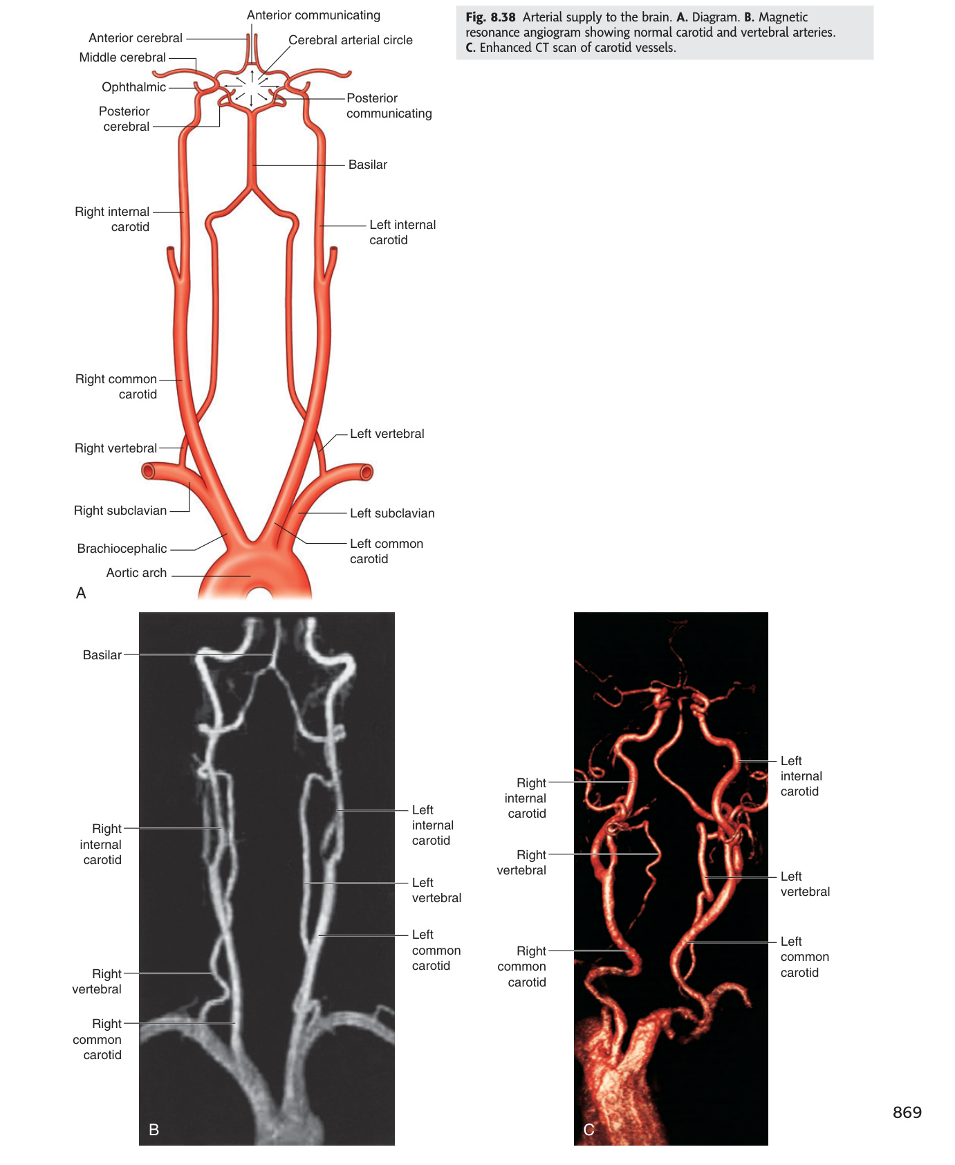
The Circle of Willis (circulus arteriosus cerebri) is an anastomotic polygon at the base of the brain formed by: 2 ACAs + AComm (anterior) + 2 ICAs + 2 PComms + 2 PCAs (from basilar tip).
| Artery | Territory | Occlusion Syndrome |
|---|---|---|
| Anterior cerebral artery (ACA) | Medial frontal + parietal cortex (leg area of homunculus); corpus callosum | Contralateral leg > arm weakness + sensory loss; urinary incontinence; abulia (frontal lobe) |
| Middle cerebral artery (MCA) | Lateral hemisphere (face + arm area > leg); Broca's area (dominant F3); Wernicke's area (dominant T1) | Contralateral face + arm > leg weakness + sensory loss; dominant = aphasia (Broca's = expressive; Wernicke's = receptive); non-dominant = neglect + apraxia; eye deviates TOWARD lesion (frontal eye field) |
| Posterior cerebral artery (PCA) | Occipital lobe (primary visual cortex); thalamus; midbrain | Contralateral homonymous hemianopia with macular sparing (MCA collaterals preserve macular cortex); thalamic pain syndrome; Weber's syndrome (ipsilateral CN III palsy + contralateral hemiplegia) |
| Anterior communicating artery (AComm) | Connects 2 ACAs; no territory | Most common site for berry aneurysm (30–35%); rupture can cause bitemporal visual field defects or ACA infarction |
| Posterior communicating artery (PComm) | Connects ICA to PCA; joins anterior + posterior circulations | Aneurysm here (25% of berry aneurysms) → compresses CN III → painful complete CN III palsy (pupil-involving = surgical emergency; mydriasis = pupillary dilation is FIRST sign) |
Sites (order of frequency): AComm (30–35%) → PComm–ICA junction (25%) → MCA bifurcation (20%) → basilar tip (5–10%) → other. Multiple aneurysms in 20%. Associated conditions: ADPKD (autosomal dominant polycystic kidney disease), Marfan's syndrome, Ehlers-Danlos, coarctation of aorta, first-degree family history.
Rupture → subarachnoid haemorrhage (SAH): "thunderclap headache" = sudden-onset worst headache of life (peak intensity in seconds); ± photophobia, neck stiffness (meningism from blood in CSF), loss of consciousness (in severe cases). Investigation: CT head (blood in basal cisterns/sulci); if negative but strong suspicion = lumbar puncture at >6 hours (xanthochromia = yellow CSF = breakdown products of blood = confirms SAH). CT angiogram or digital subtraction angiography (DSA) to find aneurysm. Treatment: neurosurgical clipping or endovascular coiling (ISAT trial: coiling = better short-term outcomes for suitable anatomy); nimodipine (calcium channel blocker → prevents vasospasm).
The posterior (vertebrobasilar) circulation supplies the brainstem, cerebellum, thalamus, and occipital cortex. Two vertebral arteries (from subclavian) → join at pontomedullary junction → basilar artery → PCAs + PComms (linking to anterior circulation via Circle of Willis).
| Vessel Occluded | Syndrome | Ipsilateral Signs | Contralateral Signs |
|---|---|---|---|
| PICA (posterior inferior cerebellar a.; largest VA branch) |
Wallenberg syndrome (lateral medullary syndrome) |
Facial pain/numbness (CN V spinal nucleus) · Horner's syndrome (descending sympathetics) · cerebellar ataxia (inf. cerebellar peduncle) · dysphagia + hoarseness (CN IX/X nuclei) · hiccups · nystagmus | Loss of pain + temperature in limbs/trunk (spinothalamic — already crossed). Proprioception + motor SPARED |
| AICA (anterior inferior cerebellar a.; from lower basilar) |
Lateral inferior pontine syndrome | Facial pain/numbness (CN V) · deafness + tinnitus (CN VIII — labyrinthine a. from AICA) · facial nerve palsy (CN VII nucleus) · Horner's · cerebellar ataxia | Loss of pain + temperature in limbs (spinothalamic) |
| Basilar artery (bilateral pontine infarct) |
Locked-in syndrome | Quadriplegia (bilateral corticospinal tracts) + loss of all voluntary movement + inability to speak. Patient is fully conscious (reticular activating system + cortex intact). Only vertical eye movements (midbrain spared) and blinking are preserved — sole means of communication. Horizontal gaze absent (PPRF in pons = infarcted). | |
| SCA (superior cerebellar a.; from upper basilar) |
Superior cerebellar syndrome | Ipsilateral cerebellar ataxia (superior cerebellar peduncle) · Horner's · CN IV palsy (trochlear) | Pain/temp loss face + limbs (spinothalamic) |
PICA infarcted = LATERAL medulla infarcted. The corticospinal tracts run medially through the medulla (pyramids = medial) → they are NOT in the lateral medullary territory → no hemiplegia in Wallenberg syndrome. This distinguishes it from medial medullary syndrome (pyramid + medial lemniscus + CN XII affected).
Ipsilateral face, contralateral body (for pain/temp) = the hallmark cross-over pattern. The spinal trigeminal nucleus (face) is in the lateral medulla ipsilaterally; the spinothalamic fibres (body) have already crossed before reaching the medulla.
Vertebral artery dissection (VAD) is an important cause of posterior circulation stroke in young adults. An intimal tear → intramural haematoma → luminal stenosis + thrombus → embolic or haemodynamic stroke in posterior circulation. Precipitants: neck manipulation (chiropractic), whiplash, vigorous exercise. Presentation: sudden-onset posterior neck or occipital pain (tearing character) followed hours to days later by posterior circulation stroke (most often Wallenberg syndrome). Investigation: MRI/MRA (shows intramural haematoma = "double lumen"). Treatment: anticoagulation (heparin → warfarin) or antiplatelet; most dissections heal within 3 months. [Gray's 4e Ch8 p527]
- CCA bifurcation level and two specialised structures at the fork? C4 (upper border thyroid cartilage). Carotid sinus = baroreceptor (CN IX) for BP regulation; carotid body = chemoreceptor (CN IX) for O₂/CO₂/pH
- Most common berry aneurysm sites in order? AComm (30–35%) → PComm–ICA junction (25%) → MCA bifurcation (20%) → basilar tip (5–10%). Associated with ADPKD, Marfan's, coarctation
- PComm aneurysm — clinical presentation and why? Painful CN III palsy (ptosis + down-and-out + mydriasis). Parasympathetic fibres run on the outside of CN III — compression by aneurysm stretches them first → pupil dilates before motor fibres fail
- Wallenberg syndrome: vessel, side of Horner's, side of limb sensory loss? PICA occlusion → lateral medullary infarct. Horner's = ipsilateral (descending sympathetics). Pain/temp loss in limbs = contralateral (spinothalamic already crossed). Motor = SPARED (pyramids are medial)
- Locked-in syndrome: vessel, consciousness, preserved movements? Basilar artery occlusion → bilateral ventral pontine infarct. Fully conscious (cortex/RAS intact). Only vertical eye movements + blinking preserved (midbrain spared); horizontal gaze absent (PPRF in pons infarcted)
Upper Limb Arteries ★★
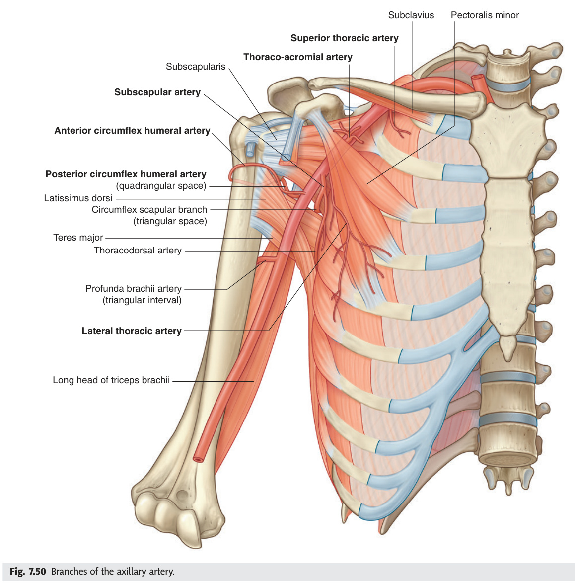
The upper limb arterial chain — subclavian → axillary → brachial → radial/ulnar → palmar arches → digital arteries — is the route surgeons trace when a hand goes cold. The subclavian becomes the axillary at the lateral border of the first rib, and the axillary becomes the brachial at the lower border of teres major — these transition points are landmarks, not anatomical changes in the vessel wall. The brachial artery runs medial to the biceps tendon in the cubital fossa, which is why you position your stethoscope there to auscultate Korotkoff sounds when measuring blood pressure. Distally, the radial artery travels through the anatomical snuffbox, whose floor is the scaphoid — so snuffbox tenderness after a fall on an outstretched hand is scaphoid fracture until proven otherwise, even with a normal X-ray. The dual palmar arches provide crucial collateral protection: Allen's test validates this collateral before you commit to an arterial line in the radial artery.
| Artery | Course | Key Branches / Clinical |
|---|---|---|
| Axillary artery | From lateral border of 1st rib (continues subclavian) → lower border of teres major. Divided by pectoralis minor into 3 parts | Part 1: superior thoracic a. | Part 2: thoracoacromial + lateral thoracic aa. | Part 3: subscapular (→ circumflex scapular + thoracodorsal) + anterior + posterior circumflex humeral aa. Posterior circumflex humeral = with axillary nerve around surgical neck of humerus → injured in surgical neck fracture |
| Brachial artery | From lower border of teres major → cubital fossa; lies medial to biceps tendon. Divides at neck of radius into radial + ulnar arteries | Profunda brachii (deep brachial — with radial nerve in spiral groove); BP measured over brachial artery; cubital fossa contents L→M: TAN = biceps Tendon, brachial Artery, median Nerve (lateral = radial nerve, outside TAN) |
| Radial artery | Lateral; runs under brachioradialis → anatomical snuffbox → deep palmar arch. Pulse at wrist lateral to flexor carpi radialis | Anatomical snuffbox (floor = scaphoid, trapezium): tenderness = scaphoid fracture; Allen's test to assess palmar arch collateral before radial arterial line; radial forearm flap surgery |
| Ulnar artery | Medial, larger than radial; Guyon's canal (with ulnar nerve at wrist) → superficial palmar arch. Pulse medial to flexor carpi ulnaris | Guyon's canal entrapment = ulnar nerve compression at wrist (cycling, hypothenar hammer syndrome); ulnar artery thrombosis → cold/pale hypothenar eminence |
| Palmar arches | Superficial arch: mainly ulnar + radial (completes) → common palmar digital → proper digital aa. Deep arch: mainly radial (enters via 1st dorsal interosseous) + ulnar (completes) → palmar metacarpal aa. | Allen's test: compress both radial + ulnar arteries at wrist → release one at a time; normal = hand flushes in <5 sec. Failure = inadequate collateral = unsafe to cannulate that artery |
Superficial palmar arch: an arterial anastomotic arch in the palm of the hand, formed mainly by the ulnar artery (its direct continuation beyond the pisiform) and completed on the radial side by the superficial palmar branch of the radial artery. It lies superficial to the long flexor tendons and the digital branches of the median and ulnar nerves. Gives off 3 common palmar digital arteries, each of which divides into 2 proper palmar digital arteries supplying adjacent sides of the fingers (2nd–5th). Mnemonic: "U comes first in Ulnar — Ulnar is superficial."
Deep palmar arch: formed mainly by the terminal part of the radial artery (entering through the 1st dorsal interosseous space / anatomical snuffbox into the palm), completed by the deep palmar branch of the ulnar artery. Lies deep to the long flexor tendons, on the bases of the metacarpals and the interossei muscles. Gives off 3 palmar metacarpal arteries which anastomose distally with the common digital branches of the superficial arch. The deep arch lies approximately 2 cm proximal to the superficial arch. Mnemonic: "Radial is deep, Ulnar is superficial" (for the major contributor to each arch).
Deep arch: mainly radial artery (enters via 1st interosseous space) + completed by deep branch of ulnar → gives 3 palmar metacarpal arteries. Lies deep to flexor tendons, 2 cm proximal to superficial arch.
Significance of dual arches: ensures digital blood supply even if one forearm artery is occluded (tested by Allen's test). Superficial arch = clinically relevant for palmar space infections, digit replantation.
- Axillary artery transition landmarks? Begins at lateral border of 1st rib (continues subclavian). Pectoralis minor divides it into 3 parts (1 branch, 2 branches, 3 branches). Ends at lower border of teres major → becomes brachial artery
- Cubital fossa contents lateral → medial (TAN)? biceps Tendon → brachial Artery → median Nerve. Radial nerve is lateral to the tendon (outside TAN). BP auscultated over brachial artery here
- Anatomical snuffbox: floor bone, artery crossing it, and clinical significance? Floor = scaphoid + trapezium. Radial artery crosses the floor. Snuffbox tenderness after FOOSH = scaphoid fracture (X-ray may be normal → repeat at 10–14 days or MRI; risk of AVN)
- Superficial vs deep palmar arch — main contributor to each? Superficial = mainly ulnar (completed by radial). Deep = mainly radial (completed by ulnar). Ulnar is Superficial, Radial is deep (U before R alphabetically = nearer surface)
- Allen's test — purpose and interpretation? Compress both radial + ulnar arteries → release one. Hand should flush in <5 sec = adequate collateral. Failure = unsafe to cannulate that artery (insufficient arch collateral)
Lower Limb Arteries & Peripheral Arterial Disease ★★
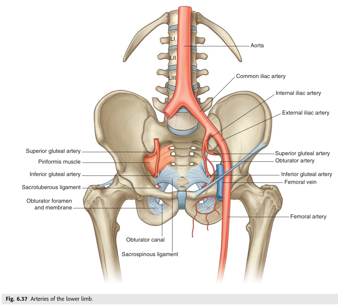
The lower limb arterial tree is the territory of peripheral vascular disease — the same atherosclerotic process that blocks coronary arteries progressively narrows the femoral, popliteal, and tibial vessels, producing the classic progression from claudication to rest pain to gangrene. The femoral artery enters the thigh at the midpoint of the inguinal ligament and travels through the adductor canal to become the popliteal artery at the adductor hiatus — knowing this anatomy explains why a tight adductor canal can compress the vessel in cyclists and runners. In the popliteal fossa the structures run deepest-to-most-superficial as PAT: popliteal Artery (deepest, against the bone), then popliteal Vein, then Tibial nerve — the reverse of the femoral triangle. Below the knee, three arteries reach the foot: anterior tibial (→ dorsalis pedis, palpable between 1st and 2nd metatarsals), posterior tibial (behind the medial malleolus, palpable in Tom Dick ANd Harry's groove), and peroneal (not palpable). Checking all five lower-limb pulses — femoral, popliteal, DP, PT, and comparing ABPI — is the minimum vascular examination every student must do fluently.
| Artery | Course | Pulse / Clinical |
|---|---|---|
| Femoral artery | Midpoint of inguinal ligament → femoral triangle (NAVEL: Nerve, Artery, Vein, Empty space, Lymphatics L→M) → adductor canal → adductor hiatus (between medial condyles) → popliteal artery | Femoral pulse at midinguinal ligament; profunda femoris (3 cm below inguinal lig) → medial + lateral circumflex femoral + 3–4 perforating aa. → posterior thigh |
| Popliteal artery | Adductor hiatus → popliteal fossa (deepest structure: artery deepest / most medial → popliteal vein → tibial nerve = PAT superficial to deep) → bifurcates at lower border of popliteus into ATA + tibioperoneal trunk | Popliteal pulse: prone, knee slightly flexed; hard to feel = deep structure. Popliteal aneurysm: most common peripheral aneurysm; 50% bilateral; thrombosis/embolism risk |
| Anterior tibial artery (ATA) | Through gap in interosseous membrane → anterior compartment between TA and EHL → becomes dorsalis pedis (DP) on dorsum of foot between extensor hallucis longus and extensor digitorum longus | Dorsalis pedis pulse: between 1st + 2nd metatarsals on dorsum. Anterior compartment syndrome: ATA + deep peroneal nerve compressed → foot drop + pain on passive toe extension → fasciotomy |
| Posterior tibial artery (PTA) | Deep posterior compartment → behind medial malleolus (with tibial nerve, Tom Dick ANd Harry = Tibialis posterior, flexor Digitorum longus, posterior tibial Artery and Nerve, flexor Hallucis longus) → divides into medial + lateral plantar aa. | Posterior tibial pulse: behind medial malleolus; plantar arches (deep plantar arch from lateral plantar + deep plantar from DP dorsalis pedis); tarsal tunnel syndrome = tibial nerve compression behind medial malleolus |
| Peroneal (fibular) artery | Tibioperoneal trunk → deep posterior compartment along fibula → lateral calcaneal branches. Not palpable clinically | May be dominant leg artery in advanced PAD; peroneal artery sparing = "peroneal runoff" — important for bypass graft planning |
0.7–0.9 = mild PAD (claudication); 0.5–0.7 = moderate PAD; <0.5 = severe / critical limb ischaemia; >1.3 = calcified vessels (medial calcinosis in diabetes — incompressible; ABPI unreliable, use toe pressures instead).
Fontaine classification: Stage I = asymptomatic; Stage II = intermittent claudication (IIa = >200 m, IIb = <200 m); Stage III = ischaemic rest pain (typically at night, hanging leg over bed relieves it by using gravity); Stage IV = tissue loss (ulceration/gangrene) = critical limb ischaemia.
Management: Exercise programme; smoking cessation (most important); antiplatelet therapy (aspirin/clopidogrel); statin; BP control. Revascularisation: percutaneous transluminal angioplasty (PTA) ± stent for short segment disease; bypass grafting (vein or PTFE) for long segment occlusion.
Inflammatory occlusive disease of small and medium vessels of the extremities; strongly associated with heavy tobacco use; predominantly young men (<45 years). Unlike atherosclerotic PAD, affects distal small vessels → severe digital ischaemia/gangrene despite minimal large vessel disease on angiography. Pathology: segmental thrombosis with inflammation (±giant cells) without atherosclerosis. Diagnosis: clinical + angiography (corkscrew collaterals). Treatment: STOP SMOKING — the only effective treatment; amputation if tissue loss. Buerger's test: elevate leg → pallor on elevation; dependent rubor on lowering = Buerger's positive (severe ischaemia).
- Popliteal fossa deep-to-superficial order (PAT)? popliteal Artery (deepest, on bone) → popliteal Vein → Tibial nerve (most superficial). Opposite of femoral triangle order. Popliteal pulse: knee slightly flexed, press deeply into the fossa
- Dorsalis pedis pulse location and parent artery? Dorsum of foot between 1st and 2nd metatarsals. Continuation of the anterior tibial artery after it crosses the ankle
- Posterior tibial pulse location and mnemonic for structures behind medial malleolus? Behind medial malleolus. Tom Dick ANd Harry = Tibialis posterior tendon, flexor Digitorum longus, posterior tibial Artery + Nerve, flexor Hallucis longus
- ABPI values: normal, claudication, critical ischaemia, calcified vessels? Normal 0.9–1.2; mild PAD/claudication 0.7–0.9; severe/critical <0.5; >1.3 = incompressible calcified vessels (diabetes) — use toe pressures instead
- Fontaine Stage III presentation and why patients hang the leg over the bed? Ischaemic rest pain at night. Hanging leg down uses gravity to improve perfusion pressure to the foot, relieving rest pain
Aortic Aneurysm & Dissection
An aneurysm is a permanent, localised dilation of an artery to more than 1.5 times its normal diameter — for the infrarenal aorta, that means ≥3 cm. AAAs grow silently and rupture without warning: the classic triad of severe tearing back pain, pulsatile abdominal mass, and hypotension is a surgical emergency with 80% overall mortality, but only 50% of patients even reach hospital. Size is the dominant rupture predictor — below 5.5 cm in men the annual risk stays low enough to watch; above it, elective repair (EVAR for most, open for complex anatomy) saves lives. Aortic dissection is a different catastrophe: an intimal tear lets blood into the media, creating a false lumen that can propagate the length of the aorta in seconds. The Stanford classification is the clinical one — Type A (ascending aorta involved) goes immediately to theatre because it can tear back into the pericardium (tamponade), shear off the coronary ostia (MI), or destroy the aortic valve (acute AR); Type B (descending only) gets β-blockers to reduce shear stress on the wall, targeting heart rate below 60 and systolic below 120 mmHg.
Dilation of infrarenal aorta to ≥3 cm (normal infrarenal aorta ~2 cm). Risk factors: male, age >65, smoking, hypertension, FH, atherosclerosis. 90% are infrarenal (below renal arteries, above bifurcation). Asymptomatic until rupture (pulsatile mass in thin patients). UK AAA Screening Programme: one-time USS at age 65 in men.
| Size | Annual rupture risk | Management |
|---|---|---|
| <4 cm | <1% | Surveillance USS every 3 years |
| 4–4.9 cm | ~1–2% | Surveillance USS every 12 months |
| 5–5.9 cm | ~5–10% | Surveillance USS every 3 months; vascular review |
| ≥5.5 cm (men) / ≥5 cm (women) | ~25%/year | Elective repair: EVAR (endovascular) or open (depends on anatomy + fitness) |
| Ruptured AAA | 80% overall mortality | Emergency (within 30 min): IV access + cross-match + theatre immediately. Classic triad: severe back/flank/abdominal pain + pulsatile abdominal mass + hypotension |
Type A: involves ascending aorta (regardless of origin) — surgical emergency. Risk: aortic regurgitation (aortic root involvement), coronary occlusion (MI — RCA most often), cardiac tamponade (haemopericardium), aortic rupture. Mortality 1–2% per hour without surgery → emergency surgical repair.
Type B: involves only descending aorta (distal to left subclavian origin) — medical management (IV β-blocker labetalol/esmolol, target SBP 100–120 mmHg, HR <60). TEVAR (thoracic endovascular aortic repair) for complicated Type B (malperfusion ischaemia, rupture, refractory pain, rapid expansion).
De Bakey Classification (anatomical, older but still examined):
Type I: dissection originates in ascending aorta, propagates through arch into descending aorta (= most extensive; includes Stanford A). Treatment: surgical.
Type II: dissection confined to ascending aorta only (= Stanford A, limited). Treatment: surgical.
Type III: dissection originates in descending aorta distal to left subclavian origin (= Stanford B). IIIa = confined to thoracic; IIIb = extends into abdominal. Treatment: medical ± TEVAR.
Mnemonic: De Bakey I + II = Stanford A (ascending involved → surgery); De Bakey III = Stanford B (descending only → medical).
Classic presentation: sudden tearing/ripping chest pain radiating to back + between scapulae; wide mediastinum on CXR (>8 cm or >1/2 chest width); unequal arm BP (>20 mmHg difference if subclavian involved); aortic regurgitation murmur (Type A). CT aortogram = gold standard. Bedside echo (TOE): useful in unstable patients.
- AAA definition, commonest site, and repair threshold? Aortic dilation ≥3 cm (normal ~2 cm). 90% infrarenal. Elective repair at ≥5.5 cm (men) / ≥5 cm (women); ruptured AAA = emergency (classic triad: tearing back pain + pulsatile mass + hypotension)
- UK AAA screening programme? One-time abdominal USS at age 65 in men. Women not routinely screened (lower incidence)
- Stanford Type A vs Type B — definition and immediate management? Type A = involves ascending aorta → emergency surgical repair (risk: tamponade, RCA occlusion, acute AR). Type B = descending aorta only → IV β-blocker (labetalol/esmolol), target SBP 100–120, HR <60; TEVAR if complicated
- De Bakey classification: which types = Stanford A, which = Stanford B? De Bakey I + II = Stanford A (ascending involved → surgery). De Bakey III = Stanford B (descending only → medical ± TEVAR)
- Classic presentation of aortic dissection and gold-standard investigation? Sudden tearing/ripping chest pain radiating to back/between scapulae; wide mediastinum on CXR (>8 cm); unequal arm BP (>20 mmHg). Gold standard = CT aortogram
Test Unit 11 knowledge
Arterial anatomy MCQs, AAA scenarios, and coronary territory questions.