The Heart
Overview, Position and Coverings
The pericardium is a fibroserous sac enclosing the heart and the roots of the great vessels, situated in the middle mediastinum. It consists of an outer fibrous layer and an inner serous layer, and functions to anchor the heart in position while allowing it to contract freely with minimal friction.
When fluid accumulates in the pericardial cavity — from pericarditis (pericardial effusion) or trauma (haemopericardium) — the inextensible fibrous pericardium cannot expand. The mounting pressure compresses the ventricles, impeding filling. This is cardiac tamponade — a medical emergency. Classic signs: distended neck veins, muffled heart sounds, falling pulse pressure (Beck's Triad). Emergency treatment: pericardiocentesis — needle inserted into the 5th/6th intercostal space adjacent to the sternum, exploiting the cardiac notch in the left lung. [Netter's 5th §22 p158]
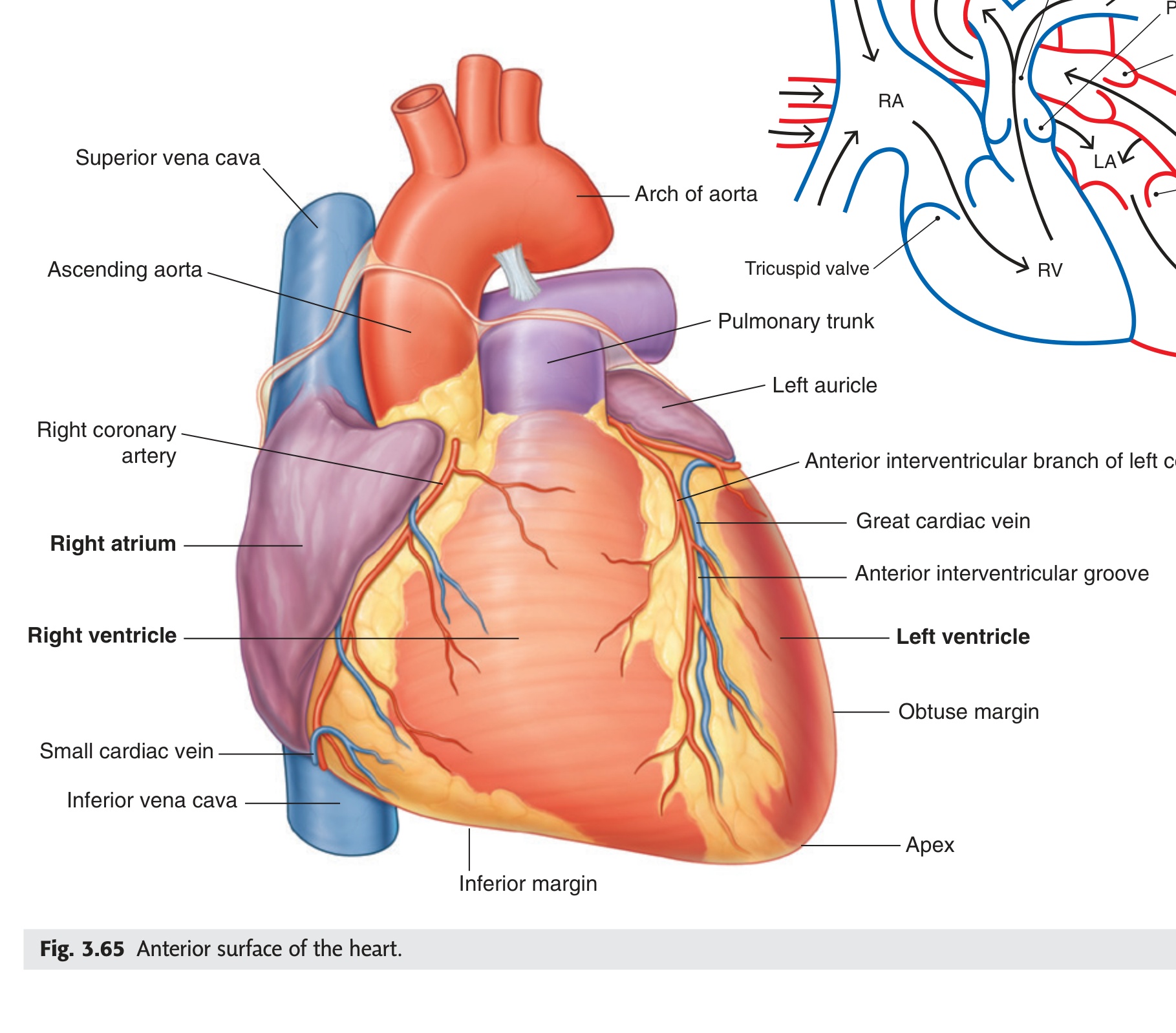
Left border — formed mainly by the left ventricle; from the 2nd left intercostal space to the apex.
Superior border — level of the 2nd costal cartilages bilaterally; great vessels emerge here.
Inferior border — from the 6th right costal cartilage to the apex.
Apex — left 5th intercostal space, midclavicular line (~9 cm from midline); formed by the left ventricle; the site of the apex beat (point of maximum impulse).
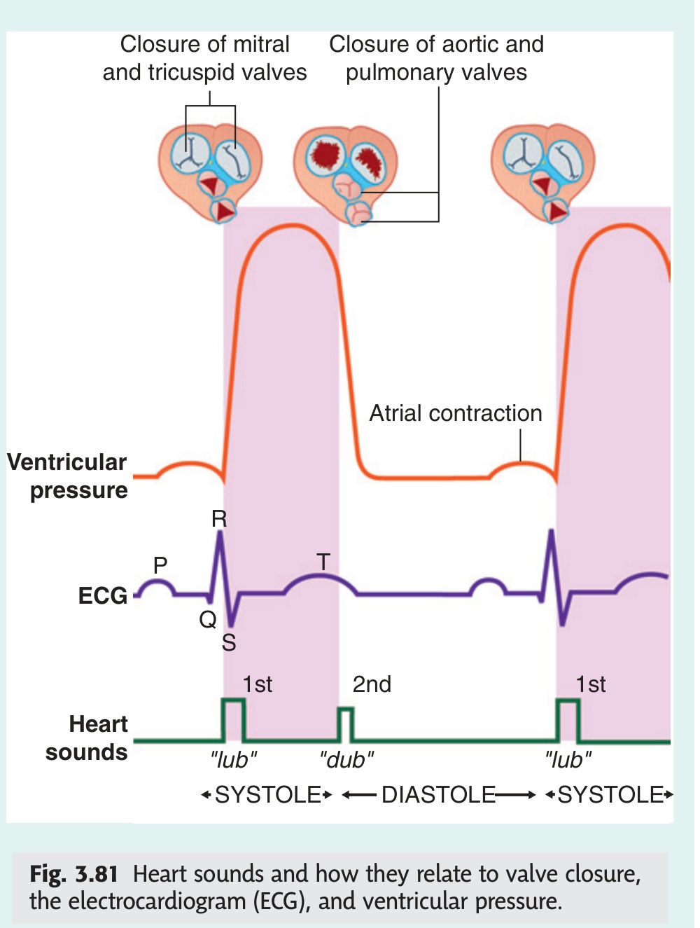
PV2L — Pulmonary valve heard over 2nd Left intercostal space
AV2R — Aortic valve heard over 2nd Right intercostal space
Tricuspid — lower left sternal edge (4th ICS) | Mitral — Apex (5th ICS MCL)
Tip: "All Patients Take Medicine" — Aortic, Pulmonary, Tricuspid, Mitral (right→left, top→bottom)
Internal Features of the Cardiac Chambers
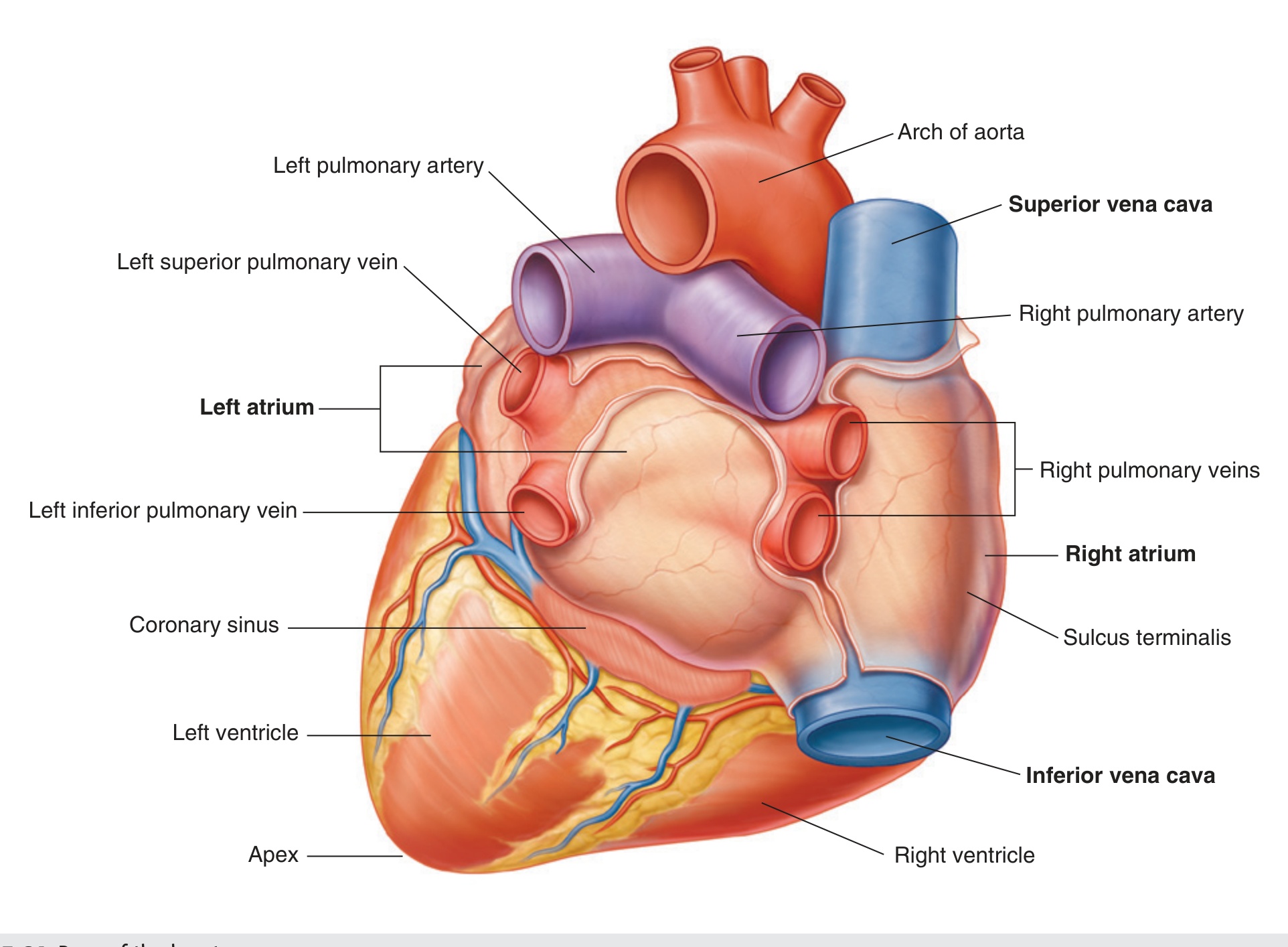
The right atrium is the thin-walled, posterosuperior chamber of the right heart, receiving deoxygenated blood from the systemic venous circulation and delivering it to the right ventricle via the right atrioventricular orifice.
- Orifice of the superior vena cava — returns deoxygenated blood from the upper half of the body (head, neck, upper limbs)
- Orifice of the inferior vena cava — returns deoxygenated blood from the lower half of the body (trunk, lower limbs, abdominal viscera)
- Orifice of the coronary sinus — returns venous blood from the myocardium (cardiac muscle) itself
The fossa ovalis is the commonest site of atrial septal defect (ASD). A patent foramen ovale (PFO) persists in ~25–30% of adults and can allow paradoxical emboli (venous clots crossing to the arterial circulation), causing cryptogenic stroke.
The right ventricle is the anterosuperior chamber of the right heart, receiving deoxygenated blood from the right atrium and pumping it through the pulmonary orifice into the pulmonary trunk for oxygenation in the lungs.
Outflowing part (conus arteriosus / infundibulum): smooth-walled, cone-shaped, directs blood upward to the pulmonary orifice, guarded by the pulmonary valve (3 semilunar cusps: anterior, right, left).
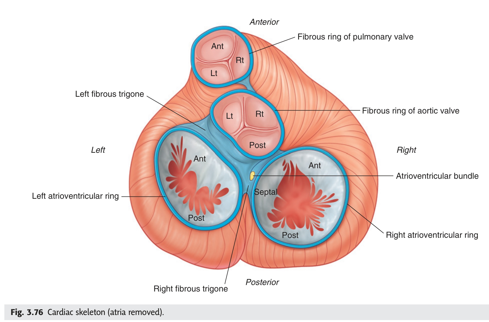
The tricuspid complex is the collective term for the four structural components that together prevent backflow of blood from the right ventricle into the right atrium during ventricular systole.
- Right atrioventricular fibrous ring — the annular fibrous skeleton anchoring the valve cusps.
- Tricuspid valve cusps — three cusps (anterior, posterior, septal) projecting into the ventricular lumen. Their free edges face downward into the ventricle.
- Chordae tendineae — strong tendinous cords attaching the free edges and ventricular surfaces of the cusps to the papillary muscles. They prevent the cusps from inverting into the atrium during systole.
- Papillary muscles — three cone-shaped muscular projections (anterior, posterior, septal) arising from the ventricular wall. They contract simultaneously with the ventricle, maintaining chordae tension throughout systole.
When the tricuspid complex fails — from right ventricular dilation stretching the annulus, or rheumatic disease — blood leaks back into the right atrium during systole. This raises right atrial and venous pressure, causing: raised jugular venous pressure (JVP), peripheral oedema, hepatomegaly, and ascites — the classic signs of right heart failure.
The left atrium is the posterosuperior chamber of the left heart, receiving oxygenated blood from the four pulmonary veins and delivering it to the left ventricle via the left atrioventricular orifice.
One outlet: the left atrioventricular orifice — guarded by the bicuspid (mitral) valve; leads blood into the left ventricle.
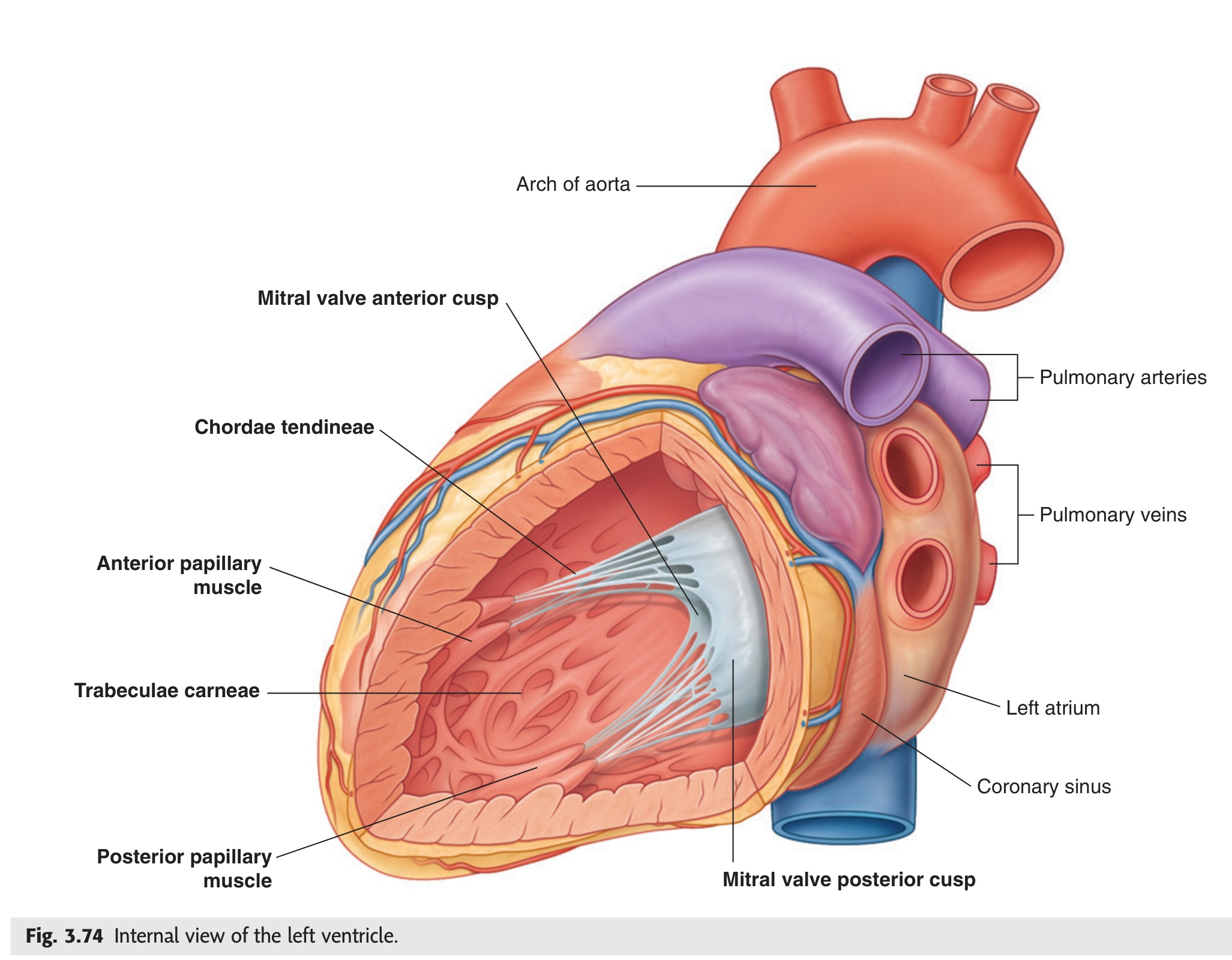
The left ventricle is the posteroinferior chamber of the left heart that receives oxygenated blood from the left atrium and pumps it into the aorta for systemic distribution. Its wall is approximately three times as thick as the right ventricle, reflecting the much higher pressures it must generate.
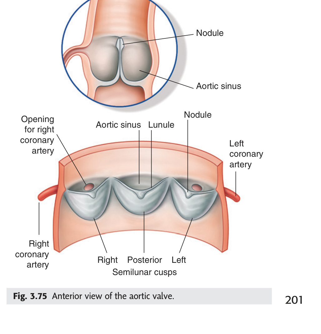
Outflowing part (aortic vestibule): smooth-walled. Leads to the aortic orifice guarded by the aortic valve (three semilunar cusps: posterior, right, left). Above the aortic valve cusps, the aortic wall bulges outward to form the aortic sinuses (sinuses of Valsalva) — the right and left aortic sinuses give rise to the right and left coronary arteries respectively.
The bicuspid complex is the set of four structural components guarding the left atrioventricular orifice that together prevent backflow of blood from the left ventricle into the left atrium during ventricular systole.
- Left atrioventricular fibrous ring — the structural anchor for the bicuspid valve cusps.
- Bicuspid valve cusps — two cusps (anterior and posterior). The mitral valve is the most frequently diseased heart valve.
- Chordae tendineae — tether the free edges of the cusps to the papillary muscles below, preventing inversion into the left atrium.
- Papillary muscles — anterior and posterior; contract simultaneously with the left ventricle to maintain chordae tension throughout systole.
The mitral valve is the most frequently diseased heart valve — particularly in rheumatic heart disease and infective endocarditis. Mitral regurgitation allows backflow into the left atrium, producing a high-pitched pansystolic murmur loudest at the apex. Longstanding mitral regurgitation leads to left atrial enlargement, pulmonary hypertension, and eventually left ventricular failure. [Netter's 5th §22 p172]
Conduction System of the Heart ★★★
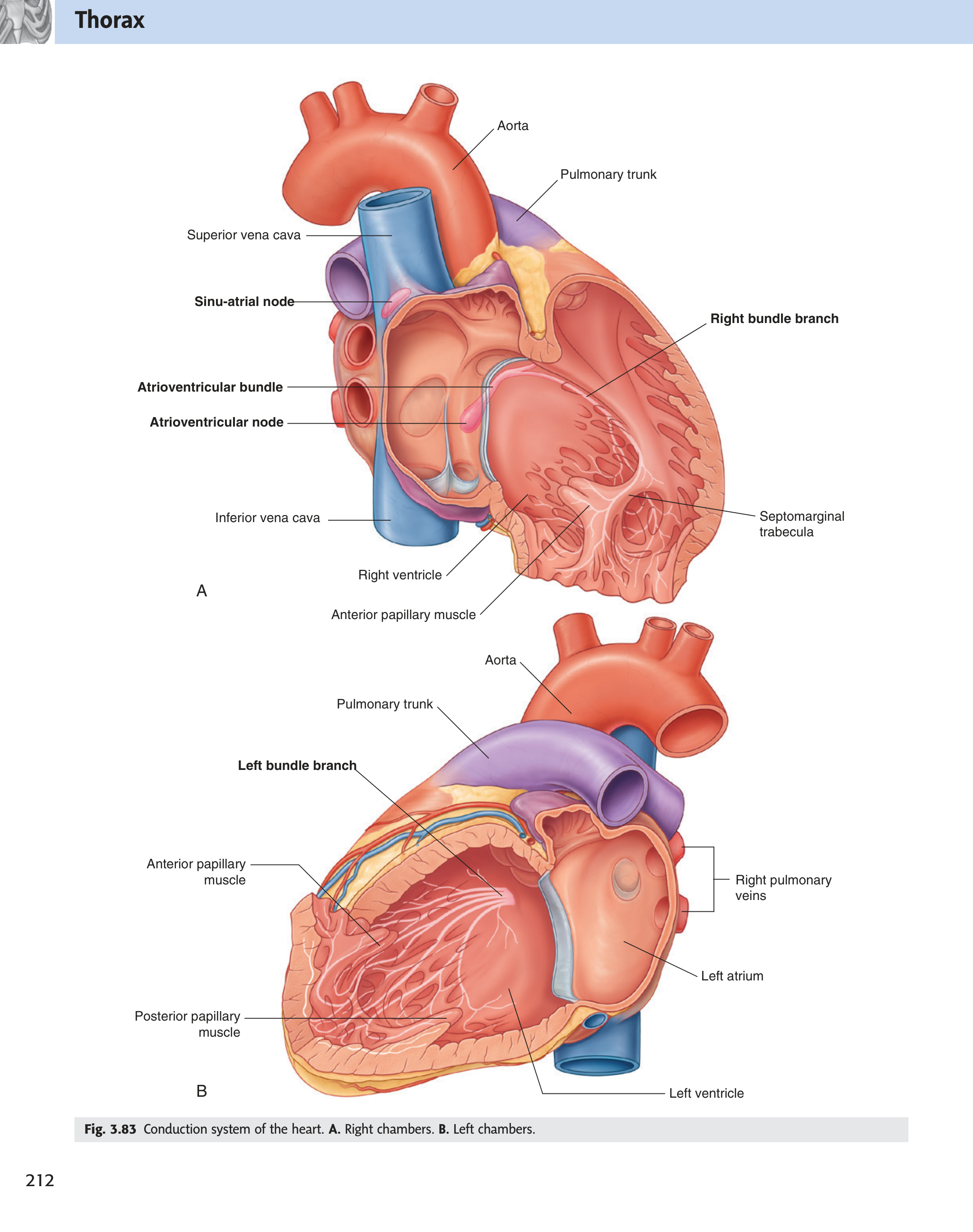
The cardiac conduction system is a network of specialised myocardial fibres that spontaneously generates and coordinates electrical impulses, ensuring the sequential, rhythmic contraction of the atria followed by the ventricles at an appropriate rate.
- Sinoatrial node (SA node) — in the wall of the right atrium, near the opening of the superior vena cava. The pacemaker of the heart — spontaneously depolarises at 60–100/min, initiating each cardiac cycle.
- Internodal tracts — three pathways (anterior, middle, posterior) conducting the impulse through the atrial walls to the AV node, causing atrial contraction en route.
- Atrioventricular node (AV node) — in the interatrial septum near the opening of the coronary sinus. Introduces a brief delay (~0.1 seconds) — allowing atrial contraction to complete and ventricular filling to occur before ventricular systole begins.
- Atrioventricular bundle (Bundle of His) with right and left bundle branches — passes from the AV node into the interventricular septum; the right bundle branch runs within the septomarginal trabecula (moderator band) to the right ventricular wall; the left bundle branch passes through the septum to the left ventricular wall.
- Subendocardial plexus of Purkinje fibres — terminal network of large, rapidly-conducting fibres spreading from the bundle branches throughout both ventricular walls. Their rapid conduction ensures near-simultaneous ventricular depolarisation, producing a single powerful coordinated contraction. [Gray's 4e Ch3 p188]
SA node (60–100/min) → AV node (40–60/min) → Purkinje fibres (30–40/min)
Each level becomes the pacemaker if the one above it fails. At Purkinje rate (30–40/min) the patient is haemodynamically compromised → requires an implanted cardiac pacemaker.
Coronary Circulation ★★★
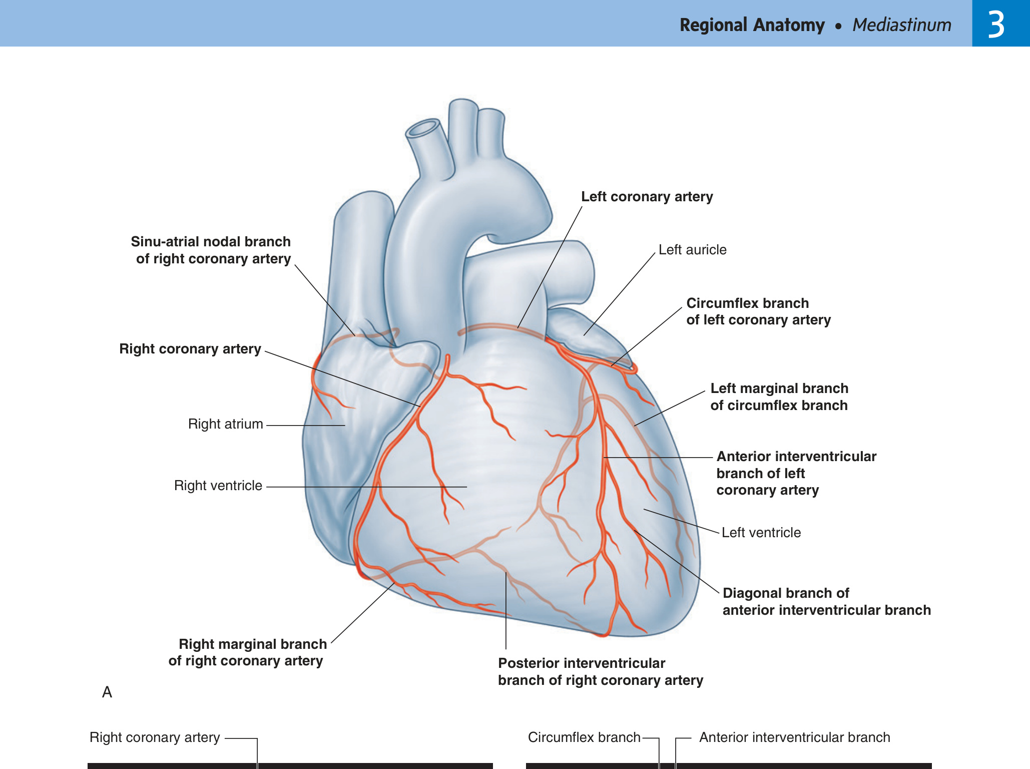
The coronary arteries are the first branches of the aorta, arising from the aortic sinuses (sinuses of Valsalva) immediately above the aortic valve cusps, and supplying the entire myocardium with oxygenated blood.
| Vessel | Origin | Main Branches | Territory Supplied |
|---|---|---|---|
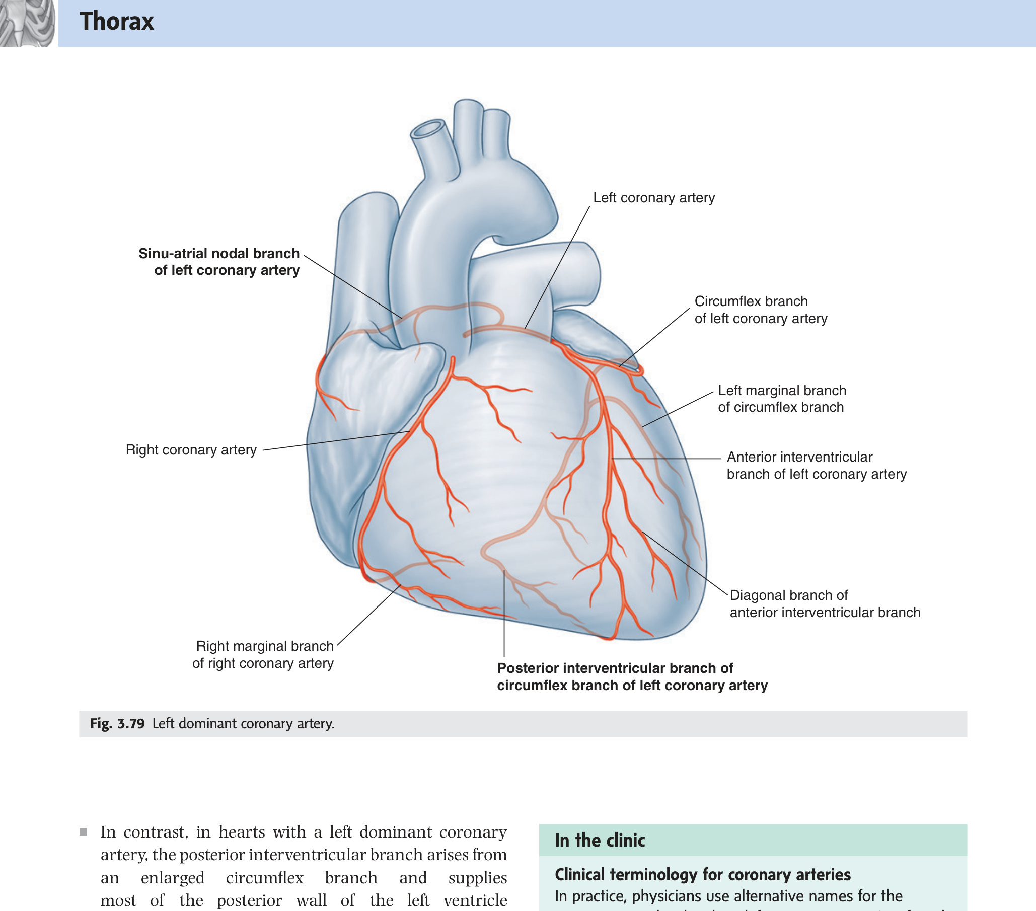
| Right Coronary Artery (RCA) | Right aortic sinus | Posterior interventricular branch (posterior descending artery); Posterior branch of left ventricle; SA nodal branch; AV nodal branch | Right atrium, right ventricle, posterior septum, posterior left ventricle, SA node (60%), AV node (80–90%) |
| Left Coronary Artery (LCA) | Left aortic sinus | Anterior interventricular branch (LAD); Circumflex branch | Anterior walls of both ventricles, anterior 2/3 of interventricular septum (LAD); left atrium, posterior left ventricle (circumflex) |
The left anterior descending artery (LAD) — the "widow maker" — supplies the largest amount of ventricular muscle. Its occlusion causes a large anterior myocardial infarction with high mortality. In CABG surgery, the internal thoracic artery or saphenous vein is grafted to bypass the occlusion, restoring distal perfusion. [Netter's 5th §22 p158]
Cardiac ischaemic pain is referred to the left pectoral region and medial left arm because visceral afferent fibres from the heart enter spinal cord segments T1–T4/T5 on the left — the same segments receiving somatic sensory input from the chest wall and arm. The brain misinterprets the cardiac signal as coming from the arm.
| Chamber | Inlets | Outlet | Key Internal Feature | Exam Note |
|---|---|---|---|---|
| Right Atrium | SVC orifice IVC orifice Coronary sinus orifice |
Right AV orifice (tricuspid valve) | Fossa ovalis; crista terminalis; pectinate muscles | 3 inlets = most tested |
| Right Ventricle | Right AV orifice (tricuspid) | Pulmonary orifice (pulmonary valve) | Septomarginal trabecula (moderator band); conus arteriosus | Moderator band = right bundle branch |
| Left Atrium | 4 pulmonary vein orifices | Left AV orifice (bicuspid/mitral valve) | Smooth posterior wall; left auricle | 4 pulmonary veins = always tested |
| Left Ventricle | Left AV orifice (bicuspid/mitral) | Aortic orifice (aortic valve) | Thickest wall; aortic vestibule; aortic sinuses → coronary arteries | Thickest wall = 3× right ventricle |