Peritoneum & Endocrine Glands
Peritoneum — Layers & Terminology
The peritoneum is a single continuous serous sheet that lines the entire abdomen — think of it as cling film draped over and around every organ, with the visceral layer kissing organ surfaces and the parietal layer hugging the walls. The critical distinction is that parietal peritoneum has somatic innervation (sharp, localisable pain), while visceral peritoneum has only autonomic supply (dull, midline, colicky pain) — which is why appendicitis starts as central periumbilical discomfort before somatic peritoneal irritation produces the classic right-iliac-fossa localisation. Organs classified as retroperitoneal sit behind this membrane; they were either always posterior (kidneys, aorta) or started intraperitoneal and fused to the posterior wall during gut rotation — a developmental trick that stabilises structures like the duodenum and ascending colon.
A single continuous sheet of serous membrane (mesothelium + connective tissue) lining the abdominal cavity and covering the viscera. Its secretion (~50 mL serous fluid) allows free movement of organs against each other.
| Term | Meaning | Examples |
|---|---|---|
| Parietal peritoneum | Lines the abdominal + pelvic walls; innervated by somatic nerves (phrenic, intercostal, lumbar) — localises pain well | Anterior abdominal wall lining; over diaphragm |
| Visceral peritoneum | Covers organs; innervated by visceral autonomic nerves — diffuse, poorly localised pain (colicky, midline referred) | Covers liver capsule, small bowel, uterus |
| Intraperitoneal organ | Almost completely covered by peritoneum, suspended by a mesentery | Stomach, jejunum, ileum, sigmoid colon, uterus (functionally), spleen |
| Retroperitoneal organ | Behind the parietal peritoneum; only anterior surface covered (or none) | Kidneys, ureters, aorta, IVC, duodenum (2nd–4th), pancreas (body/tail), ascending + descending colon |
| Secondarily retroperitoneal | Originally intraperitoneal; fused to posterior abdominal wall during development | Duodenum, pancreas, ascending + descending colon |
| Mesentery | Double fold of peritoneum attaching intraperitoneal organ to abdominal wall; contains vessels, lymphatics, nerves | Mesentery of small bowel (root = 15 cm diagonal, L2 → RIJF); transverse mesocolon; sigmoid mesocolon |
| Omentum | Peritoneal fold attaching stomach to another structure | Greater omentum (4-layered; stomach → transverse colon → hangs down); lesser omentum (stomach/duodenum → liver) |
| Ligament (peritoneal) | Double fold connecting organ to wall or organ to organ; may contain vessels | Falciform (liver → anterior abdominal wall); coronary ligament; gastrosplenic; splenorenal |
Suprarenal glands (adrenals) · Aorta & IVC · Duodenum (2nd–4th parts) · Pancreas (head, body — tail usually intraperitoneal) · Ureters · Colon (ascending & descending) · Kidneys · Esophagus (thoracic) · Rectum (middle + lower third)
Primary retroperitoneal (always were): suprarenal glands, aorta/IVC, kidneys, ureters, gonads.
Secondary retroperitoneal (fused during development): duodenum D2–D4, pancreas, ascending colon, descending colon, rectum. These were initially intraperitoneal but their mesenteries fused posteriorly — therefore the anterior surface has peritoneum but the posterior does not.
Peritonitis: inflammation of peritoneum. Primary: spontaneous bacterial (ascites + E. coli/Klebsiella; paracentesis + neutrophils >250/mm³ → treat empirically). Secondary: perforated viscus (appendix, DU, diverticulitis) — suddenly diffuse abdominal pain, board-like rigidity, guarding, absent bowel sounds. Rebound tenderness = parietal peritoneum inflamed (Blumberg sign). Erect CXR: sub-diaphragmatic free gas = perforation until proven otherwise. Tx: IV antibiotics + emergency laparotomy/laparoscopy.
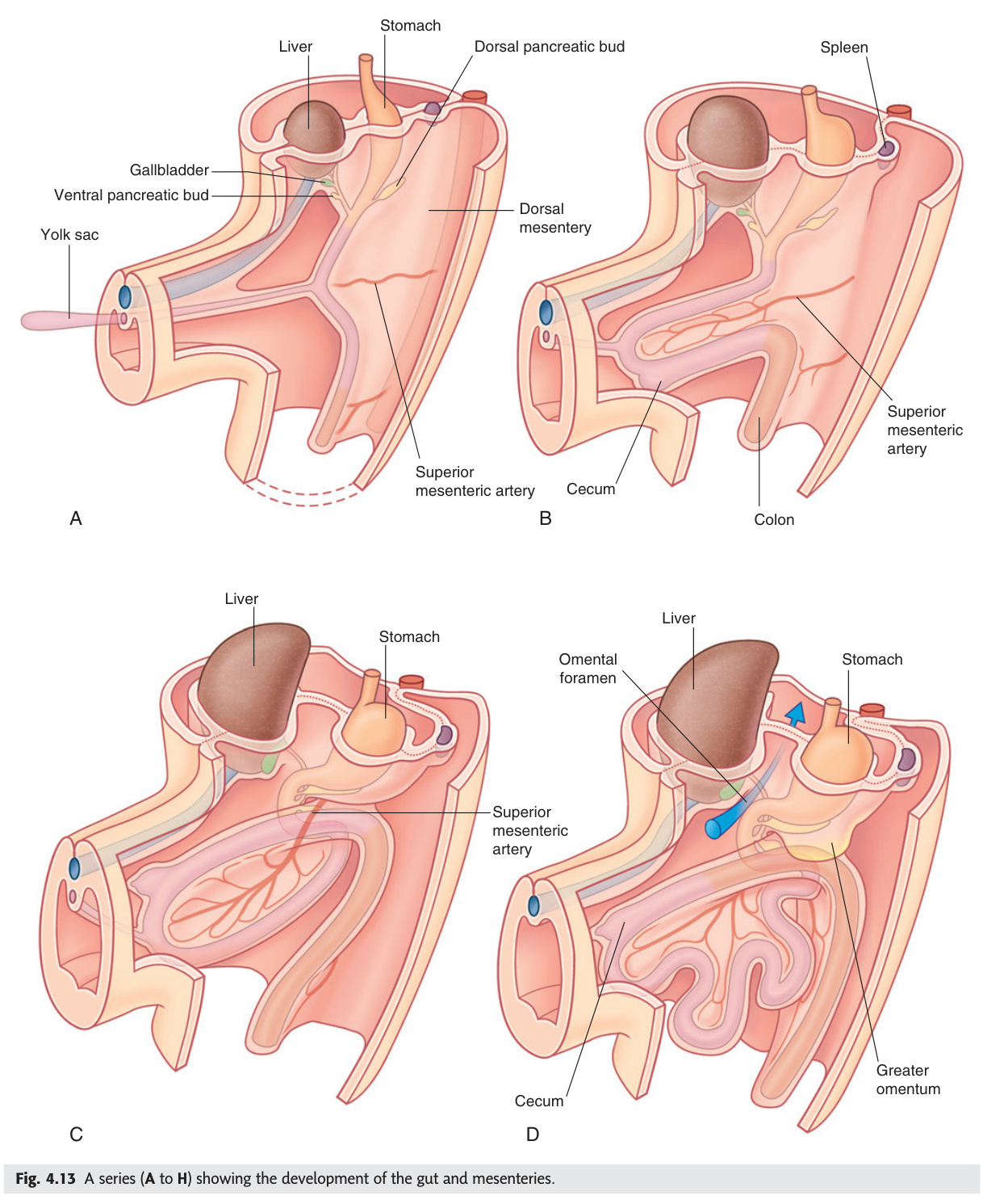
Understanding embryological gut rotation explains the adult layout of the peritoneum, mesenteries, and retroperitoneal organs.
| Stage | Weeks | Events |
|---|---|---|
| Herniation | 6–10 | Midgut elongates rapidly → herniates through umbilical ring (physiological umbilical hernia); rotates 270° anticlockwise around the superior mesenteric artery (SMA) axis as it returns |
| First 90° rotation | 6–8 (outside) | Pre-arterial (small bowel) limb rotates to the left; post-arterial (large bowel) limb rotates to the right |
| Return + next 180° | 10–11 | Small bowel re-enters abdomen first (displaced to right then final left position); caecum starts high, descends to right iliac fossa; total = 270° anticlockwise rotation |
| Peritoneal fixation | After return | Ascending colon + descending colon fuse to posterior abdominal wall → secondarily retroperitoneal; transverse colon + sigmoid remain intraperitoneal; mesentery of small bowel = 15 cm diagonal root (duodenojejunal flexure L2 left → right iliac fossa) |
Malrotation: failure of complete 270° rotation → duodenojejunal flexure stays to the right of midline (should be left); caecum stays subhepatic rather than descending to right iliac fossa. Ladd's bands: fibrous peritoneal bands from abnormally positioned caecum → cross and compress duodenum → neonatal/infant bilious vomiting. Upper GI contrast = diagnostic (DJ flexure position). Midgut volvulus: the unrotated midgut has a narrow mesenteric pedicle around the SMA → prone to axial twist → catastrophic ischaemia of almost all small bowel (SMA occlusion). Presents neonates as bilious vomiting + abdominal distension → emergency Ladd's procedure (detorsion, division of Ladd's bands, appendicectomy, caecum placed left, duodenum right).
- Parietal vs visceral peritoneum pain? Parietal: somatic (intercostal/phrenic/lumbar) → sharp, localised. Visceral: autonomic → dull, midline, referred (e.g., appendicitis starts periumbilical)
- Name three secondarily retroperitoneal structures. Duodenum D2–D4, pancreas (head/body), ascending + descending colon — originally intraperitoneal, mesenteries fused posteriorly during gut rotation
- Mesenteric root of small bowel and its significance? 15 cm diagonal from D-J flexure (L2 left) to right iliac fossa; narrow pedicle around SMA = the axis of midgut volvulus in malrotation
- "SAD PUCKER" stands for? Retroperitoneal: Suprarenal, Aorta/IVC, Duodenum, Pancreas, Ureters, Colon (ascending/descending), Kidneys, Esophagus, Rectum
- Rebound tenderness (Blumberg sign) indicates? Parietal peritoneal irritation — somatic nerves localise the pain; guarding + rigidity confirm peritonism (parietal, not visceral, is inflamed)
Peritoneal Sacs & Epiploic Foramen (of Winslow)
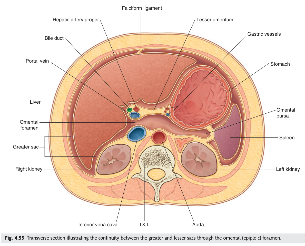
The peritoneal cavity is not one uniform space — it has a hidden compartment called the lesser sac (omental bursa), tucked behind the stomach and connected to the greater sac through only one narrow door: the epiploic foramen of Winslow. Knowing the four boundaries of this foramen is surgically vital, because the hepatoduodenal ligament — carrying the portal triad of portal vein, hepatic artery, and bile duct — forms its anterior wall, and squeezing it between finger and thumb (the Pringle manoeuvre) instantly controls haemorrhage from the liver. The greater omentum, a four-layered fat-rich apron hanging from the stomach, earns its nickname "abdominal policeman" by migrating toward inflamed or perforated viscera to wall off infection and prevent generalised peritonitis.
| Structure | Boundaries / Description |
|---|---|
| Greater sac | Main peritoneal cavity — all of it except the lesser sac |
| Lesser sac (omental bursa) | Potential space posterior to stomach + lesser omentum, anterior to pancreas. Communicates with greater sac via epiploic foramen only |
| Epiploic foramen (foramen of Winslow) | Opening between greater and lesser sac. Anteriorly: free edge of lesser omentum (hepatoduodenal ligament — contains portal triad: portal vein, hepatic artery, bile duct) Posteriorly: IVC Superiorly: caudate lobe of liver Inferiorly: first part of duodenum |
| Greater omentum | 4-layered peritoneal apron from greater curvature of stomach → hangs down → folds back up to transverse colon. Contains fat + immune cells (macrophages). "Abdominal policeman" — seals off perforations |
| Lesser omentum | Hepatogastric (liver→stomach) + hepatoduodenal (liver→duodenum) ligaments. Free right edge = hepatoduodenal ligament (portal triad). Forms anterior boundary of epiploic foramen |
Pancreatic pseudocyst: walled-off fluid collection in lesser sac following acute pancreatitis; pancreatic enzymes leak → collections form behind stomach. Presents as epigastric mass + persistent pain after acute pancreatitis. USS/CT confirms. Most resolve; large ones → endoscopic or surgical drainage. Penetrating peptic ulcer: posterior gastric ulcer erodes through stomach wall directly into pancreas/lesser sac → severe back pain + sudden relief of dyspeptic symptoms (ulcer now sealed by pancreas).
- Four boundaries of the epiploic foramen? Anterior: hepatoduodenal ligament (portal triad); Posterior: IVC; Superior: caudate lobe; Inferior: 1st part of duodenum
- What is the Pringle manoeuvre? Compress hepatoduodenal ligament between finger (through foramen) and thumb → occludes portal vein + hepatic artery → controls hepatic haemorrhage (safe ~15–20 min)
- Pancreatic pseudocyst: where and why? Walled-off collection in lesser sac after acute pancreatitis; enzyme leak → collection behind stomach; treat large ones by endoscopic drainage
- Contents of the free edge of the lesser omentum? Portal vein (posterior), hepatic artery proper (left), common bile duct (right) — the portal triad in the hepatoduodenal ligament
- Greater vs lesser omentum? Greater: 4-layered apron from greater curvature → transverse colon, "abdominal policeman". Lesser: hepatogastric + hepatoduodenal ligaments connecting stomach/duodenum to liver
Peritoneal Pouches & Clinically Important Spaces
Gravity and posture determine which peritoneal pouch fills first with blood or pus, making knowledge of these spaces directly actionable in resuscitation. In the supine patient, Morison's pouch — the hepatorenal recess between the right liver lobe and right kidney — is the most dependent point of the entire peritoneal cavity, so the FAST ultrasound exam targets it first in abdominal trauma. When the patient sits or stands, the rectouterine pouch of Douglas becomes the lowest point in females (accessible by needle culdocentesis for a ruptured ectopic), while the rectovesical pouch serves the same role in males, and the subphrenic spaces communicate diaphragmatic irritation as referred right-shoulder-tip pain via the C3–C5 phrenic nerve.
| Space | Location | Clinical Significance |
|---|---|---|
| Hepatorenal recess (Morison's pouch) | Between right lobe of liver + right kidney; most dependent part of peritoneal cavity in the SUPINE position | Fluid collects here first in supine patient — USS FAST exam looks for fluid here in trauma; ruptured ectopic + appendicitis abscess can track here |
| Rectouterine pouch (pouch of Douglas) | Between uterus + rectum in females — most dependent part of peritoneal cavity in the ERECT female | Free fluid accumulates here (ruptured ectopic, PID, ovarian cyst rupture); accessible via culdocentesis/transvaginal USS |
| Rectovesical pouch | Between bladder + rectum in males — most dependent part in ERECT male | Fluid accumulates here in erect male trauma patients; tumours can seed here (Blumer's shelf — rectal shelf palpated on PR) |
| Vesicouterine pouch | Between uterus + bladder in females | Less deep than pouch of Douglas; rarely accumulates significant fluid |
| Left subphrenic space | Between diaphragm + left lobe of liver / stomach / spleen | Abscess here after perforated gastric ulcer or splenectomy; gas under left hemidiaphragm |
| Right subphrenic space | Between diaphragm + right lobe of liver; anterior to coronary ligament | Abscess after perforated appendix/DU — referred right shoulder tip pain (diaphragm irritation → C3–C5 phrenic) |
Supine: Morison's pouch (right hepatorenal) fills first. Erect female: pouch of Douglas. Erect male: rectovesical pouch. Knowing posture = knowing where to look on USS FAST.
- Which space fills first in a supine trauma patient on FAST? Morison's pouch (hepatorenal recess, right) — most dependent in supine; first target of FAST ultrasound
- Pouch of Douglas: location and clinical access? Between uterus and rectum (female); most dependent erect; accessed by culdocentesis through posterior fornix or transvaginal USS
- What is Blumer's shelf? Tumour seedlings in the rectovesical pouch felt on PR — classically gastric or rectal carcinoma metastasis palpated as a "shelf"
- Right shoulder-tip pain post-abdominal surgery suggests? Right subphrenic abscess or free gas under right diaphragm → diaphragmatic irritation → referred via phrenic nerve (C3–C5)
- Most dependent space in an erect male? Rectovesical pouch (between bladder and rectum)
Suprarenal (Adrenal) Glands
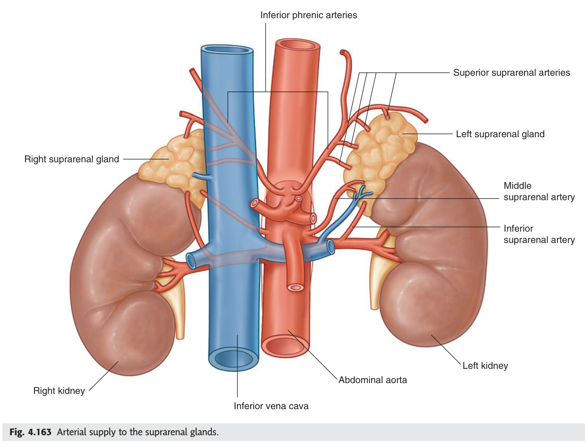
The adrenal glands sit atop both kidneys but are anatomically and functionally distinct from them — the right is pyramidal and drains via an extremely short vein directly into the IVC (a hazard in right adrenalectomy), while the left is crescent-shaped and drains more forgivingly into the left renal vein. Each gland is really two organs fused together: the outer cortex (mesodermal, zoned GFR — producing aldosterone, cortisol, and androgens) and the inner medulla (neural crest–derived chromaffin cells releasing adrenaline and noradrenaline as the body's emergency catecholamine reservoir). When the cortex fails, as in Addison's disease, the absence of cortisol feedback drives ACTH sky-high, and because ACTH shares sequence with melanocyte-stimulating hormone (both cleaved from POMC), the hallmark sign is hyperpigmentation — the patient's skin and mucous membranes darkening even as they collapse from hypotension and hypoglycaemia.
| Feature | Right | Left |
|---|---|---|
| Shape | Pyramidal | Crescent/semilunar |
| Position | Anteromedial to right kidney; posterior to IVC | Anteromedial to left kidney; related to pancreas + splenic vessels |
| Vein drainage | Short right suprarenal vein → directly into IVC (IMPORTANT — very short!) | Left suprarenal vein → left renal vein → IVC (longer) |
| Arterial supply | Superior (inferior phrenic a.), middle (abdominal aorta at T12), inferior (renal artery) — all three on each side | |
| Zone (outer → inner) | Mnemonic layer | Hormone |
|---|---|---|
| Zona glomerulosa (outer) | GFR (like kidney): Glomerulosa → Fasciculata → Reticularis | Aldosterone (mineralocorticoid) — regulated by RAAS + K⁺ |
| Zona fasciculata (middle) | Cortisol (glucocorticoid) — regulated by ACTH | |
| Zona reticularis (inner) | Androgens (DHEA, androstenedione) — regulated by ACTH | |
| Medulla | Chromaffin cells | Adrenaline (80%) + noradrenaline — sympathetic preganglionic → chromaffin cells (modified postganglionic neurones) |
Phaeochromocytoma: catecholamine-secreting tumour of adrenal medulla (or extra-adrenal paraganglia). "10% rule": 10% bilateral, 10% extra-adrenal, 10% malignant, 10% in children, 10% familial (MEN2, NF1, VHL). Presents with paroxysmal hypertension + headache + sweating + palpitations. Diagnose: 24-hour urinary metanephrines. Block α first (phenoxybenzamine) BEFORE β to avoid unopposed α → hypertensive crisis.
Conn's syndrome (primary hyperaldosteronism): adrenal adenoma → excess aldosterone → hypertension + hypokalaemia + metabolic alkalosis. RAAS suppressed. Treat with laparoscopic adrenalectomy.
Addison's disease: primary adrenocortical insufficiency — failure of the cortex to produce cortisol and aldosterone. Distinguished from secondary insufficiency (pituitary ACTH deficiency) by hyperpigmentation (↑ACTH/MSH from POMC processing) and mineralocorticoid deficiency.
| Feature | Primary (Addison's) | Secondary (ACTH deficiency) |
|---|---|---|
| Cause | Autoimmune (80%), TB, haemorrhage (Waterhouse-Friderichsen), metastases | Pituitary adenoma, Sheehan's syndrome, steroid withdrawal |
| ACTH | ↑↑ (no cortisol feedback) | ↓↓ |
| Hyperpigmentation | Yes — buccal mucosa, skin creases, scars | No (ACTH is low) |
| Mineralocorticoid | Deficient → hyponatraemia + hyperkalaemia | Preserved (aldosterone via RAAS) |
| Diagnosis | Short Synacthen test (cortisol <550 nmol/L at 30 min) | ITT or glucagon stimulation |
| Treatment | Hydrocortisone + fludrocortisone (lifelong) | Hydrocortisone only |
Addisonian crisis: triggered by physiological stress (infection, surgery, trauma) in undertreated Addison's. Features: severe hypotension, vomiting, hypoglycaemia, collapse. Management: immediate IV hydrocortisone 100 mg bolus + IV 0.9% NaCl + dextrose. Do NOT delay for investigation results.
Waterhouse-Friderichsen syndrome: bilateral adrenal haemorrhage in overwhelming septicaemia — classically Neisseria meningitidis in children. DIC co-exists. Causes acute primary adrenal insufficiency. Rapidly fatal without IV steroids.
Cushing's Syndrome — Full Differential ★★★
| Cause | ACTH | Key Features | High-dose Dex Test |
|---|---|---|---|
| Pituitary adenoma (Cushing's disease, ~70%) | ↑ | Bilateral adrenal hyperplasia; female predominance | Suppresses ≥50% |
| Adrenal adenoma/carcinoma (~15%) | ↓↓ (suppressed) | Unilateral adrenal mass; carcinoma: virilisation + rapid onset | No suppression |
| Ectopic ACTH (~15%) | ↑↑↑ (very high) | SCLC most common; rapid onset; severe hypokalaemia + hyperpigmentation | No suppression |
| Iatrogenic (most common overall) | ↓ (exogenous suppresses axis) | Long-term steroid therapy; adrenal atrophy; abrupt withdrawal → crisis | N/A |
Investigation sequence: 24-h urinary free cortisol (or late-night salivary cortisol) → overnight 1 mg dexamethasone suppression screen → low-dose 2-day DST (confirm Cushing's) → ACTH level (dependent vs independent) → high-dose DST or CRH stimulation → bilateral inferior petrosal sinus sampling (BIPSS) if equivocal.
Congenital Adrenal Hyperplasia (CAH) — 21-Hydroxylase Deficiency ★★
95% of cases. Blocks 17-hydroxyprogesterone → 11-deoxycortisol and progesterone → 11-deoxycorticosterone. Result: cortisol ↓ + aldosterone ↓ → ACTH rises uninhibited → adrenal androgens accumulate.
| Form | Biochemistry | Presentation |
|---|---|---|
| Classic salt-wasting (75%) | Cortisol ↓↓, Aldosterone ↓↓, Androgens ↑↑ | Neonatal crisis — vomiting, hyponatraemia, hyperkalaemia, shock; ambiguous genitalia in 46,XX females |
| Classic simple virilising | Cortisol ↓, Aldosterone ≈normal, Androgens ↑↑ | Virilisation of 46,XX; precocious puberty in males; no salt-wasting |
| Non-classic (late-onset) | Mild enzyme deficiency | Female: hirsutism, acne, oligomenorrhoea (mimics PCOS); ↑17-OHP on ACTH stimulation |
Key marker: elevated 17-hydroxyprogesterone (17-OHP). Newborn screening (heel prick) detects raised 17-OHP. Treatment: hydrocortisone (suppresses ACTH) + fludrocortisone (salt-wasting form).
- Why must α-blockade precede β-blockade in phaeochromocytoma? β-blockade alone removes vasodilatory β2 effect → unopposed α vasoconstriction → hypertensive crisis. Give phenoxybenzamine first, then propranolol
- GFR zones of adrenal cortex and products? Glomerulosa (outer) → aldosterone (RAAS); Fasciculata (middle) → cortisol (ACTH); Reticularis (inner) → androgens (DHEA); Medulla → adrenaline/noradrenaline
- Neonate with vomiting, hyponatraemia, hyperkalaemia, elevated 17-OHP — diagnosis? Classic salt-wasting CAH; 21-hydroxylase deficiency; treat with IV hydrocortisone + saline/dextrose acutely, then hydrocortisone + fludrocortisone long-term
- ACTH level to distinguish Cushing's disease vs ectopic ACTH vs adrenal adenoma? Cushing's disease: ACTH ↑, suppresses on high-dose dex. Ectopic ACTH: ACTH ↑↑↑, no suppression. Adrenal adenoma: ACTH ↓↓ (suppressed by autonomous cortisol)
- What is Waterhouse-Friderichsen syndrome? Bilateral adrenal haemorrhage in meningococcal septicaemia → acute adrenal crisis; DIC co-exists; treat immediately with IV hydrocortisone
Thyroid Gland
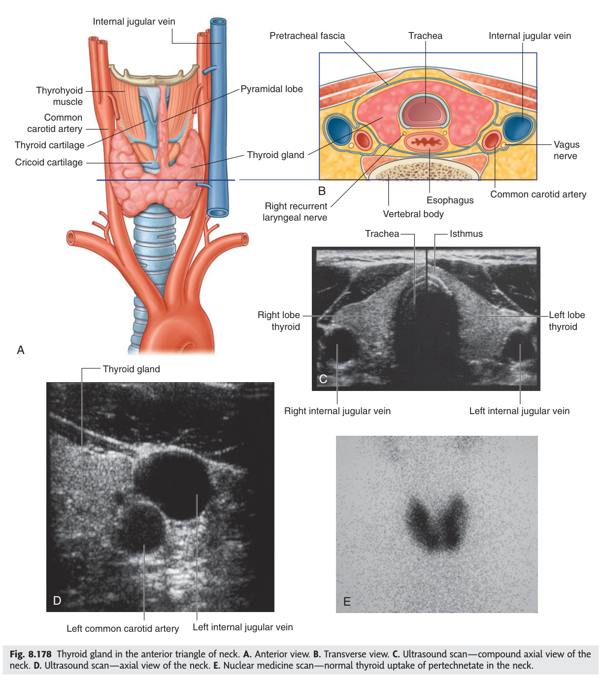
The thyroid gland wraps around the trachea at C5–T1, anchored to the cricoid by Berry's ligament — which is why it bobs up on swallowing, a clinical sign that differentiates a thyroid mass from every other neck lump. Two nerves run in dangerous proximity during thyroid surgery: the recurrent laryngeal nerve (RLN), which passes posterior to Berry's ligament and supplies all intrinsic laryngeal muscles except cricothyroid, and the external branch of the superior laryngeal nerve (EBSLN), the "singer's nerve" that tunes pitch via cricothyroid — injuring either causes hoarseness or loss of high notes respectively. The gland originates embryologically from the foramen caecum at the tongue base, descends through the thyroglossal duct, and leaves C cells (from neural crest via the ultimobranchial body) scattered among its follicles — these parafollicular cells make calcitonin and are the cell of origin for medullary thyroid carcinoma.
| Feature | Detail |
|---|---|
| Position | Anterior neck, C5–T1. Two lateral lobes connected by isthmus (over 2nd–4th tracheal rings). Pyramidal lobe (30%): extends upward from isthmus, remnant of thyroglossal duct |
| Capsule & fascia | True capsule (periosteum-like) + false capsule (pre-tracheal fascia). Berry's ligament: posterior fibrous thickening anchoring the thyroid to cricoid + upper tracheal rings → thyroid moves on swallowing (distinguishes thyroid mass from other neck masses) |
| Blood supply | Superior thyroid artery (1st branch of external carotid) + inferior thyroid artery (thyrocervical trunk of subclavian). Thyroidea ima (10%): aorta or innominate → isthmus |
| Venous drainage | Superior + middle → internal jugular; inferior → brachiocephalic veins |
| Critical nerves nearby | Recurrent laryngeal nerve (RLN): loops under arch of aorta (left) or right subclavian (right) → ascends in tracheo-oesophageal groove → enters larynx posterior to Berry's ligament. External branch of superior laryngeal nerve (EBSLN): runs with superior thyroid artery → supplies cricothyroid (voice pitch) |
2. External branch of superior laryngeal nerve (EBSLN): "high note nerve" — runs close to superior thyroid artery and superior pole. Injury → loss of cricothyroid function → loss of high-pitched voice (singers' nerve). Ligate superior thyroid artery as close to thyroid capsule as possible to preserve it.
3. Parathyroid glands: inadvertent removal → hypocalcaemia → tetany (Chvostek + Trousseau signs). See 9.6.
Development: arises from floor of primitive pharynx at the foramen caecum (junction of anterior 2/3 and posterior 1/3 of tongue, visible as blind pit in adults). Descends in the midline as the thyroglossal duct → normal position by week 7; duct normally obliterates. C cells (parafollicular, calcitonin-producing) arise from neural crest via the ultimobranchial body (5th pharyngeal pouch).
Histology: Follicles (spheres of simple cuboidal/columnar epithelium; lumen filled with colloid = thyroglobulin). Follicular cells: make T3/T4 via iodination of tyrosine. C cells (parafollicular): scattered between follicles; make calcitonin (lowers Ca²⁺, opposes PTH — clinically minor in adults but site of medullary thyroid carcinoma).
Thyroglossal duct cyst: most common midline neck mass in children; usually at or below hyoid bone; moves upward on swallowing AND on tongue protrusion (attached to hyoid via thyroglossal tract — hyoid moves with tongue). Lined by pseudostratified respiratory epithelium; may contain thyroid tissue or become infected. Treatment: Sistrunk's procedure = excise cyst + central segment of hyoid bone + tract to foramen caecum (prevents recurrence; simple excision leaves tract remnant). Lingual thyroid: failure of descent → thyroid tissue at tongue base; may be the only functioning thyroid tissue — always do radionuclide scan before surgery to confirm eutopic thyroid present.
Hyperthyroidism
| Cause | Mechanism | Key Features | Treatment |
|---|---|---|---|
| Graves' disease | TSH-receptor stimulating antibodies (TRAb/LATS) → autonomous stimulation | Diffuse goitre; exophthalmos (proptosis — TSH receptors in retroorbital fat → GAG deposition → orbital pressure); pretibial myxoedema; thyroid bruit; thyroid acropachy; ↑T3/T4, ↓TSH | Carbimazole (blocks TPO, iodination) → radioiodine or thyroidectomy (euthyroid first) |
| Toxic multinodular goitre (Plummer's) | Multiple autonomous nodules; no autoimmunity | Elderly patients; irregular goitre; no eye signs; risk of compressive symptoms (dysphagia, stridor) | Radioiodine (preferred); thyroidectomy if large/compressive |
| Toxic adenoma | Single autonomously functioning nodule; suppresses remaining gland | "Hot" nodule on radionuclide scan; rest of gland cold (suppressed) | Radioiodine or hemi-thyroidectomy |
Life-threatening exacerbation of hyperthyroidism; precipitants: surgery, infection, iodine load, trauma. Features: high fever, severe tachycardia/AF, hypertension, psychosis, cardiac failure, vomiting. Treatment (ABCDE + specific): propylthiouracil (PTU — blocks synthesis AND peripheral T4→T3 conversion) + Lugol's iodine (blocks release — give AFTER PTU) + propranolol (controls HR; also blocks T4→T3) + dexamethasone (reduces peripheral conversion) + IV fluids + cooling. Mortality 10–30% even with treatment.
Hypothyroidism
| Cause | Key Features |
|---|---|
| Hashimoto's thyroiditis (most common in developed world) | Anti-TPO antibodies (+ anti-thyroglobulin); lymphocytic infiltration + germinal centres + Hürthle cell change; firm rubbery goitre; initial "Hashitoxicosis" (follicle destruction → T4 release) → eventual permanent hypothyroidism; increased risk of B-cell thyroid lymphoma |
| Iodine deficiency | Worldwide most common cause; endemic goitre (diffuse, TSH-driven); cretinism (hypothyroidism in foetus/neonate = irreversible cognitive impairment + growth retardation; prevent with iodised salt) |
| Post-thyroidectomy / Post-RAI | Iatrogenic; lifelong levothyroxine replacement |
Thyroid Carcinoma ★★★
| Type | % | Origin | Key Features | Prognosis |
|---|---|---|---|---|
| Papillary (PTC) | 80% | Follicular cells | "Orphan Annie eye" nuclei (optically clear, empty-looking); nuclear grooves; nuclear pseudoinclusions; psammoma bodies (calcified concentric rings); BRAF V600E or RET/PTC rearrangements; cervical lymph node spread (but good prognosis!); Hashimoto's = risk factor; all ages; cold nodule on scan | Excellent (>95% 10-year); treat: total thyroidectomy + RAI + TSH suppression |
| Follicular (FTC) | 10% | Follicular cells | FNA cannot diagnose (cannot see capsular/vascular invasion on cytology — only on histology); RAS mutations; haematogenous spread to lung + bone; solitary "warm" nodule on scan | Good with surgery; treat: total thyroidectomy + RAI |
| Medullary (MTC) | 5% | C cells (neural crest) | Calcitonin = tumour marker; amyloid stroma on histology; 25% familial (MEN2A, MEN2B — RET mutations); screen family with RET sequencing; pre-op 24 h urinary catecholamines to exclude phaeochromocytoma before surgery | Intermediate; treat: total thyroidectomy + central neck dissection; unresponsive to RAI (non-follicular) |
| Anaplastic | 1–2% | Follicular cells (de-differentiated) | Elderly; rapidly enlarging hard fixed neck mass; compresses trachea (stridor) + oesophagus (dysphagia); no follicular differentiation; no RAI uptake; p53 mutations; may arise in pre-existing goitre | Median survival 6 months; palliative chemoradiotherapy |
"P-F-M-A" in decreasing frequency. Papillary = most common + best prognosis ("paper is light"). Follicular = FNA false negative (need histology). Medullary = Calcitonin + MEN. Anaplastic = worst (rapidly fatal). All except medullary + anaplastic take up RAI.
- Why does a thyroid mass move on swallowing? Berry's ligament anchors thyroid to cricoid/upper tracheal rings; swallowing elevates larynx → thyroid moves with it; distinguishes thyroid from other neck masses
- Which nerve injury causes hoarseness vs loss of high-pitched voice? Hoarseness = RLN (vocal cord paralysis). Loss of high notes = EBSLN ("singer's nerve", cricothyroid denervated); ligate superior thyroid artery close to capsule to preserve EBSLN
- Midline neck mass moving on tongue protrusion — diagnosis and operation? Thyroglossal duct cyst; tract runs through hyoid to foramen caecum. Operation = Sistrunk's procedure (cyst + central hyoid segment + tract to foramen caecum)
- Four thyroid carcinomas in order of frequency with key feature each? Papillary 80% (orphan Annie nuclei, psammoma bodies, BRAF); Follicular 10% (FNA cannot diagnose — needs histology, haematogenous spread); Medullary 5% (calcitonin, MEN2, amyloid stroma); Anaplastic 1–2% (rapidly fatal, no RAI uptake)
- Initial treatment of thyroid storm? PTU first (blocks synthesis + T4→T3 conversion) → then Lugol's iodine (blocks release) → propranolol + dexamethasone + cooling + IV fluids
Parathyroid Glands
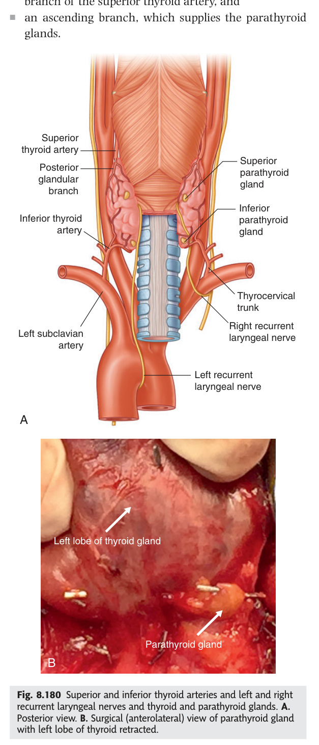
Most people have four parathyroid glands, but their embryological origins explain why they are so hard to find: the superior pair descend from the 4th pharyngeal pouch and settle reliably behind the upper thyroid poles, while the inferior pair migrate with the thymus from the 3rd pouch and may end up anywhere from the carotid bifurcation to the anterior mediastinum. All four glands usually share their blood supply from the inferior thyroid artery — which means that preserving this vessel close to the thyroid capsule during thyroidectomy is the single most important manoeuvre for avoiding post-operative hypocalcaemia. PTH from chief cells is the master regulator of serum calcium: it mobilises bone, enhances renal calcium reabsorption, and activates vitamin D to promote gut absorption — three levers that primary hyperparathyroidism pulls simultaneously, producing the classic "Bones, Stones, Groans, and Moans" syndrome.
| Feature | Superior pair | Inferior pair |
|---|---|---|
| Number (usual) | 2 (occasionally more; supernumerary glands may be in thymus, mediastinum) | 2 |
| Embryology | 4th pharyngeal pouch | 3rd pharyngeal pouch (same as thymus — they migrate together, accounting for ectopic parathyroid in thymus) |
| Position | Posterior to upper third of lateral lobe, outside true capsule | Variable — posterior to lower thyroid or within thymic tissue |
| Blood supply | Inferior thyroid artery (both superior + inferior parathyroids usually supplied by inferior thyroid artery branches) | |
| Function | Chief cells secrete PTH → ↑ Ca²⁺ (bone resorption + renal reabsorption + 1,25-(OH)₂D activation). Oxyphil cells — function uncertain | |
The inferior thyroid artery supplies BOTH superior and inferior parathyroid glands. During thyroidectomy, preserve the inferior thyroid artery close to the thyroid capsule (not at its origin) to maintain parathyroid blood flow. Post-op hypocalcaemia: Chvostek's sign — tapping facial nerve → ipsilateral facial twitching. Trousseau's sign — inflating BP cuff above systolic pressure for 3 min → carpal spasm (main d'accoucheur). Treat: IV calcium gluconate → oral calcium + calcitriol. "Inadvertent parathyroidectomy" is the most common cause of post-thyroidectomy hypoparathyroidism.
| Type | Cause | Ca²⁺ | PTH | PO₄³⁻ | Treatment |
|---|---|---|---|---|---|
| Primary | Adenoma 85%, hyperplasia 14%, carcinoma <1% | ↑↑ | ↑ (inappropriately normal) | ↓ | Surgical parathyroidectomy (sestamibi scan + USS to localise); medical: cinacalcet (calcimimetic) |
| Secondary | CKD → ↓1,25-(OH)₂D → ↓Ca²⁺ → compensatory ↑PTH; all 4 glands hyperplastic | ↓ or normal | ↑↑ | ↑ | Treat CKD + Vit D + phosphate binders + cinacalcet |
| Tertiary | Prolonged secondary → glands become autonomous (after renal transplant or long-standing CRF) | ↑ | ↑↑ | ↓ | Subtotal parathyroidectomy |
| Syndrome | Gene | Tumours (in order of frequency) | Mnemonic |
|---|---|---|---|
| MEN1 (Wermer's) | MEN1 (menin, chromosome 11q13) — tumour suppressor | 1. Parathyroid hyperplasia (95%, often first; hypercalcaemia) 2. Pituitary adenoma (prolactinoma most common) 3. Pancreatic tumours (gastrinoma = Zollinger-Ellison syndrome; insulinoma; VIPoma; glucagonoma) | "3 Ps" — Parathyroid, Pituitary, Pancreas |
| MEN2A (Sipple's) | RET proto-oncogene (chromosome 10) — gain of function | 1. Medullary thyroid carcinoma (95%, nearly always; earliest; calcitonin ↑) 2. Phaeochromocytoma (50%; bilateral; exclude pre-op with metanephrines; α-block first) 3. Parathyroid hyperplasia (20%) | "2 Ps + MTC" — Phaeochromocytoma, Parathyroid, Medullary Thyroid Carcinoma |
| MEN2B | RET proto-oncogene (different codon from MEN2A) | 1. Medullary thyroid carcinoma (most aggressive; presents earliest, even infancy) 2. Phaeochromocytoma 3. Marfanoid habitus + mucosal neuromas (lips/tongue/GI) + ganglioneuromas — no parathyroid | MEN2B = MEN2A minus parathyroid, plus marfanoid + neuromas |
In MEN2A/2B: always screen for phaeochromocytoma first before any surgery (thyroidectomy or parathyroidectomy) — an undiagnosed phaeochromocytoma under general anaesthesia = hypertensive crisis + death. RET gene carriers in MEN2 families offered prophylactic thyroidectomy (timing depends on codon mutation severity — highest risk = before age 6 months).
- Why do inferior parathyroids end up ectopic? They descend from the 3rd pharyngeal pouch alongside the thymus → may migrate into thymic tissue or anterior mediastinum; superior pair (4th pouch) are more predictable
- Ca²⁺ ↑, PTH ↑ inappropriately, PO₄³⁻ ↓ — diagnosis and commonest cause? Primary hyperparathyroidism; commonest cause = solitary parathyroid adenoma (85%); treat with surgical parathyroidectomy
- Chvostek's and Trousseau's signs? Both indicate hypocalcaemia (post-thyroidectomy hypoparathyroidism). Chvostek = facial twitch on tapping facial nerve. Trousseau = carpal spasm (main d'accoucheur) with inflated BP cuff for 3 min
- Distinguish MEN1 from MEN2A. MEN1 (menin gene, 11q13): 3 Ps — Parathyroid + Pituitary + Pancreatic tumours. MEN2A (RET proto-oncogene): MTC + Phaeochromocytoma + Parathyroid. Always exclude phaeochromocytoma before any MEN2 surgery
- Why is PTH high in secondary hyperparathyroidism? CKD → reduced 1α-hydroxylase → ↓1,25-(OH)₂D → ↓gut Ca²⁺ absorption → compensatory PTH rise; all 4 glands hyperplastic; Ca²⁺ low or normal
Pituitary Gland (Hypophysis Cerebri)
The pituitary sits in the bony sella turcica of the sphenoid bone, directly beneath the optic chiasm, which means that an expanding macroadenoma compresses the crossing nasal fibres and destroys temporal vision in both eyes — bitemporal hemianopia — before any other symptom develops. The anterior lobe (adenohypophysis) is an island of gut-derived epithelium that receives no direct nerve supply but is controlled entirely through the hypothalamo-hypophyseal portal system, while the posterior lobe (neurohypophysis) is literally a downward extension of the hypothalamus, storing and releasing ADH and oxytocin made in supraoptic and paraventricular nuclei. Prolactin is unique among anterior pituitary hormones in that it is under tonic dopamine inhibition — meaning any lesion that compresses the pituitary stalk disconnects the inhibition and paradoxically raises prolactin, a "disconnection hyperprolactinaemia" that mimics a prolactinoma.
| Feature | Detail |
|---|---|
| Location | Hypophyseal fossa of sella turcica (sphenoid bone). Covered by diaphragma sellae (dura). Optic chiasm lies above and anterior |
| Lobes | Anterior (adenohypophysis, 75%): derived from Rathke's pouch (oral ectoderm — craniopharyngioma arises here). Posterior (neurohypophysis, 25%): outgrowth of hypothalamus; stores + releases ADH + oxytocin made in hypothalamic nuclei |
| Hypothalamo-hypophyseal portal system | Hypothalamic releasing/inhibiting hormones secreted into primary capillary plexus → portal veins → secondary capillary plexus in anterior pituitary → regulate anterior pituitary hormones (GH, TSH, ACTH, FSH, LH, prolactin). No direct nerve supply to anterior pituitary |
| Superior hypophyseal arteries | From internal carotid + anterior/posterior communicating arteries (Circle of Willis) → form portal system |
| Venous drainage | Into surrounding cavernous sinus → petrosal sinuses → jugular vein |
FSH · LH · ACTH · TSH · Prolactin · (i) · GH. Prolactin is under tonic inhibition by dopamine → dopamine agonists treat prolactinoma; dopamine antagonists (metoclopramide, antipsychotics) → ↑prolactin → galactorrhoea/amenorrhoea.
| Hormone | Cell type | Target organ / action | Hypothalamic regulator | Key clinical point |
|---|---|---|---|---|
| GH (somatotrophin) | Somatotroph (50%) | Liver → IGF-1 → growth of long bones; protein synthesis, lipolysis, anti-insulin | GHRH (+) · somatostatin (−) | Excess → acromegaly (adults) / gigantism (children); screen with IGF-1 → confirm with OGTT (GH fails to suppress below 1 µg/L) |
| TSH (thyrotrophin) | Thyrotroph (5%) | Thyroid gland → T3/T4 synthesis and release | TRH (+) · T3/T4 negative feedback | TSH ↓ in primary hyperthyroidism; TSH ↑ in primary hypothyroidism; TSH normal/low in secondary (pituitary) hypothyroidism |
| ACTH (corticotrophin) | Corticotroph (20%) | Adrenal cortex → cortisol + androgens (not aldosterone, which is RAA-regulated) | CRH (+) · cortisol negative feedback | Cushing's disease = pituitary ACTH excess; Nelson's syndrome = ACTH-secreting tumour after bilateral adrenalectomy |
| FSH | Gonadotroph (10%) | Ovary → follicle growth, oestrogen; Testis → Sertoli cells, spermatogenesis | GnRH (pulsatile +) · oestrogen/inhibin negative feedback | FSH + LH ↑ in gonadal failure (primary hypogonadism); ↓ in hypothalamic/pituitary failure |
| LH | Gonadotroph (10%) | Ovary → ovulation + corpus luteum (progesterone); Testis → Leydig cells → testosterone | GnRH (pulsatile +) | LH surge triggers ovulation; continuous GnRH analogues paradoxically suppress LH (downregulation of GnRH receptors) |
| Prolactin | Lactotroph (15–20%) | Breast → milk production (lactation after delivery) | Dopamine (−) is dominant; TRH + oestrogen stimulate | Most common secretory pituitary tumour; dopamine agonist (cabergoline) first-line treatment; stalk compression by any pituitary mass → ↑ prolactin (disconnection hyperprolactinaemia) |
| MSH (melanotrophin) | Corticotroph (POMC cleavage) | Melanocytes → skin pigmentation | CRH (+) · dopamine (−) | POMC → ACTH + β-LPH; also cleaved to α-MSH. Hyperpigmentation in Addison's and Nelson's due to ↑ ACTH sharing MSH sequences |
| Tumour | % | Presentation | Key Diagnostic Test | Treatment |
|---|---|---|---|---|
| Prolactinoma | 40% | Women: galactorrhoea + amenorrhoea + infertility. Men: impotence + infertility (often macro at presentation). Headache/bitemporal hemianopia if macro. | ↑↑ serum prolactin (>5000 mU/L for macro); MRI | Dopamine agonist first-line (cabergoline/bromocriptine — shrinks tumour medically); trans-sphenoidal surgery if medical failure |
| GH-secreting | 20% | Adults: acromegaly (coarse features, prognathism, enlarged extremities, hyperhidrosis, carpal tunnel, diabetes, sleep apnoea, cardiomegaly). Children: gigantism | IGF-1 elevated; failure to suppress GH on OGTT (<1 µg/L normal); MRI | Trans-sphenoidal surgery; octreotide/lanreotide (somatostatin analogues) if residual; radiosurgery |
| ACTH-secreting | 15% | Cushing's disease (pituitary ACTH → bilateral adrenal hyperplasia → ↑↑cortisol). Central obesity, moon face, buffalo hump, purple striae, easy bruising, proximal myopathy, diabetes, osteoporosis | 24-h urinary cortisol ↑; low-dose dexamethasone fails to suppress; high-dose suppresses (ectopic ACTH = small cell lung cancer = does NOT suppress on high dose); IPSS (inferior petrosal sinus sampling) confirms pituitary source | Trans-sphenoidal surgery; bilateral adrenalectomy if fails (risk Nelson's syndrome = residual ACTH tumour enlarges → hyperpigmentation) |
| Non-functioning | 25% | Mass effect: bitemporal hemianopia, headache, hypopituitarism (order of loss: GH first → FSH/LH → TSH → ACTH last) | MRI; no hormone excess; ↓ sex steroids + gonadotrophins | Trans-sphenoidal surgery; radiotherapy for residual |
| Craniopharyngioma | Suprasellar | Children/young adults; Rathke's pouch remnant; bitemporal hemianopia + growth failure + DI + raised ICP | Calcification 80% on CT (suprasellar); MRI = solid-cystic + "machine oil" cyst fluid | Surgery + RT; high recurrence |
Central DI: ADH (vasopressin) deficiency; causes = hypothalamic/posterior pituitary destruction (head trauma, surgery, tumour, sarcoidosis, LCH). Responds to desmopressin (DDAVP) → urine osmolality rises >50%.
Nephrogenic DI: renal resistance to ADH; causes = lithium (most common drug), hypercalcaemia, hypokalaemia, familial (V2 or AQP2 mutation). Does NOT respond to desmopressin.
Water deprivation test: deprive fluid → urine fails to concentrate (plateau) → give desmopressin → central DI concentrates >50%; nephrogenic DI does not. Normal: concentrates during dehydration alone without needing desmopressin.
Sheehan's syndrome: pituitary infarction from postpartum haemorrhage (pituitary enlarged in pregnancy → more ischaemia-prone when blood pressure falls acutely). First sign = failure to lactate (prolactin first lost). Subsequent: amenorrhoea (FSH/LH), fatigue/cold intolerance (TSH), hypotension/hypoglycaemia (ACTH). Order of pituitary hormone loss in hypopituitarism: GH → FSH/LH → TSH → ACTH (most critical). Treat: lifelong hormone replacement (hydrocortisone first — life-threatening without it; then thyroxine, then oestrogen, ± GH, ± DDAVP if DI co-exists).
Empty sella syndrome: herniation of subarachnoid space through diaphragma sellae → CSF fills sella → pituitary flattened. Primary = idiopathic (obese parous women; IIH association). Secondary = post-Sheehan's/surgery/RT. MRI: CSF signal fills sella; pituitary tissue along floor. Usually asymptomatic; pituitary function may be preserved.
- Why does a pituitary macroadenoma cause bitemporal hemianopia? Expands superiorly → compresses decussating nasal fibres of optic chiasm → loss of temporal visual fields bilaterally; central vision preserved initially
- First-line treatment for prolactinoma? Dopamine agonist (cabergoline or bromocriptine) — shrinks tumour medically; surgery (trans-sphenoidal) reserved for medical failure or visual compromise
- Water deprivation test: how to distinguish central from nephrogenic DI? Both fail to concentrate urine on deprivation → give desmopressin → central DI: urine osmolality rises >50%; nephrogenic DI: no response (renal resistance to ADH)
- What is Nelson's syndrome? Enlargement of residual ACTH-secreting pituitary tumour after bilateral adrenalectomy for Cushing's disease → severe hyperpigmentation (↑↑ ACTH/MSH) + bitemporal hemianopia
- Order of hormone loss in hypopituitarism? GH first → FSH/LH → TSH → ACTH last; replace hydrocortisone first (Addisonian crisis is life-threatening without it), then thyroxine, then oestrogen/testosterone
Thymus & Pineal Gland
The thymus is the primary lymphoid organ that programs T-cell identity: in its cortex, T cells are tested for MHC recognition (positive selection), then shuffled to the medulla where those that attack self-antigens are deleted (negative selection), with Hassall's corpuscles — whorls of keratinised epithelium — marking the medulla uniquely on histology. Because the thymus shares its 3rd pharyngeal pouch origin with the inferior parathyroid glands, a failure of that pouch to develop (DiGeorge syndrome) simultaneously abolishes T-cell immunity, wipes out the inferior parathyroids causing hypocalcaemia, and disrupts pharyngeal arch heart development — a trio of deficits linked by one embryological event. The pineal gland, tucked behind the superior colliculi in the epithalamus, translates light/dark cycles into melatonin pulses; its clinical relevance is that a germinoma compressing the colliculi produces Parinaud's syndrome (paralysis of upgaze), and its post-pubertal calcification on skull X-ray serves as a midline reference — a shift suggests a space-occupying lesion.
| Feature | Detail |
|---|---|
| Position | Anterior mediastinum, posterior to manubrium and sternal body, anterior to great vessels. In children: extends into the neck |
| Embryology | 3rd pharyngeal pouch (bilateral); migrates inferiorly → same as inferior parathyroid glands (explains ectopic parathyroid tissue in thymus) |
| Histology | Cortex: dense T-lymphocytes (positive selection). Medulla: Hassall's corpuscles (thymic epithelial whorls — unique identifier); less dense lymphocytes (negative selection). Both positive + negative selection must occur for self-tolerant, MHC-restricted T cells |
| Involution | Involutes after puberty; replaced by fat. Active throughout childhood (critical period for T-cell development) |
| Clinical | Thymoma: associated with myasthenia gravis (70% of thymomas). DiGeorge syndrome: failure of 3rd pharyngeal pouch development → absent thymus + absent inferior parathyroids → T-cell immunodeficiency + hypocalcaemia + cardiac defects |
Small endocrine gland in the epithalamus (posterior diencephalon), in the groove between the two superior colliculi. Secretes melatonin (from serotonin) — regulates circadian rhythms + sleep-wake cycle; secretion peaks at night in darkness. Paediatric pineal germinoma: compresses superior colliculi → Parinaud's syndrome (paralysis of upward gaze + light-near dissociation + convergence-retraction nystagmus). Calcifies after puberty (visible on skull X-ray — useful landmark for midline shift in trauma).
- Histological feature unique to thymic medulla? Hassall's corpuscles — whorls of keratinised thymic epithelial cells; absent in cortex and every other organ; confirm medulla on histology
- DiGeorge syndrome: which pouch, and what triad? 3rd pharyngeal pouch failure → absent thymus (T-cell immunodeficiency) + absent inferior parathyroids (hypocalcaemia) + cardiac defects (conotruncal)
- What is Parinaud's syndrome and what causes it? Paralysis of upward conjugate gaze + light-near dissociation + convergence-retraction nystagmus; caused by compression of superior colliculi (e.g., pineal germinoma)
- Clinical significance of pineal calcification on skull X-ray? Normal after puberty; provides midline reference landmark — a shift of the calcified pineal suggests mass effect from a space-occupying lesion
- Thymoma is associated with which autoimmune condition? Myasthenia gravis — 70% of thymomas associated; antibodies against acetylcholine receptors at NMJ; thymectomy improves symptoms
Test Unit 09 knowledge
Peritoneum + endocrine gland MCQs and clinical scenarios.