The Female Genital System
Ovaries
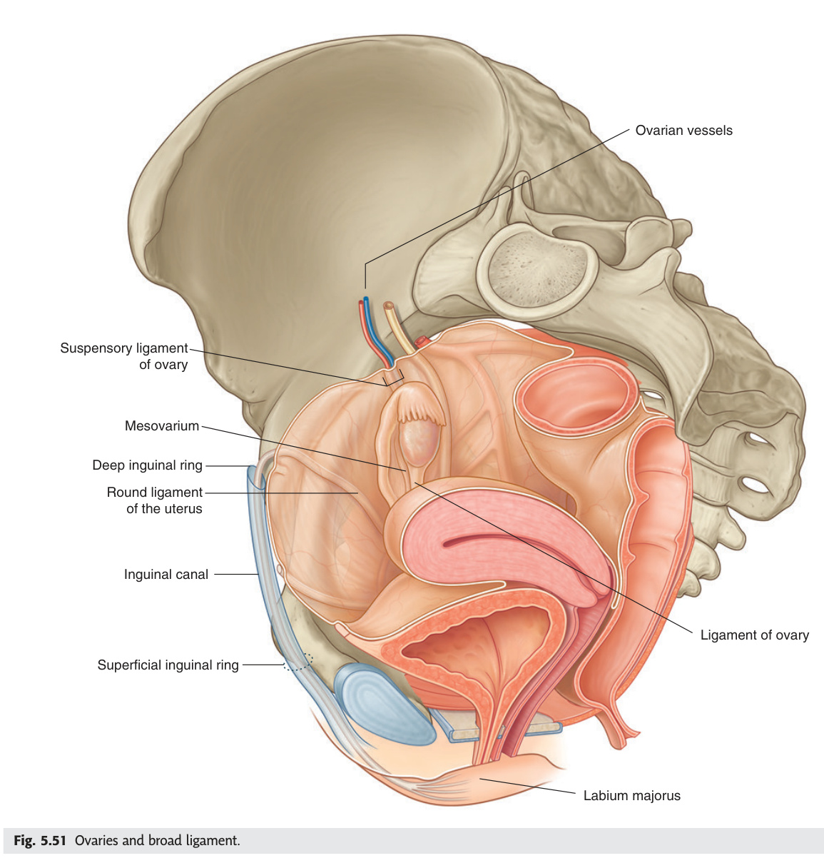
The ovary's developmental origin at L1–L2 determines its entire neurovascular anatomy — just like the testes, the ovarian artery comes from the aorta (not the internal iliac), lymphatics drain to para-aortic nodes (not pelvic), and pain is referred to the umbilicus (T10). The ovary is attached to the broad ligament by the mesovarium but is the only intraperitoneal organ NOT covered by peritoneum — its surface is a layer of germinal epithelium (a misnomer; it is coelomic mesothelium, and most ovarian carcinomas actually arise from the fallopian tube fimbriae). The suspensory ligament (infundibulopelvic ligament) carries the ovarian vessels and must be identified and ligated early in salpingo-oophorectomy to avoid bleeding — it runs over the pelvic brim lateral to the ureter, which must be protected at that point.
| Feature | Detail |
|---|---|
| Position | On the lateral pelvic wall in the ovarian fossa (bounded by external iliac vessels + ureter + obturator nerve + internal iliac). Attached posteriorly to broad ligament by mesovarium |
| Ligaments | Ovarian ligament (ligament of ovary): ovary → uterus (deep to broad ligament). Round ligament: uterus → labium majus via inguinal canal (homologue of gubernaculum testis). Suspensory ligament of ovary (infundibulopelvic ligament): lateral pelvic wall → ovary; contains ovarian vessels |
| Blood supply | Ovarian artery from aorta at L2 (same developmental origin as testicular). Venous: right ovarian vein → IVC; left ovarian vein → left renal vein |
| Lymphatics | Para-aortic/retroperitoneal nodes (L1–L2) — same as testes |
| Nerve | Aortic plexus (T10 sympathetic) → referred ovarian pain at umbilicus (T10) |
Ovarian torsion: sudden onset severe unilateral lower abdominal pain + N&V; ovary (usually with cyst >5 cm) rotates on its pedicle cutting off vascular supply. USS: absent Doppler flow. Emergency laparoscopic detorsion. Ovarian carcinoma: CA-125 elevated; insidious presentation (bloating, early satiety); peritoneal seeding → ascites ('omental cake' on CT). BRCA1/2 mutations increase lifetime risk. Salpingo-oophorectomy ± chemotherapy.
| Phase | Days | Hormones | Ovarian Events |
|---|---|---|---|
| Follicular | 1–14 | FSH rises → oestradiol rises | Cohort of primordial follicles recruited; one dominant (Graafian, 18–20 mm); theca interna (LH → androgens) + granulosa cells (FSH + aromatase → oestradiol); antrum forms (secondary → Graafian follicle) |
| Ovulation | Day 14 | Oestradiol surge → LH surge (peaks 36–40 h before ovulation) | Stigma rupture → secondary oocyte + cumulus oophorus released; swept by fimbriae; oocyte viable 12–24 h; sperm viable 48–72 h; fertilisation in ampulla |
| Luteal | 15–28 | LH → progesterone + oestradiol from corpus luteum | Granulosa + theca cells luteinised (yellow, lipid-rich = corpus luteum); if no fertilisation → luteolysis day 24 → corpus albicans; if fertilisation → trophoblast hCG maintains corpus luteum until placenta takes over (weeks 8–10) |
Primordial → Primary → Secondary (antrum forms) → Graafian (dominant, 18–20 mm) → Ovulation → Corpus luteum → Corpus albicans. Mittelschmerz = mid-cycle pelvic pain at ovulation (peritoneal irritation by follicular fluid). LH surge detected by ovulation predictor kits (urine LH test).
Most common endocrine disorder of reproductive-age women (5–10%). Rotterdam criteria — 2 of 3: (1) oligo/anovulation (cycles >35 days or <8/year); (2) clinical or biochemical hyperandrogenism (acne, hirsutism, elevated testosterone/DHEAS); (3) polycystic ovaries on USS (≥20 follicles per ovary OR ovarian volume >10 mL). Pathophysiology: elevated LH:FSH ratio (>2:1 in classic PCOS) → excess LH → theca cell androgen excess → impaired follicular maturation; insulin resistance → hyperinsulinaemia → further theca stimulation. Long-term risks: type 2 diabetes (50%), cardiovascular disease, endometrial carcinoma (anovulation → unopposed oestrogen → hyperplasia). Management: COCP (cycle regulation + anti-androgen effect); metformin (insulin sensitiser); clomifene citrate or letrozole (ovulation induction for fertility); lifestyle modification (5–10% weight loss restores ovulation in obese PCOS).
- Q: Why does ovarian pain refer to the umbilicus, not the groin? — Ovarian nerve supply is from the aortic plexus (T10 sympathetic) — same dermatome as the umbilicus. The ovary develops at T10 level retroperitoneally, acquiring its nerve supply before descending into the pelvis.
- Q: Ovarian cancer lymph drainage — why does this matter for staging? — Para-aortic/retroperitoneal nodes (L1–L2), following the ovarian vessels. Staging CT must include the para-aortic chain; pelvic lymphadenopathy alone does not represent the primary drainage, unlike cervical cancer.
- Q: PCOS Rotterdam criteria — list the three criteria and how many are needed. — (1) Oligo/anovulation; (2) Hyperandrogenism (clinical or biochemical); (3) Polycystic ovaries on USS. Diagnosis requires 2 of 3.
- Q: What triggers ovulation on day 14 and what is the LH surge timeline? — Oestradiol rise from the dominant Graafian follicle triggers positive feedback → LH surge; ovulation ~36–40 hours after the LH peak. The oocyte is viable for only 12–24 hours post-release.
- Q: Corpus luteum rescue — what does it and when does the placenta take over? — Trophoblast hCG (structurally identical to LH) rescues the corpus luteum, maintaining progesterone until the luteo-placental shift at weeks 8–10.
Uterine (Fallopian) Tubes
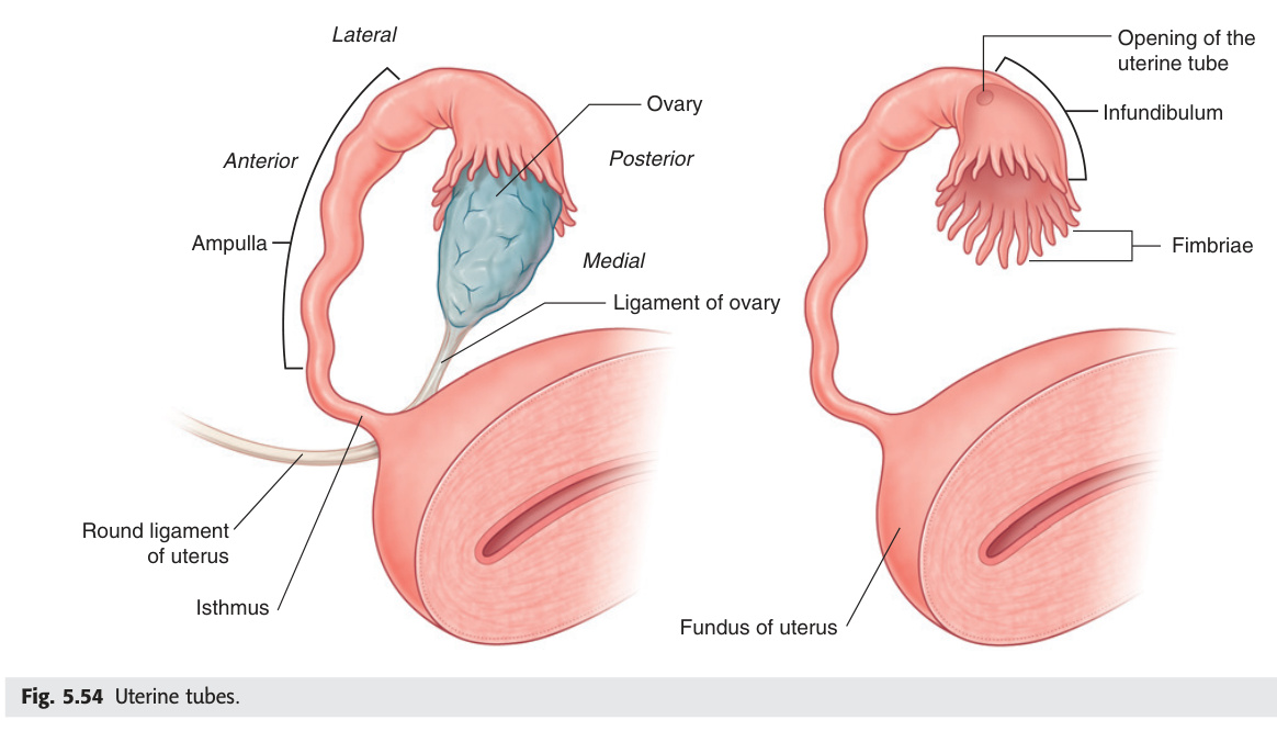
The uterine tube is not a passive conduit — it is an active transport system. Cilia beat toward the uterus, and the ampulla provides the environment for fertilisation. The tube's four segments have dramatically different wall thicknesses and luminal widths, and this anatomy directly predicts which ectopic site ruptures first: the isthmus is narrow and ruptures early (thin lumen cannot distend), the ampulla is wide and ruptures later, while the interstitial/cornual segment ruptures latest but catastrophically because it is surrounded by the vascular uterine wall (rupture at 10–12 weeks, massive haemorrhage). Every ectopic pregnancy is a surgical emergency until proven otherwise — the posterior fornix is separated from the pouch of Douglas by only the vaginal wall, so ruptured ectopic blood pools there and is detectable by culdocentesis or USS.
| Part | Description | Clinical Note |
|---|---|---|
| Infundibulum | Funnel-shaped lateral end with fimbriae; opens into peritoneal cavity. One fimbria (ovarian fimbria) attaches to ovary | Ectopic pregnancy most common in ampulla (the widest part just medial to infundibulum) |
| Ampulla | Widest, longest part (~5 cm); normal site of fertilisation; thin wall | 90% of ectopic pregnancies in ampulla → rupture when embryo outgrows it |
| Isthmus | Narrow, thick-walled part joining uterus | Ectopic here = rare but ruptures early (narrow); site of tubal ligation (clips or rings at isthmus) |
| Intramural (uterine) | Passes through uterine wall at the cornu | Interstitial/cornual ectopic = rare, high mortality (ruptures at 10–12 weeks, rich blood supply) |
- Q: Where do 90% of ectopic pregnancies occur, and why does this site rupture later than the isthmus? — Ampulla (70%); isthmus (12%). The ampulla is wider with a thinner wall → can accommodate more growth before rupture. The isthmus is narrow and thick-walled → ruptures early (smaller embryo).
- Q: Classic clinical triad of ectopic pregnancy. — Amenorrhoea + unilateral pelvic/adnexal pain + vaginal bleeding. β-hCG positive. TVUSS shows empty uterus + adnexal mass (or free fluid in pouch of Douglas).
- Q: Criteria for methotrexate treatment of ectopic pregnancy (vs surgery). — Unruptured, β-hCG <3000–5000 IU/L (depending on protocol), no fetal cardiac activity, haemodynamically stable, compliant for follow-up. Methotrexate inhibits dihydrofolate reductase → stops trophoblast proliferation.
- Q: Which tubal site is rarest but most dangerous for ectopic pregnancy? — Interstitial/cornual (2–3%). Ruptures at 10–12 weeks (surrounded by thick vascular myometrium → tolerates growth longer) then causes catastrophic haemorrhage. Mortality 2–2.5× higher than other sites.
- Q: Site of normal fertilisation? — Ampulla of the uterine tube. Sperm is viable 48–72 hours; oocyte viable 12–24 hours. Fertilised embryo reaches the uterine cavity at blastocyst stage (~day 5–6) for implantation.
Uterus
The uterus has two anatomical relationships that generate more surgical disasters than any other single fact in gynaecology: (1) the uterine artery crosses over the ureter 2 cm lateral to the cervix ("water under the bridge") — inadvertent ureteric ligation during hysterectomy is the most common serious intraoperative urological injury; (2) the cervical transformation zone (TZ), where squamocolumnar epithelium meets, is the origin of >95% of cervical cancers because HPV preferentially infects the metaplastic cells of the TZ. The uterus's position (anteverted/anteflexed = normal; retroverted = may indicate endometriosis/adhesions) determines how it is palpated bimanually. The lower uterine segment — the isthmus of the non-pregnant uterus — is the site of lower segment Caesarean section because it is avascular and thin, meaning less bleeding and easier repair than the thick vascular upper segment.
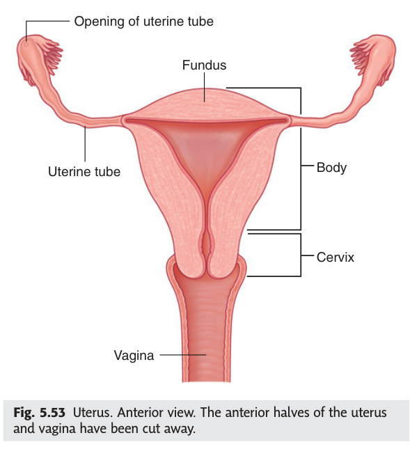
| Part | Description |
|---|---|
| Fundus | Dome above the uterine tubes. Perimetrium (peritoneum) covers it completely |
| Body | Main bulk. Uterine cavity is triangular. Walls: perimetrium (outer) + myometrium (middle, smooth muscle) + endometrium (inner, cyclic changes) |
| Isthmus | Narrow junction between body and cervix. Lower uterine segment (LUS) in pregnancy = site of lower segment Caesarean section (LSCS) |
| Cervix | Cylindrical lower part. Supravaginal + vaginal portions. External os opens into vagina. Ectocervix = stratified squamous. Endocervix = columnar. Transformation zone = junction (site of CIN + cervical carcinoma) |
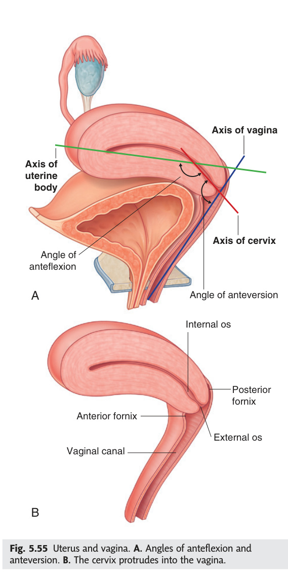
| Position | Description | Significance |
|---|---|---|
| Anteversion | Uterus tilted anteriorly relative to vaginal axis (cervix + uterus form ~90° angle) — NORMAL (80%) | Normal position; anteverted + anteflexed most common |
| Anteflexion | Body of uterus flexed forward on cervix — NORMAL | Augments the normal forward tilt |
| Retroversion + retroflexion | Uterus tilts + flexes posteriorly (toward sacrum) — 20% of women | Associated with endometriosis, pelvic adhesions; posterior fornix becomes accessible for culdocentesis; may complicate catheterisation in labour |
The endometrium has a functional layer (shed at menstruation; regenerates each cycle) supplied by spiral arteries (progesterone-sensitive) and a basal layer (permanent; regenerates the functional layer) supplied by straight basal arteries (oestrogen-dependent, do NOT constrict at menstruation).
| Phase | Days | Driving Hormone | Histological Changes |
|---|---|---|---|
| Menstrual | 1–4 | Progesterone + oestrogen withdrawal → PGF2α → spiral artery spasm → ischaemia | Functional layer sheds; ~50 mL blood loss; basal layer intact; endometrial thickness ~1 mm |
| Proliferative | 5–14 | Rising oestrogen (FSH → follicle → oestradiol) | Mitosis; endometrium thickens 1–10 mm; straight tubular glands; spindle-shaped stromal cells; thin watery cervical mucus (ferning pattern, high spinnbarkeit = sperm-friendly) |
| Secretory (early) | 15–17 | Progesterone from corpus luteum | Sub-nuclear vacuoles (glycogen deposits beneath nuclei, day 16–17 = earliest histological sign of ovulation); glands become tortuous |
| Secretory (peak) | 18–24 | Progesterone (peak) | Saw-tooth gland lumens; secretory products (glycogen, glycoprotein) peak day 22; "window of implantation" = days 20–24; thick sticky cervical mucus (blocks sperm = post-ovulation) |
| Secretory (late) | 25–28 | Corpus luteum regresses; progesterone falls | Stromal predecidualisation (preadaptation for implantation); if no implantation → luteolysis → progesterone falls → PGF2α → spiral artery vasoconstriction → ischaemia → menstruation |
Leiomyomas (Fibroids)
Most common benign uterine tumour; smooth muscle origin; oestrogen-dependent (grow in reproductive years, shrink post-menopause; enlarge rapidly in pregnancy). Fibroid uterus feels irregular and firm on bimanual examination.
| Type | Location | Primary Symptoms | Treatment Implications |
|---|---|---|---|
| Submucosal | Projects into uterine cavity; distorts endometrium | Heaviest menorrhagia; implantation failure + recurrent miscarriage (most likely to cause subfertility) | Hysteroscopic resection (type 0 = fully intracavitary, type I–II = partial intramural component) |
| Intramural | Within myometrium; does not distort cavity | Bulk symptoms, dysmenorrhoea, pelvic pressure, urinary frequency (if large) | Myomectomy (open/laparoscopic, fertility-preserving); hysterectomy if complete |
| Subserosal | Projects outward from serosal surface | Pressure symptoms; if pedunculated → can tort (acute pain mimicking torsion) | Laparoscopic myomectomy; UAE (uterine artery embolisation) |
Red (carneous) degeneration: rapid growth during pregnancy → central infarction → acute localized uterine pain + fever + leukocytosis; management = analgesia + hydration (conservative). Leiomyosarcoma: malignant transformation is RARE (<0.5%); suspect in post-menopausal women with rapidly enlarging uterine mass. Medical treatment: GnRH analogues shrink fibroids ~50% (temporary; used pre-operatively); UAE = embolisation of uterine arteries → infarction of fibroid (preserves uterus, not suitable if future pregnancy desired).
Endometriosis
Ectopic endometrial glands + stroma outside the uterus; responds cyclically to hormones → bleeding at ectopic site → inflammation → fibrosis → adhesions.
| Site (order of frequency) | Specific Features |
|---|---|
| Ovaries (most common) | Chocolate cysts (endometriomata) — dark thick old blood; may rupture → acute abdomen + chemical peritonitis |
| Posterior cul-de-sac + uterosacral ligaments | Nodularity palpable on PV/PR examination; fixed retroverted uterus |
| Rectovaginal septum + rectosigmoid | Dyschezia; rectal bleeding at menstruation; deep dyspareunia |
| Bladder + ureter | Cyclical haematuria; ureteric obstruction → hydronephrosis |
| Diaphragm / pleura (rare) | Catamenial haemothorax / pneumothorax (collapse + haemoptysis at menstruation) |
Adenomyosis
Endometrial glands + stroma within the myometrium (compare endometriosis = outside uterus). Results in globular, bulky uterus ("boggy" on palpation). Typically multiparous perimenopausal women. Symptoms: heavy painful periods (secondary dysmenorrhoea + menorrhagia); uterus tender and uniformly enlarged. USS shows heterogeneous myometrium + sub-endometrial cysts; MRI is the gold standard imaging (JEZ = junctional zone >12 mm). Treatment: medical (progestogens, LNG-IUS/Mirena = most effective); hysterectomy = definitive.
- Q: "Water under the bridge" — what exactly crosses what, and where? — The uterine artery (branch of internal iliac) crosses OVER the ureter at the lateral fornix, approximately 2 cm lateral to the cervix. Surgically the ureter must be identified and retracted before clamping the uterine artery during hysterectomy.
- Q: Earliest histological sign of ovulation on endometrial biopsy? — Sub-nuclear vacuoles (glycogen deposits beneath glandular cell nuclei), appearing days 16–17. They confirm progesterone secretion from a corpus luteum and therefore confirm ovulation.
- Q: Submucosal vs intramural vs subserosal fibroid — which most causes subfertility? — Submucosal (projects into cavity) — distorts the endometrium, impairs implantation, causes heaviest menorrhagia. Treated hysteroscopically.
- Q: Endometriosis "three Ds" — name them. — Dysmenorrhoea (secondary, worsening cyclically); Dyspareunia (deep); Dyschezia/Dysuria (bowel/bladder involvement). Gold standard diagnosis = laparoscopy + biopsy.
- Q: Adenomyosis vs endometriosis — key distinguishing feature. — Adenomyosis = endometrial glands/stroma WITHIN the myometrium (intrinsic to uterus) → boggy, uniformly enlarged uterus, MRI junctional zone >12 mm. Endometriosis = ectopic outside the uterus → often normal-sized uterus with adnexal/peritoneal deposits.
Broad Ligament & Uterine Ligaments
The broad ligament is a double fold of peritoneum — it is not truly a ligament but a mesentery that gives the uterus and tubes their peritoneal covering and contains their neurovascular supply. Its three subdivisions each contain a specific structure: the mesosalpinx carries the tube in its free upper edge, the mesovarium suspends the ovary posteriorly, and the mesometrium is the main body lateral to the uterus containing the uterine vessels and ureter at its base. The truly load-bearing supports of the uterus are not the peritoneal folds but the condensations of endopelvic fascia — the cardinal (Mackenrodt's) ligament resists descent, the uterosacral ligaments resist forward displacement, and deficiency of these (multiparous, post-menopausal, oestrogen-deficient) causes prolapse. The round ligament merely maintains anteversion and provides no support against descent.
• Uterine tube (in the free upper edge = mesosalpinx)
• Round ligament (runs anterolaterally in mesometrium → inguinal canal → labium majus)
• Ovarian ligament (connects ovary to uterus, under the mesovarium)
• Uterine + ovarian vessels and lymphatics
• Ureter (runs in the base of broad ligament, under the uterine artery)
• Parametrium: loose connective tissue in base
Sub-divisions: mesosalpinx (above tube), mesovarium (behind; suspends ovary), mesometrium (main part, lateral to uterus)
| Ligament | From → To | Function / Notes |
|---|---|---|
| Round ligament | Uterine fundus → labium majus (via inguinal canal) | Maintains anteversion; may carry hernias alongside it; gubernaculum homologue |
| Uterosacral ligament | Cervix → sacrum (S2–S4) | Strongest support ligament for uterus; feels thick on DRE; nodularity in endometriosis |
| Cardinal (transverse cervical) ligament of Mackenrodt | Cervix/vaginal vault → lateral pelvic wall | Main support against uterine prolapse; cut during hysterectomy |
| Pubocervical ligament | Cervix → pubic symphysis | Supports anterior vaginal wall; deficiency → cystocele |
Cardinal + uterosacral ligament laxity (multiparous, post-menopausal) → uterine descent. Graded 1st (cervix above hymen) → 2nd (cervix at hymen) → 3rd (procidentia, whole uterus outside). Cystocele (anterior wall) + rectocele (posterior wall) often co-exist. Treatment: pelvic floor physiotherapy → ring pessary → surgical repair (Manchester repair/hysterectomy + pelvic floor reconstruction).
- Q: Name the three sub-divisions of the broad ligament and what structure each contains. — Mesosalpinx (free upper edge) = uterine tube; mesovarium (posterior layer) = ovary (attached by it); mesometrium (main body) = uterine vessels, ureter, parametrium.
- Q: Which ligament is the main support against uterine prolapse? — Cardinal (transverse cervical) ligament of Mackenrodt — runs from cervix/vaginal vault to lateral pelvic wall. The uterosacral ligament adds posterior support. Together, their laxity = uterine prolapse.
- Q: What does the round ligament do, and what is its embryological homologue? — Maintains uterine anteversion only — it does NOT prevent descent. Homologue of gubernaculum testis. Travels through the inguinal canal to the labium majus; may conduct hernial sac alongside it.
- Q: Where is the ureter in relation to the broad ligament and the uterine artery? — Runs in the base (parametrium) of the broad ligament, below and lateral to the uterine artery. At the lateral fornix, the uterine artery crosses over it ("water under the bridge"). The ureter then dips under the artery to enter the bladder.
- Q: Nodularity of the uterosacral ligaments on DRE — what diagnosis does it suggest? — Endometriosis (deposits on the uterosacral ligaments are one of the most common sites of deep infiltrating endometriosis; palpable as firm nodular thickening, often tender, best felt mid-cycle or during menstruation).
Vagina
The posterior fornix of the vagina is one of the most clinically exploited spaces in the pelvis: it is separated from the pouch of Douglas — the most dependent part of the peritoneal cavity when standing — by only the posterior vaginal wall. This means that free intraperitoneal fluid (blood from ruptured ectopic, pus from a tubo-ovarian abscess) collects there first and is accessible by needle (culdocentesis) or incision (colpotomy). The vaginal lymphatic drainage has a sharp anatomical boundary at the hymen level: upper two-thirds drain to internal iliac nodes, lower one-third and vulva drain to superficial inguinal nodes. This boundary determines the radiotherapy field for vaginal carcinoma and the sentinel node sampling strategy for vulval carcinoma.
Fibromuscular tube (~8 cm), runs upward and backward from vestibule to cervix. Axis is perpendicular to the uterine axis (since uterus is anteverted). The cervix projects into the vaginal vault, creating the fornices: anterior (shortest), posterior (deepest, related to pouch of Douglas), and lateral fornices (× 2, related to ureter and uterine artery).
| Relation | Structure |
|---|---|
| Anterior | Base of bladder (upper vagina) + urethra (lower vagina) |
| Posterior | Pouch of Douglas (rectouterine pouch) via posterior fornix (upper) + perineal body + anal canal (lower) |
| Lateral | Ureter (passes close to lateral fornix), uterine artery, levator ani |
The posterior fornix is separated from the pouch of Douglas (rectouterine pouch = most dependent part of the peritoneal cavity in the erect female) by only the posterior vaginal wall. Culdocentesis: needle through posterior fornix to aspirate fluid from pouch of Douglas (blood in ectopic rupture, pus in PID). Colpotomy: incision through posterior fornix for drainage of pelvic abscess. Blood in the pouch of Douglas on USS = ruptured ectopic until proven otherwise.
- Q: What is the pouch of Douglas and what is its clinical importance? — Rectouterine pouch — the most dependent part of the female peritoneal cavity in the erect position, separated from the posterior fornix by the vaginal wall only. Free intraperitoneal blood (ruptured ectopic) or pus (tubo-ovarian abscess) collects here first; aspirated by culdocentesis through the posterior fornix.
- Q: Upper 2/3 vs lower 1/3 vaginal lymph drainage — clinical implication. — Upper 2/3 → internal iliac nodes. Lower 1/3 + vulva → superficial inguinal nodes. Vaginal carcinoma spanning both zones needs combined pelvic + inguinal field RT; vulval carcinoma sentinel node is always inguinal.
- Q: Which fornix is deepest and why? — Posterior fornix, because the uterus is anteverted (tilted anteriorly). The cervix therefore projects more posteriorly, making the posterior fornix deeper and directly related to the pouch of Douglas.
- Q: What structures are related to the lateral fornix? — Ureter and uterine artery (the "water under the bridge" relation) — 2 cm lateral to the cervix. The ureter runs beneath the uterine artery at exactly this level, making the lateral fornix the danger zone during hysterectomy.
- Q: What is the normal vaginal pH and what maintains it? — pH 3.8–4.5 (acidic). Maintained by Lactobacillus fermentation of glycogen from oestrogen-stimulated squamous epithelium → lactic acid. Post-menopausal atrophy (low oestrogen) → glycogen loss → alkaline pH → increased susceptibility to BV and urinary infections.
Vulva & Female Perineum
The pudendal nerve (S2, S3, S4) is the sensory and motor nerve of the entire perineum — its course from greater sciatic foramen, around the ischial spine and sacrospinous ligament, through Alcock's canal on the lateral ischiorectal fossa wall, and into the perineum is one of the most tested nerve pathways in obstetric anatomy. The ischial spine is the landmark for pudendal nerve block: palpate it transvaginally and inject just medial and posterior to it. Critically, the pudendal block does NOT cover the anterior labia (ilioinguinal + genitofemoral territory) and does NOT block uterine pain (T10–L1) — so it works for second-stage pain and perineal repair but not contractions. The Bartholin's glands open at 4 and 8 o'clock in the vaginal vestibule; blockage causes cyst/abscess in the posterior labia majora, treated by marsupialization not simple I&D (which recurs).
| Structure | Description |
|---|---|
| Mons pubis | Fibrofatty mound over pubic symphysis |
| Labia majora | Homologue of scrotum; contain round ligament termination + fat + Bartholin's gland duct. Fused posteriorly at posterior commissure |
| Labia minora | Thin folds; no fat; converge anteriorly to form clitoris hood (prepuce + frenulum). Posterior frenulum = fourchette |
| Clitoris | Homologue of penis; erectile tissue (corpora cavernosa); highly innervated; supplied by dorsal nerve of clitoris (pudendal nerve) |
| Vestibule | Space between labia minora; contains urethral orifice (anterior), vaginal orifice (posterior), Bartholin's gland ducts (4 o'clock + 8 o'clock) |
| Bartholin's glands | Greater vestibular glands. Secrete lubricant during arousal. Duct occlusion → Bartholin's cyst/abscess (tender mass in posterior labia majora). Treat by marsupialization |
Episiotomy: incision of posterior vaginal wall + perineum during delivery. Right mediolateral episiotomy preferred (avoids external anal sphincter + Bartholin's duct). Perineal tears (obstetric): 1° = skin only; 2° = perineal body muscles; 3° = external anal sphincter (EAS); 4° = EAS + internal anal sphincter + rectal mucosa. 3rd + 4th degree tears → faecal incontinence if not repaired correctly.
Technique: Palpate the ischial spine transvaginally or transperineally → inject 10 mL local anaesthetic (lignocaine 1%) medial and posterior to the ischial spine (just medial to the sacrospinous ligament, where the nerve passes). Bilateral block required. Landmarks: ischial spine is palpable in the posterolateral vaginal wall; the nerve lies just behind (posterior to) and slightly medial to the tip of the spine.
Indications: second stage pain relief (forceps/ventouse delivery); repair of perineal/vaginal lacerations; episiotomy repair when central block has worn off.
What it does NOT block: does NOT cover the perineum above the ischial spine (ilioinguinal + genitofemoral nerve territory = anterior labia) — supplementary local infiltration needed for complete anaesthesia. Does not block the cervix or uterus (uterine pain is T10–L1 via uterine plexus).
- Q: Pudendal nerve course — from pelvis to perineum. — Exits greater sciatic foramen (below piriformis) → curves around ischial spine + sacrospinous ligament → re-enters via lesser sciatic foramen → travels in Alcock's canal (lateral ischiorectal fossa wall) → branches: inferior rectal nerve, perineal nerve, dorsal nerve of clitoris.
- Q: Pudendal nerve block — landmark and what it does NOT cover. — Inject medial and posterior to the ischial spine (palpated transvaginally). Does NOT cover anterior labia (ilioinguinal/genitofemoral) or uterine pain (T10–L1). Used for second-stage analgesia and perineal repair.
- Q: Bartholin's glands — location of ducts and treatment of abscess. — Duct openings at 4 o'clock and 8 o'clock in vaginal vestibule. Abscess = marsupialization (creates permanent opening) not simple I&D (recurs).
- Q: 3rd degree obstetric perineal tear — definition and consequence. — Involves the external anal sphincter (EAS). 4th degree adds internal anal sphincter + rectal mucosa. Both risk faecal incontinence if not repaired correctly. Test pudendal nerve terminal motor latency (PNTML) before sphincter repair.
- Q: Right mediolateral episiotomy — why right, and what does it avoid? — Angled right at 45° from midline. Avoids the external anal sphincter (directly posterior) and Bartholin's gland duct (4 o'clock = directly mediolateral on right side). Midline episiotomy extends more easily into the sphincter.
Pelvic Floor Muscles
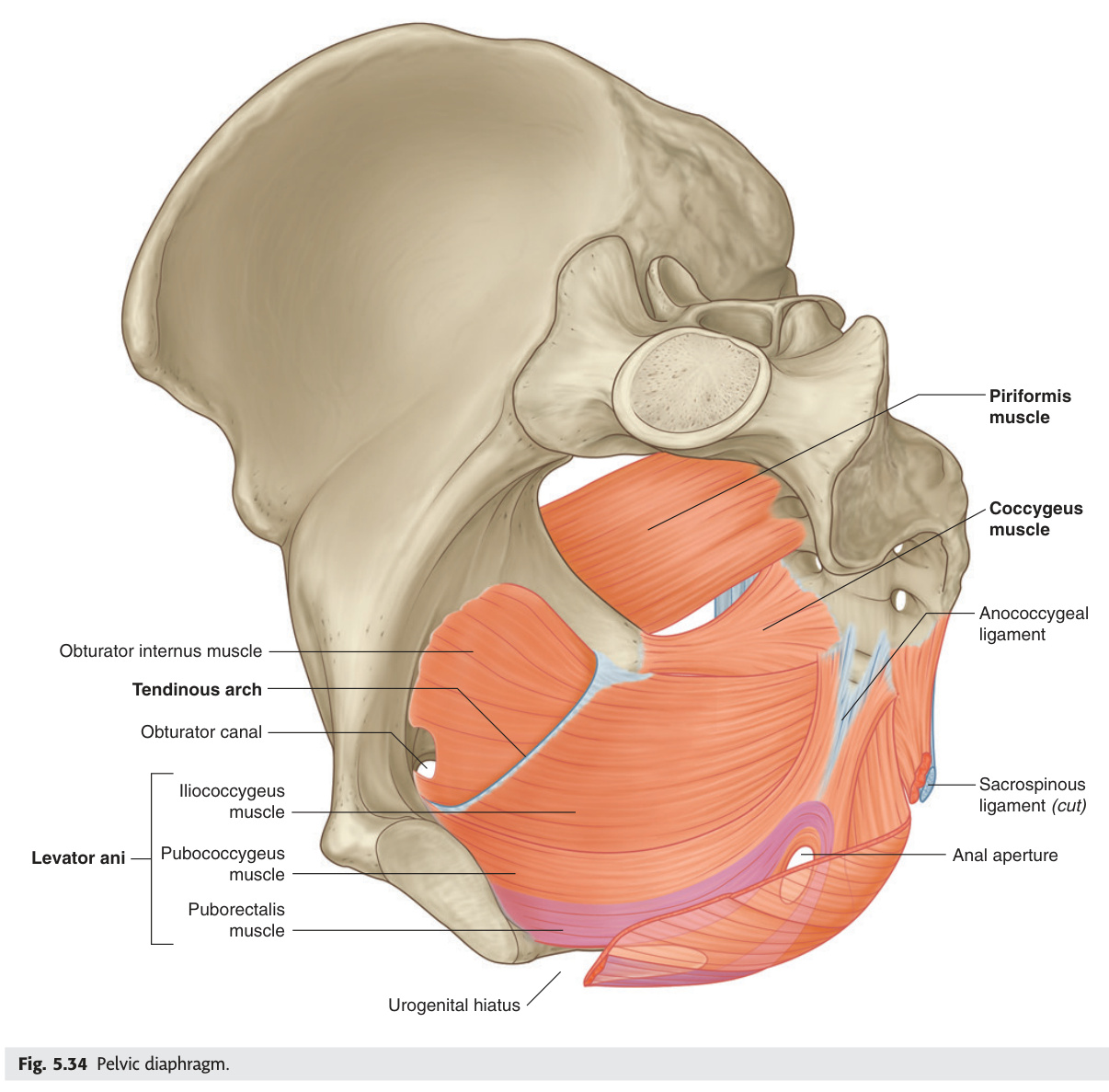
The pelvic floor is a funnel-shaped muscular diaphragm — its key functional insight is that it simultaneously supports the pelvic viscera against gravity and intra-abdominal pressure spikes AND maintains continence through an active mechanical mechanism. The puborectalis creates the anorectal angle (~90°) by sling-looping behind the anorectal junction: this angle is a flap valve that prevents rectal contents entering the anal canal. During defaecation, puborectalis voluntarily relaxes, the angle opens to ~135°, and stool can pass. Injury to puborectalis (obstetric third/fourth degree tears, posterior pelvic floor repair) destroys this angle and causes faecal incontinence. Understanding pelvic diameters is critical for obstetric management: the obstetric conjugate (sacral promontory to posterior pubic symphysis) is the shortest AP diameter the fetal head must pass, and the ONLY diameter measurable clinically is the diagonal conjugate (promontory to inferior symphysis), from which you subtract 1.5 cm.
| Muscle | Origin → Insertion | Action / Notes |
|---|---|---|
| Levator ani (main pelvic floor) | Three parts: Pubococcygeus (including puborectalis), Iliococcygeus, Ischiococcygeus. Pubic bone → coccyx + perineal body + anococcygeal raphe | Supports pelvic viscera; maintains continence; puborectalis creates the anorectal angle (~90°) which is critical for faecal continence. Nerve: S3–S4 + perineal branch of pudendal |
| Puborectalis | Pubis → loops behind anorectal junction | Creates anorectal angle. Defaecation: relaxes puborectalis → angle straightens → stool can pass. If hypertonic → anismus (obstructed defaecation) |
| Coccygeus | Ischial spine → coccyx | Supports pelvic viscera; flexes coccyx |
| Urogenital diaphragm | Deep transverse perineal + external urethral sphincter. Between ischiopubic rami | External urethral sphincter = voluntary (pudendal nerve S2–S4). Contains Cowper's glands in male. Female urethra + vagina pass through it |
The anorectal angle (~90° at rest) is maintained by constant puborectalis contraction — this creates a flap valve that prevents rectal contents from entering the anal canal. During defaecation, puborectalis relaxes → angle widens to ~135° → rectal contents can pass. Injury to puborectalis (obstetric, surgery) → loss of anorectal angle → faecal incontinence. This is why pudendal nerve integrity is critical — test with pudendal nerve terminal motor latency (PNTML) before sphincter repair.
(1) Anatomical (true) conjugate: promontory of sacrum → top of pubic symphysis. ~11 cm. NOT clinically used (cannot measure directly).
(2) Obstetric (conjugata vera) conjugate: promontory of sacrum → posterior surface of pubic symphysis (the shortest AP distance the fetal head traverses). Normal ≥10 cm. If <10 cm → contracted pelvis → obstructed labour.
(3) Diagonal conjugate: promontory of sacrum → inferior border of pubic symphysis. Normal ≥12.5 cm. This is the only diameter measured clinically (by vaginal examination — middle finger tip reaches promontory while index finger marks the subpubic angle). Obstetric conjugate = diagonal conjugate − 1.5 cm.
Pelvic outlet diameters: AP outlet = tip of coccyx (displaced back) to inferior symphysis (~11.5 cm with coccyx flexed); transverse outlet (intertuberous diameter) = 10.5 cm (measured with closed fist between ischial tuberosities externally).
Caldwell-Moloy pelvis types:
• Gynaecoid (50%): round inlet, wide subpubic arch — most favourable for delivery.
• Android (30%): heart-shaped (narrow anterior), narrow subpubic arch — common cause of obstructed labour; OP position.
• Anthropoid (20%): oval inlet (AP > transverse) — favours OP presentation but delivers if head flexed.
• Platypelloid (rare): flat inlet (transverse >> AP) — transverse arrest; most likely to need CS.
- Q: What is the anorectal angle, what creates it, and what happens if it is lost? — ~90° at rest, created by constant puborectalis contraction (sling behind anorectal junction). Lost when puborectalis is injured → angle straightens → faecal incontinence. Relaxation during defaecation opens angle to ~135°.
- Q: Nerve supply of the external urethral sphincter vs levator ani. — EUS = pudendal nerve (S2–S4). Levator ani = direct perineal branches of S3–S4 + pudendal nerve.
- Q: Obstetric conjugate — define and how estimated clinically? — Sacral promontory to posterior surface of pubic symphysis = shortest AP pelvic inlet diameter (~10 cm). Estimated as diagonal conjugate minus 1.5 cm. Diagonal conjugate = promontory to inferior symphysis, the only clinically measurable one.
- Q: Which pelvis type is most favourable for vaginal delivery and which most likely needs CS? — Gynaecoid (round inlet, wide subpubic arch) = most favourable. Platypelloid (flat, transverse >> AP) = most likely CS (transverse arrest). Android (heart-shaped) = obstructed labour + OP position.
- Q: Why does bilateral internal iliac ligation not cause pelvic ischaemia? — Because ovarian and lumbar arteries provide anastomotic collateral supply. Ligation converts pulsatile high-pressure arterial flow to venous-pressure flow, reducing bleeding from pelvic vessels while collaterals maintain organ perfusion.
Blood Supply Summary
All pelvic organs are ultimately supplied by the internal iliac artery (hypogastric artery), and this has one crucial surgical implication: bilateral internal iliac ligation — used as a last resort for catastrophic postpartum haemorrhage — converts pulsatile arterial flow to venous-pressure flow because anastomoses from ovarian and lumbar arteries maintain organ viability. The uterine artery comes from the anterior division; its descending branch supplies the cervix and upper vagina. The ovarian artery is the exception — it comes directly from the aorta at L2, following the developmental origin of the gonad. Lymphatic drainage follows the arterial supply: ovary → para-aortic (follows ovarian artery to aorta), cervix → pelvic nodes (iliac chain), vulva → inguinal nodes. This three-way lymphatic split determines which CT fields are included in gynaecological cancer staging.
| Structure | Artery | Origin |
|---|---|---|
| Ovary | Ovarian artery | Abdominal aorta L2 |
| Uterine tube | Ovarian + uterine arteries (anastomosis) | Aorta + internal iliac |
| Uterus (body) | Uterine artery | Anterior division of internal iliac |
| Cervix + upper vagina | Uterine artery descending branch + vaginal artery | Internal iliac |
| Lower vagina + vulva | Internal pudendal artery | Internal iliac → pudendal canal → perineum |
| Clitoris | Dorsal artery of clitoris (internal pudendal) | Internal iliac |
The internal iliac artery (hypogastric) is the main pelvic artery. Anterior division supplies most pelvic organs: uterine, vaginal, inferior vesical, middle rectal, internal pudendal, inferior gluteal, obturator. Posterior division: superior gluteal, iliolumbar, lateral sacral. Bilateral internal iliac ligation used for intractable pelvic haemorrhage (postpartum haemorrhage) — converts pulsatile arterial to venous-pressure flow; ovarian + lumbar anastomoses ensure viability.
Uterine fundus/body → internal iliac nodes; some vessels follow round ligament → superficial inguinal (clinically minor).
Cervix → internal iliac → external iliac → obturator nodes (important for Wertheim's pelvic node dissection).
Upper vagina → internal iliac nodes.
Lower vagina + vulva → superficial inguinal nodes → deep inguinal → external iliac.
Exam tip: ovarian cancer spreads para-aortically (explains why staging CT scans para-aortic nodes); cervical cancer spreads to pelvic nodes first; vulval cancer always gets inguinal sentinel node biopsy.
- Q: Which artery supplies the ovary and why not the internal iliac? — Ovarian artery directly from the aorta at L2, because the ovary develops retroperitoneally at L1–L2 and acquires its blood supply before descending into the pelvis (same principle as the testicular artery).
- Q: Lymph nodes for ovarian cancer vs cervical cancer vs vulval cancer. — Ovarian → para-aortic (L1–L2). Cervical → internal iliac → external iliac → obturator nodes. Vulval → superficial inguinal → deep inguinal → external iliac. Determines staging CT fields and node dissection targets.
- Q: Bilateral internal iliac ligation for PPH — mechanism and safety. — Reduces pulse pressure in pelvic vessels → haemostasis. Safe because ovarian arteries (from aorta) and lumbar arteries anastomose with pelvic vessels, maintaining perfusion at venous-level pressure.
- Q: What does the uterine artery descending branch supply? — Cervix and upper vagina (via vaginal artery anastomosis). The ascending branch supplies the uterine body and tube. The vaginal artery (also from internal iliac anterior division) supplies the middle and lower vagina.
- Q: Which pelvis artery supplies the clitoris? — Dorsal artery of the clitoris — a terminal branch of the internal pudendal artery (itself from the internal iliac), which travels through Alcock's canal in the ischiorectal fossa.
Test Unit 08 knowledge
Female genital system MCQs, T/F and clinical essays.
Gynaecological Malignancies ★★★
Three gynaecological cancers dominate the MBBS curriculum: cervical, endometrial, and ovarian — each with distinct aetiology, screen-ability, and anatomical spread pattern. Cervical carcinoma arises at the transformation zone (squamocolumnar junction of the ectocervix) and is caused by HPV 16/18 — it is the only gynaecological cancer with a national screening programme and a vaccine. Endometrial carcinoma (most common in developed-world women) is driven by unopposed oestrogen and presents with postmenopausal bleeding — a 10% cancer risk that mandates endometrial biopsy. Ovarian carcinoma is the killer: 75% present at stage III–IV because peritoneal spread is silent and early. High-grade serous ovarian cancer (HGSOC) actually originates in the fallopian tube fimbriae (not the ovary), explaining why risk-reducing salpingectomy is now recommended for BRCA carriers even without oophorectomy in younger women. For all three, lymph node spread follows the arterial supply: cervical → pelvic nodes, ovarian → para-aortic nodes.
HPV 16 + 18 cause ~70% of squamous cell carcinomas (SCC = 80% of cervical cancers); HPV 18 also causes most adenocarcinomas. SCC arises at the transformation zone (TZ) — junction where ectocervical stratified squamous epithelium meets endocervical columnar epithelium. TZ shifts outward (ectropion) with oestrogen (menarche, pregnancy, COCP) → increased exposure to HPV.
| CIN Grade | Histology | Management |
|---|---|---|
| CIN 1 | Mild dysplasia; basal 1/3 of epithelium; 60% regress spontaneously | Surveillance colposcopy (repeat smear/HPV test at 12 months) |
| CIN 2 | Moderate dysplasia; basal 2/3; 40% regress; 20% progress | LLETZ (large loop excision of transformation zone) or ablation |
| CIN 3 / CIS | Severe dysplasia / full-thickness involvement; 30% progress to invasive carcinoma over 10–15 years if untreated | LLETZ (excision preferred — provides histological specimen); cone biopsy if glandular involvement |
Stage II — beyond cervix but not pelvic wall: IIA (upper 2/3 vagina, no parametrium) = surgery or CRT; IIB (parametrial involvement) = chemoradiotherapy (cisplatin + pelvic EBRT + brachytherapy).
Stage III — pelvic wall or lower vaginal third or hydronephrosis = CRT.
Stage IV — IVA (bladder/rectum) = CRT; IVB (distant mets) = palliative chemotherapy.
Wertheim's complications: ureteric fistula (vesicovaginal/ureterovaginal), de novo bladder dysfunction (pelvic autonomic nerve injury → voiding difficulty/urgency), lymphoedema (pelvic lymphadenectomy), sexual dysfunction.
UK screening: Liquid-based cytology + primary HPV testing; ages 25–64; every 3 years (25–49), 5 years (50–64). Gardasil 9: protects against HPV types 6, 11 (genital warts), 16, 18, 31, 33, 45, 52, 58 (cancer). Given at age 12–13 in school programme; 2-dose schedule; most effective before sexual debut. Adenocarcinoma arises from endocervical columnar cells (not TZ squamous), so harder to detect on smear → colposcopy if HPV 18 positive.
Most common gynaecological malignancy in the developed world. Postmenopausal bleeding = endometrial carcinoma until proven otherwise (10% risk of cancer with PMB).
| Type 1 (Endometrioid) | Type 2 (Serous / Clear Cell) | |
|---|---|---|
| Proportion | 80% | 20% |
| Hormone | Oestrogen-driven (unopposed oestrogen) | Not oestrogen-related |
| Precursor | Endometrial hyperplasia (simple → complex → atypical hyperplasia → carcinoma) | No recognised precursor lesion |
| Grade / prognosis | Low grade; generally good prognosis (80% stage I at diagnosis) | High grade; aggressive; peritoneal spread even stage I; worse prognosis |
| Mutations | PTEN, microsatellite instability, KRAS; Lynch syndrome | TP53 mutations (serous = "serous endometrial = p53-driven") |
Risk factors for Type 1: obesity (peripheral aromatisation of androstenedione → oestrone), anovulation/PCOS (chronic unopposed oestrogen), nulliparity, late menopause, oestrogen-only HRT (without progestogen), tamoxifen (agonist on endometrium), oestrogen-secreting tumours (granulosa cell), Lynch syndrome (HNPCC — 40–60% lifetime risk; screen with MLH1/MSH2 immunohistochemistry on tumour). Investigation: TVUSS (endometrial thickness — post-menopausal threshold = >4 mm warrants biopsy); outpatient Pipelle biopsy (10% false-negative); hysteroscopy + directed biopsy = gold standard. MRI for staging (myometrial invasion depth = IA vs IB). Treatment: laparoscopic TAH + BSO + pelvic node assessment; high-risk = adjuvant vaginal vault brachytherapy or pelvic EBRT; metastatic = progestogen therapy (MPA), carboplatin + paclitaxel. Fertility-sparing: high-dose progestogen + hysteroscopic surveillance (only for young women with grade 1, stage IA, no myometrial invasion, desire pregnancy).
Third most common gynaecological malignancy but highest mortality — 75% present at stage III–IV because it spreads silently along the peritoneum before causing symptoms.
| Histological Type | Proportion | Key Features |
|---|---|---|
| High-grade serous (HGSOC) | ~70% | Most common; arises from fallopian tube fimbriae (not ovary per se); BRCA1/2; TP53; peritoneal spread; best response to platinum |
| Mucinous | ~10% | Large multilocular cysts; can reach enormous size; must exclude appendiceal/GI primary (pseudomyxoma peritonei) |
| Endometrioid | ~10% | Associated with endometriosis; better prognosis; ARID1A mutations |
| Clear cell | ~5% | Associated with endometriosis; chemotherapy-resistant; poorer prognosis |
| Brenner (transitional) | <2% | Benign usually; urothelial-like cells; incidental finding |
FIGO Staging: Stage I = one/both ovaries, capsule intact, no ascites; Stage II = pelvic extension (uterus/tubes); Stage III = peritoneal implants or retroperitoneal nodes (most common at diagnosis); Stage IV = pleural effusion with malignant cells or parenchymal liver/spleen mets.
Treatment: Primary cytoreductive (debulking) surgery = TAH + BSO + omentectomy + peritoneal stripping (aim for <1 cm residual disease = optimal debulking) → adjuvant carboplatin + paclitaxel × 6 cycles. Neoadjuvant chemotherapy → interval debulking if poor surgical candidate. PARP inhibitors (olaparib, niraparib) = maintenance therapy in BRCA-mutated or HRD-positive cases after platinum response (significantly extends PFS). Bevacizumab (anti-VEGF) = added in high-risk stage III/IV.
Germ cell tumours (young women): dysgerminoma (most common malignant GCT; LDH + β-hCG elevated; radiosensitive = excellent prognosis); teratoma (mature = dermoid cyst, commonest ovarian tumour in reproductive age — teeth, hair, Rokitansky protuberance; immature = malignant); yolk sac tumour (AFP elevated); choriocarcinoma (β-hCG).
Sex cord-stromal: granulosa cell tumour (oestrogen → precocious puberty in children, PMB in adults; inhibin marker; Call-Exner bodies); Sertoli-Leydig cell tumour (testosterone → virilisation).
Benign tumours: serous cystadenoma (most common benign, thin-walled); mucinous cystadenoma (largest ovarian tumour — can fill entire abdomen); fibroma (Meigs' syndrome = ovarian fibroma + ascites + right pleural effusion).
- Q: HPV types in cervical carcinoma — which cause SCC vs adenocarcinoma? — HPV 16 + 18 cause ~70% overall. HPV 16 = predominantly SCC (80% of cervical cancers). HPV 18 = predominantly adenocarcinoma (endocervical columnar cells; harder to detect on smear; accounts for rising incidence despite screening).
- Q: Postmenopausal bleeding — what risk does it carry and what is the investigation? — 10% risk of endometrial carcinoma. Investigate with TVUSS (endometrial thickness >4 mm post-menopause = biopsy threshold) + outpatient Pipelle biopsy. Hysteroscopy + directed biopsy = gold standard.
- Q: Why does ovarian cancer have such high mortality despite being less common than endometrial? — 75% present at stage III–IV (peritoneal spread is silent). No effective early-detection screen. HGSOC arises from fallopian tube fimbriae → shed cells seed the peritoneum early before causing symptoms.
- Q: BRCA1 vs BRCA2 — ovarian cancer lifetime risk and recommended intervention. — BRCA1 (chr 17q): 40–60% lifetime risk. BRCA2 (chr 13q): 10–30%. Risk-reducing bilateral salpingo-oophorectomy (RRBSO) recommended after childbearing (~40 for BRCA1, ~45 for BRCA2). PARP inhibitors (olaparib) = maintenance therapy after platinum response.
- Q: Krukenberg tumour — define, primary site, and histological hallmark. — Ovarian metastasis from GI primary (most often stomach diffuse-type). Bilateral ovarian masses + signet ring cells (mucin-filled, nucleus pushed to periphery) on histology. Any bilateral ovarian mass in known GI malignancy = Krukenberg until proven otherwise.
Anal Canal & Pectinate Line
The pectinate (dentate) line is the single most tested anatomical line in MBBS surgery because every clinical feature above and below it differs completely — and these differences follow directly from embryology. Above = endodermal hindgut: columnar epithelium, portal venous drainage (internal haemorrhoids drain to portal system → vary in size with portal pressure), autonomic innervation (painless), internal iliac lymphatics. Below = ectodermal proctodeum: squamous epithelium, systemic venous drainage (inferior rectal vein → internal iliac → IVC), somatic innervation (inferior rectal nerve — exquisitely painful), inguinal lymphatics. The clinical pearl that follows: internal haemorrhoids above the line are painless and grade by prolapse; external haemorrhoids below are painful and non-reducible; cancer of the lower anal canal spreads to inguinal nodes (presents as inguinal lymphadenopathy), while upper canal and rectal cancers spread to pelvic/mesenteric nodes.
The pectinate line (dentate line) is a serrated line at the base of the anal columns, formed by the anal valves. It marks the embryological junction between endodermal hindgut (upper anal canal) and ectodermal proctodeum (lower anal canal) — one of the highest-yield anatomical lines in MBBS examinations.
| Feature | Above Pectinate Line (Endodermal) | Below Pectinate Line (Ectodermal) |
|---|---|---|
| Epithelium | Columnar (transitional at immediate junction) | Stratified squamous (non-keratinised; keratinised at anal margin) |
| Arterial supply | Superior rectal a. (branch of IMA) | Inferior rectal a. (branch of internal pudendal) |
| Venous drainage | Superior rectal v. → portal circulation | Inferior rectal v. → systemic (internal iliac) |
| Lymphatic drainage | Internal iliac nodes | Superficial inguinal nodes |
| Nerve supply | Autonomic (visceral) — painless | Somatic (inferior rectal n.) — pain-sensitive |
| Haemorrhoids | Internal (painless; graded I–IV by prolapse) | External (painful; not reducible) |
Above = PAVE: Portal vein drainage; Autonomic (visceral, painless); Visceral lymphatics (internal iliac); Endodermal epithelium.
Below = SIGN: Systemic venous drainage; Inguinal lymph nodes; Gross pain (somatic); Non-keratinised squamous.
Clinical pearl: anorectal carcinoma above the line metastasises to pelvic nodes; below the line → inguinal lymphadenopathy.
Internal anal sphincter (IAS): smooth muscle; thickening of the circular muscularis externa; involuntary; autonomic supply (sympathetic = contraction; parasympathetic = relaxation). Contributes ~80% of resting anal tone. Divided in lateral internal sphincterotomy for chronic anal fissure.
External anal sphincter (EAS): striated (skeletal) muscle arranged in three loops; voluntary; supplied by the pudendal nerve (S2–S4) and perineal branch of S4. Damaged in 3rd/4th degree obstetric tears → faecal incontinence to solid stool.
Puborectalis: part of levator ani; creates the anorectal angle (~80°) by forming a sling around the anorectal junction. Maintains faecal continence by creating a mechanical kink. Relaxes during defaecation to straighten the angle.
| Anorectal Space | Location | Clinical Significance |
|---|---|---|
| Perianal space | Immediately around anus below levator ani | Most common site of anorectal abscess; portal of entry for fistula-in-ano |
| Ischioanal (ischiorectal) fossa | Lateral to external sphincter, medial to ischium | Horseshoe abscess — pus tracks posteriorly around both sides of sphincter complex |
| Intersphincteric space | Between IAS and EAS | Commonest route (70%) of fistula-in-ano tract; intersphincteric abscess |
| Supralevator space | Above levator ani, lateral to rectum | Rare abscess; may indicate pelvic source (Crohn's, diverticular) |
Parks' classification of fistula-in-ano (relation to sphincter complex): Intersphincteric (70%, most common, low risk); Transsphincteric (25%, passes through EAS); Suprasphincteric (rare, loops over EAS); Extrasphincteric (rarest, outside sphincters entirely). Higher-level fistulae risk incontinence if laid open without sphincter preservation techniques (seton).
Goodsall's rule: draw a transverse line through the anus. External openings posterior to this line track in a curved path to a single posterior midline internal opening. External openings anterior to this line track directly (radially) inward to the nearest crypt. Useful for predicting internal opening site at examination under anaesthesia (EUA).
- Q: Above vs below pectinate line — list epithelium, venous drainage, nerve, and lymphatics. — Above: columnar, portal (superior rectal vein), autonomic (painless), internal iliac nodes. Below: squamous, systemic (inferior rectal → internal iliac → IVC), somatic (inferior rectal nerve, painful), superficial inguinal nodes.
- Q: Why are internal haemorrhoids painless but external are painful? — Internal haemorrhoids lie above the pectinate line = autonomic (visceral) innervation = no pain unless prolapse + strangulation. External = below the line = somatic innervation (inferior rectal nerve) = exquisitely painful.
- Q: Parks classification of fistula-in-ano — name 4 types in order of frequency. — Intersphincteric (70%, between IAS and EAS, low risk), Transsphincteric (25%, through EAS), Suprasphincteric (rare, loops over EAS), Extrasphincteric (rarest, outside sphincters entirely). Higher types need seton to avoid incontinence.
- Q: Goodsall's rule — posterior vs anterior external openings. — Posterior external openings track in a curve to a single posterior midline internal opening. Anterior external openings track radially straight to the nearest crypt. Used to predict internal opening location at EUA.
- Q: Why does LIS not cause faecal incontinence, but dividing the EAS would? — LIS cuts the IAS (smooth muscle, provides ~80% resting tone) while leaving the EAS (striated, voluntary, pudendal nerve) intact. EAS controls voluntary continence to solid/liquid stool — its division causes incontinence. Hence seton insertion for high fistulae.