Urinary & Male Genital System
Kidneys
The kidneys are retroperitoneal at T12–L3, and their three fascial layers explain a clinically important phenomenon: Gerota's fascia is closed superiorly but open inferiorly, so perinephric collections (pus, blood, extravasated urine) track downward toward the iliac fossa, never upward. Renal vascular anatomy has two high-yield asymmetries: the longer left renal vein crosses the aorta under the superior mesenteric artery — the "nutcracker" anatomy that explains why left-sided varicoceles are common and why new adult left varicocele demands renal imaging to exclude RCC tumour thrombus. The hilum mnemonic (VAP: Vein anterior, Artery middle, Pelvis posterior) generates correct answers across anatomy and radiology questions. Lymph from the kidney drains to para-aortic nodes, not pelvic nodes — which is where testicular cancers go too, for the same embryological reason.
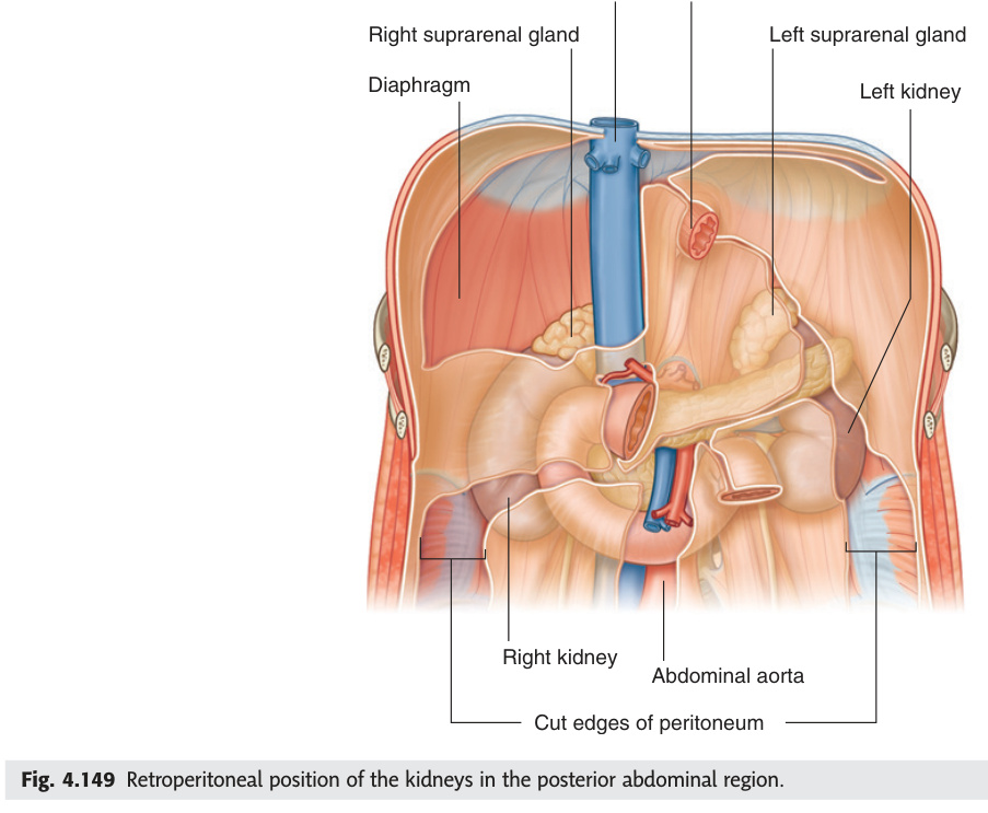
Retroperitoneal organs on the posterior abdominal wall, at the level of T12–L3. Right kidney is ~1 cm lower than the left (liver). Each is surrounded by: perinephric fat → Gerota's (renal) fascia → paranephric fat. The right renal hilum is at L1; the left is at L1/L2.
| Surface | Right Kidney Relations | Left Kidney Relations |
|---|---|---|
| Anterior | Right suprarenal, liver (upper 2/3), hepatic flexure, 2nd part duodenum (retroperitoneal), small intestine | Left suprarenal, stomach, spleen, tail of pancreas, splenic flexure, jejunum |
| Posterior | Diaphragm (12th rib), quadratus lumborum, psoas major, transversus abdominis aponeurosis. Subcostal + iliohypogastric + ilioinguinal nerves | Same muscles + 11th + 12th ribs (left kidney extends higher) |
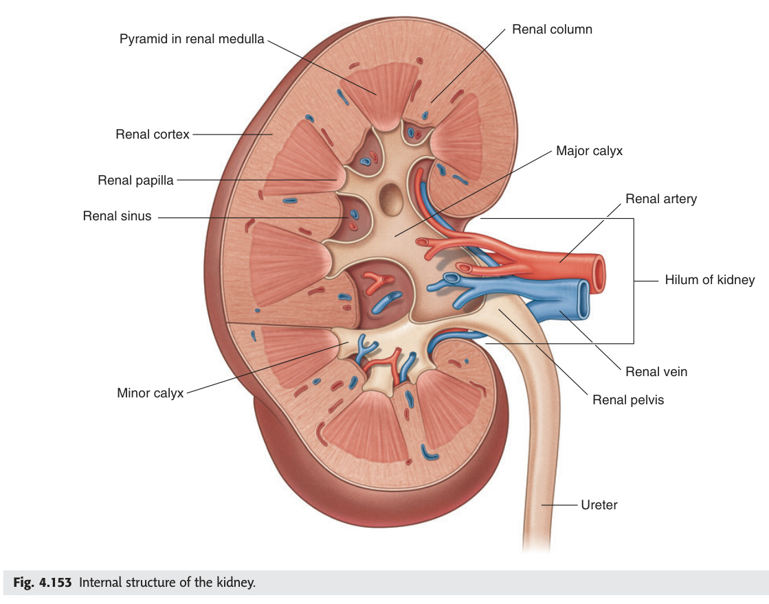
| Structure | Description | Clinical Note |
|---|---|---|
| Renal cortex | Outer granular zone; contains glomeruli, proximal + distal convoluted tubules; columns of Bertin extend between the pyramids | Site of diabetic nephropathy, glomerulonephritis — cortical thinning on USS = chronic kidney disease |
| Renal medulla | 8–18 renal pyramids; striated appearance (loops of Henle, collecting ducts); apex = renal papilla (projects into minor calyx) | Renal papillary necrosis: analgesic nephropathy (NSAIDs), sickle cell, diabetes, TB → sloughed papillae in urine |
| Minor calyces (8–18) | Cup-shaped structures receiving each renal papilla | Obstruction → calyceal dilatation (hydronephrosis) |
| Major calyces (2–3) | Formed by fusion of minor calyces; upper, middle, lower groups | Staghorn calculus fills the entire renal pelvis and calyces (struvite stones, urease-producing bacteria) |
| Renal pelvis | Funnel-shaped; receives major calyces; narrows to form ureter at PUJ (L1) | PUJ obstruction = most common cause of hydronephrosis in infants; pyeloplasty treatment |
Classic triad: haematuria + loin pain + palpable mass (only 10% have all three). Clear cell type (75%, from proximal tubule cells) most common. Risk: smoking, von Hippel-Lindau mutation (bilateral multifocal), adult polycystic kidney disease. Paraneoplastic: polycythaemia (EPO), hypercalcaemia (PTHrP), hypertension (renin). Left varicocele in adult: RCC tumour thrombus in left renal vein obstructs drainage. Staging: radical nephrectomy (stage I–II) ± targeted therapy (sunitinib, everolimus for advanced). Direct invasion into renal vein/IVC is characteristic of RCC.
Renal arteries arise from the aorta at L1–L2. Right renal artery is longer (passes posterior to IVC, right renal vein, head of pancreas, descending duodenum). The hilum (medial concave border) contains (from front to back): renal vein → renal artery → renal pelvis (mnemonic: VAP).
Atherosclerotic (usually ostial, elderly males) or fibromuscular dysplasia (mid-artery, young females). Renovascular hypertension: renin-angiotensin system activation. Renal artery stenosis murmur = continuous bruit in flank/epigastrium. Managed by renal artery angioplasty + stenting. Bilateral stenosis + ACE inhibitor → acute kidney injury (removes efferent arteriolar tone that maintains GFR). Horseshoe kidney: fused lower poles across midline — lower than normal (L3–L5), hooked by IMA. Risk of hydronephrosis, stones, UTI, Wilms' tumour.
- Q: Why does Gerota's fascia allow inferior spread of perinephric collections? — Gerota's fascia is closed superiorly, medially, and laterally but open inferiorly — no fusion at the lower pole, so gravity directs blood/pus/urine toward the iliac fossa.
- Q: Why is the left renal vein longer and what crosses it anteriorly? — Left kidney is further from the IVC (which lies right of midline). Left renal vein (~7 cm) crosses anterior to the aorta, passing under the SMA — this "nutcracker" can compress it, causing left varicocele.
- Q: Hilum contents front to back — mnemonic? — VAP: Vein (anterior), Artery (middle), Pelvis/ureter (posterior).
- Q: Why does testicular/renal carcinoma metastasise to para-aortic nodes, not inguinal? — Both structures developed at L1–L2 and their lymphatics follow the gonadal vessels back to para-aortic nodes — the embryological origin determines lymphatic drainage regardless of final anatomical position.
- Q: What drug class is contraindicated in bilateral renal artery stenosis and why? — ACE inhibitors; they remove efferent arteriolar tone (angiotensin II maintains efferent constriction), causing acute loss of GFR in kidneys already dependent on this to maintain filtration pressure.
Ureters
Three physiological narrowings create three predictable sites where ureteric calculi lodge — the PUJ, the pelvic brim (where the ureter crosses the common iliac artery bifurcation), and the VUJ (narrowest, where 70% of stones finally impact). The ureter's pelvic course creates two surgical traps: it lies anterior to the common iliac artery bifurcation during pelvic dissection, and in the female it runs beneath the uterine artery in the cardinal ligament ("water under the bridge"), making it the most common site of iatrogenic injury during hysterectomy. Ureteric colic pain radiates from loin to groin via T11–L2 dermatomes, often with testicular/labial referral — the same nerve roots that supply the ureter and the genitalia.
Retroperitoneal muscular tubes (~25–30 cm). Run on the tips of lumbar transverse processes, cross the bifurcation of the common iliac arteries at the pelvic brim, then curve forward in the pelvis to enter the bladder obliquely at the trigone.
2. Pelvic brim: where ureter crosses the bifurcation of common iliac artery.
3. Vesicoureteric junction (VUJ): where ureter enters the bladder — narrowest point; most common site of stone impaction (70% of ureteric stones pass from here).
Ureteric colic: severe colicky loin-to-groin pain (T11–L2 distribution). Haematuria (>90%). Plain KUB film (90% of stones radio-opaque), low-dose CT KUB = gold standard.
The ureter crosses the pelvic brim anterior to the common iliac artery bifurcation — a key landmark during pelvic lymph node dissection and colectomy. At risk during hysterectomy: the ureter passes under the uterine artery ('water under the bridge') — must be identified before ligating the uterine artery. Also at risk during ligation of the inferior mesenteric artery and during posterior dissection for rectal cancer.
- Q: Three physiological constrictions of the ureter — where and clinical significance? — (1) PUJ: most common congenital obstruction. (2) Pelvic brim: crosses common iliac bifurcation. (3) VUJ: narrowest — most common site of stone impaction (70% of ureteric stones).
- Q: "Water under the bridge" — what structure passes under what? — The ureter passes under the uterine artery in the base of the broad ligament (cardinal ligament). Risk of ureteric injury if the uterine artery is ligated without clearly identifying the ureter first.
- Q: Ureteric colic pain distribution and nerve roots? — Loin to groin, T11–L2. May radiate to testis/labia via genitofemoral nerve (L1–L2). Associated haematuria in >90%.
- Q: Most radio-opaque ureteric stones on plain film? — Calcium oxalate and calcium phosphate (most common, >80% radio-opaque). Uric acid stones are radiolucent. Gold standard = low-dose CT KUB (unenhanced).
- Q: At what level does the ureter cross the pelvic brim and what does it cross? — At the bifurcation of the common iliac artery, anterior to it, at the level of the sacroiliac joint.
Urinary Bladder
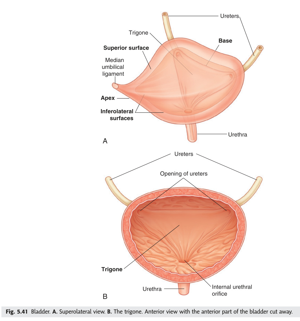
The full bladder rises above the pubic symphysis and strips the peritoneum off its anterior wall, creating the retropubic (space of Retzius) extraperitoneal approach for suprapubic catheterisation — the anatomical basis of a procedure that avoids peritoneal entry. The trigone is the one smooth area in an otherwise rugose bladder: its different embryological origin (mesonephric duct tissue, not urogenital sinus) explains its lack of rugae, its resistance to distension, and why it is the earliest site visible for tumour surveillance on cystoscopy. Three neurotransmitter systems control micturition in a hierarchy: parasympathetics (S2–S4) contract the detrusor to void; sympathetics (L1–L2) relax the detrusor and close the bladder neck during storage; somatic pudendal (S2–S4) provides voluntary control of the external sphincter, which is the last line of continence.
| Feature | Detail |
|---|---|
| Empty vs full | Empty: entirely in pelvis, pyramidal. Full: rises above pubic symphysis into abdomen — peritoneum is stripped off anteriorly → suprapubic catheter can be placed without entering peritoneum |
| Surfaces | Apex (umbilicus via median umbilical ligament = obliterated urachus); Superior surface (covered by peritoneum); Two inferolateral; Posterior (base/fundus) |
| Trigone | Smooth triangular area on posterior bladder wall between 2 ureteric orifices and internal urethral orifice. Mucosa closely adherent (no rugae) → earliest site of bladder tumour detection |
| Muscle | Detrusor muscle (3 layers of smooth muscle); internal urethral sphincter = involuntary thickening of detrusor at bladder neck |
| Nerve supply | Parasympathetic (S2–S4) = detrusor contraction; Sympathetic (L1–L2) = relaxes detrusor + contracts internal sphincter; Somatic (pudendal S2–S4) = external sphincter |
Transitional cell carcinoma (urothelial carcinoma) = 90%. Risk factors: smoking (most important), aniline dye exposure (industrial), cyclophosphamide, schistosomiasis (squamous cell type). Painless haematuria in an adult = bladder carcinoma until proven otherwise. Cystoscopy + biopsy gold standard. Superficial (Ta/T1): TURBT + intravesical BCG. Muscle-invasive (T2+): radical cystectomy ± neoadjuvant chemo.
- Q: Why can a suprapubic catheter be inserted without entering the peritoneum? — When the bladder is full it rises above the pubic symphysis, stripping the peritoneum off its anterior wall → an extraperitoneal window exists for safe suprapubic access.
- Q: What is the trigone, why is it smooth, and why is it clinically important? — Smooth triangle between the two ureteric orifices and the internal urethral orifice; smooth because mesonephric duct origin (no rugae); earliest site for detecting urothelial carcinoma on cystoscopy.
- Q: Three nerve supplies to the bladder and their functions? — Parasympathetic S2–S4: detrusor contraction (voiding); Sympathetic L1–L2: detrusor relaxation + internal sphincter closure (storage); Somatic pudendal S2–S4: external sphincter (voluntary control).
- Q: Most important risk factor for bladder carcinoma and what type? — Smoking (most important). Transitional cell (urothelial) carcinoma = 90%. Painless haematuria = bladder carcinoma until proven otherwise.
- Q: Obliterated urachus — what is it and what congenital anomaly results if it remains patent? — Median umbilical ligament connecting bladder apex to umbilicus; patent = urachal fistula (urine draining from umbilicus at birth).
Urethra
The male urethra's three parts have distinct clinical significances that map onto their anatomy: the prostatic urethra is the widest (BPH compresses it from the transition zone outward), the membranous is the narrowest and least dilatable (pelvic fracture tears it here — retrograde urethrogram before any catheterisation), and the spongy penile urethra runs in the corpus spongiosum and is the site of strictures from gonococcal infection. The female urethra at 4 cm is mechanically equivalent to the male membranous urethra alone — its brevity is the single most important anatomical reason for the dramatically higher UTI prevalence in women, since ascending bacteria have far less distance to travel. Blood at the urethral meatus after trauma is a sign that must never be ignored: it contraindicates blind catheterisation until a urethrogram rules out disruption.
| Part (Male, ~20 cm) | Description | Clinical |
|---|---|---|
| Prostatic (3–4 cm) | Widest and most dilatable part. Runs through the prostate. Seminal colliculus (verumontanum) on posterior wall with ejaculatory duct openings | Enlarged prostate = most common obstruction. TURP (transurethral resection of prostate) |
| Membranous (1–2 cm) | Shortest + narrowest + least dilatable. Pierces urogenital diaphragm. External urethral sphincter here (voluntary) | Most commonly injured in pelvic fractures → urethral disruption → urethrogram before catheter |
| Spongy (penile) (~15 cm) | Runs in corpus spongiosum. Dilated at fossa navicularis (glans). External opening = external urethral meatus | Urethral stricture post-gonococcal infection or instrumentation. Hypospadias = abnormal opening on ventral surface |
Female urethra: only ~4 cm; much shorter → more susceptible to UTI. Passes anterior to the vagina, opens into vestibule. External urethral sphincter present.
Posterior urethral injury (membranous, pelvic fracture): classic signs = blood at urethral meatus, perineal bruising, high-riding prostate on DRE (if prostate displaced superiorly), inability to void. Do retrograde urethrogram before catheterisation. Anterior urethral injury (bulbar, straddle injury): urine extravasates into superficial perineal pouch → Colles' fascia limits spread → scrotum + lower anterior abdominal wall (Scarpa's fascia).
- Q: Which part of the male urethra is most commonly injured in pelvic fractures and why? — Membranous urethra — shortest, narrowest, least dilatable; fixed by the urogenital diaphragm which shears when the pelvis fractures.
- Q: Signs of posterior urethral injury — when is blind catheterisation contraindicated? — Blood at meatus, perineal bruising, high-riding prostate on DRE, inability to void. Always do retrograde urethrogram first.
- Q: Anterior urethral injury (bulbar, straddle) — where does urine extravasate? — Into the superficial perineal pouch → limited by Colles' fascia → spreads into scrotum and up anterior abdominal wall under Scarpa's fascia (NOT into thighs — Scarpa's fuses with fascia lata).
- Q: Hypospadias vs epispadias — which is more common and which is associated with bladder exstrophy? — Hypospadias more common (1:300 male births, ventral meatus opening). Epispadias rarer (1:30,000, dorsal opening); epispadias is associated with bladder exstrophy.
- Q: Why are women much more susceptible to UTI than men? — Female urethra is only ~4 cm (vs ~20 cm male); much shorter distance for bacteria to ascend to the bladder; no prostatic secretions with antibacterial properties.
Testes & Epididymis
Testicular development at L1–L2 explains two anatomical rules that appear repeatedly in clinical medicine: arterial supply comes from the abdominal aorta (not the internal iliac) because the testes acquired their blood supply before descending, and lymphatic drainage goes to the para-aortic/retroperitoneal nodes (not inguinal) — testicular carcinoma metastasises to the retroperitoneum. Testicular torsion is the only true urological emergency with a time window (6 hours for orchid salvage) and must be treated on clinical suspicion alone — the bell-clapper deformity allows the testis to rotate freely on the spermatic cord, strangling its own blood supply, and absent cremasteric reflex is the most reliable sign. Distinguishing torsion from epididymitis clinically saves testes: torsion = sudden severe pain, young male, absent cremasteric reflex, absent Doppler flow; epididymitis = gradual onset, older male, tender posterior epididymis, leucocytes in urine.
Descent: testes develop retroperitoneally on posterior abdominal wall at L1 level, guided by the gubernaculum, descending through the inguinal canal at ~28 weeks, reaching the scrotum by birth. They carry with them layers of anterior abdominal wall (scrotal layers).
The epididymis (head = superior, body, tail = inferior) caps the posterior-lateral surface of the testis. The ductus epididymis is ~6 m long, tightly coiled. Sperm maturation and storage occurs here. The tail of the epididymis is attached to the inferior pole of the testis by the gubernaculum remnant.
Testicular torsion (emergency): sudden onset severe testicular pain in young males (peak 12–18 years). "Bell-clapper deformity" (high attachment of tunica vaginalis → testis hangs freely). Cremasteric reflex absent. Prehn's sign: elevation makes pain worse (vs epididymitis: elevation relieves). Doppler USS: absent blood flow. 6-hour window for orchidopexy — do not wait for USS if clinically suspicious. Bilateral orchidopexy required. Epididymitis: older males, gradual onset, tender epididymis, leucocytes in urine, Chlamydia/Gonorrhoea or E. coli (elderly).
- Q: Why does the testicular artery arise from the aorta, not the internal iliac? — Testes develop retroperitoneally at L1–L2 and acquire blood supply before descending; they retain this aortic origin even after reaching the scrotum.
- Q: Testicular cancer lymph node drainage — why not inguinal? — Lymphatics follow the gonadal vessels back to para-aortic nodes (L1–L2 level). Inguinal node involvement only occurs if the scrotum or scrotal skin (not testis proper) is involved.
- Q: Testicular torsion — time window, key sign, and management? — 6-hour window for >90% salvage. Absent cremasteric reflex = most reliable sign. Immediate surgical exploration without waiting for USS; bilateral orchidopexy at the same time.
- Q: Left varicocele — why is it more common than right, and what must you exclude in an adult? — Left testicular vein drains to left renal vein at right angle (no valve); right drains to IVC at acute angle with a valve. Sudden adult left varicocele = exclude RCC invading the left renal vein.
- Q: Bell-clapper deformity — what is it? — High attachment of tunica vaginalis (peritoneum) around the spermatic cord rather than the posterior testis → testis hangs freely and can rotate on its vascular pedicle → predisposes to torsion.
Spermatic Cord & Inguinal Canal
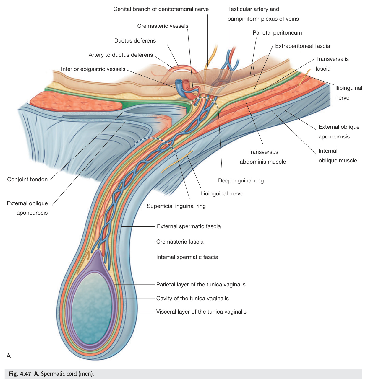
The spermatic cord is the retroperitoneal contents of the testis pulled into the scrotum during descent — each layer of cord fascia is a direct continuation of a layer of the anterior abdominal wall. The pampiniform plexus — the venous drainage network surrounding the testicular artery — has a left-right asymmetry that matters clinically: the left testicular vein joins the left renal vein at a right angle without a valve, while the right joins the IVC at an acute angle with a competent valve. This explains the overwhelming left-sided predominance of varicoceles. A varicocele appearing for the first time in an adult man (rather than a teenager) is a red flag for RCC tumour thrombus blocking the left renal vein — USS kidneys is mandatory.
"3 arteries, 3 nerves, 3 other"
Arteries: testicular artery (from aorta) + cremasteric artery (from inferior epigastric) + artery to ductus deferens (from inferior vesical)
Veins: pampiniform plexus (→ testicular vein), lymphatics
Nerves: genital branch of genitofemoral nerve (L1–L2, cremaster reflex) + autonomic fibres + ilioinguinal nerve (runs in the inguinal canal but NOT in the cord)
Other: ductus deferens, processus vaginalis remnant
Varicocele: dilated pampiniform plexus veins. Left side (90%) because left testicular vein enters left renal vein at a right angle (no valve). Sudden onset left varicocele in adult man = renal cell carcinoma until proven otherwise (tumour thrombus obstructs left renal vein). "Bag of worms" on palpation. Can impair fertility (elevated scrotal temperature inhibits spermatogenesis). Embolisation or surgical ligation.
- Q: Name the three arteries in the spermatic cord and their origins. — Testicular artery (aorta, L2); cremasteric artery (inferior epigastric artery); artery to ductus deferens (inferior vesical artery).
- Q: Why is varicocele predominantly left-sided? — Left testicular vein joins left renal vein at a right angle with no valve; right testicular vein drains to IVC at an acute angle with a competent valve — so left-side venous back-pressure is higher.
- Q: New-onset left varicocele in a 50-year-old man — what must you exclude? — Renal cell carcinoma invading the left renal vein with tumour thrombus. Order renal USS immediately.
- Q: Which nerve runs in the inguinal canal but NOT inside the spermatic cord? — Ilioinguinal nerve (L1). It travels between the internal and external oblique then exits the superficial ring; the genital branch of the genitofemoral nerve (inside the cord) mediates the cremaster reflex.
- Q: What is the genital branch of the genitofemoral nerve responsible for? — The efferent limb of the cremasteric reflex. L1–L2 is the reflex arc; absent in testicular torsion (useful distinguishing sign).
Prostate
The prostate is architecturally divided into zones (McNeal), and the zone determines the disease — BPH grows in the transition zone (squeezing the urethra), while 70–80% of prostate cancers originate in the peripheral zone (palpable as a hard nodule on posterior DRE, because the peripheral zone is immediately anterior to the rectal wall). The posterior boundary of the prostate is Denonvilliers' fascia, a condensed layer of peritoneum-derived fibrous tissue that forms a surgical plane between prostate and rectum. Prostate cancer staging hinges on whether this plane is intact: T3b = seminal vesicle invasion; bilateral involvement = loss of curative intent for radical surgery. The autonomic nerve supply runs as the neurovascular bundles at 5 o'clock and 7 o'clock lateral to the prostate — nerve-sparing prostatectomy tries to preserve erection by protecting these bundles.
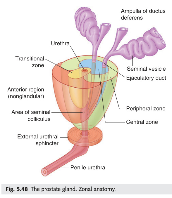
| Zone | Volume | Disease |
|---|---|---|
| Peripheral zone | 70% | Prostate carcinoma arises here (70–80% of carcinomas) — palpable on DRE as hard nodule posterior-laterally |
| Transition zone | 5–10% (increases with age) | Benign prostatic hyperplasia (BPH) arises here — surrounds urethra → compresses it |
| Central zone | 25% | Surround ejaculatory ducts; relatively resistant to disease |
Relations: Anterior = symphysis pubis (puboprostatic ligaments); Posterior = rectum (separated by Denonvilliers' fascia/rectovesical septum — felt on DRE); Superior = bladder neck; Inferior = urogenital diaphragm; Lateral = levator ani.
| Feature | Detail |
|---|---|
| Position | Paired saccular glands posterior to the bladder base; lateral to each ductus deferens; lie on the posterior surface of the prostate base |
| Function | Produce ~60–70% of seminal fluid; high in fructose (energy substrate for sperm) + prostaglandins + fibrinogen (coagulates semen) |
| Duct | Duct of seminal vesicle + ductus deferens → ejaculatory duct → opens into prostatic urethra at verumontanum (seminal colliculus) |
| Nerve supply | Sympathetic (L1–L2) via hypogastric plexus → ejaculation. Parasympathetic (S2–S4) → erection |
| Clinical | Prostate carcinoma invades seminal vesicles (T3b stage) — reduces chance of curative resection. On MRI: loss of normal triangular shape + restricted diffusion. Checked with PSA and mpMRI pre-prostatectomy |
BPH (transition zone): urinary outflow obstruction — frequency, nocturia, hesitancy, poor stream, incomplete emptying. Medical: alpha-blockers (relax smooth muscle) + 5-alpha reductase inhibitors (shrink gland). TURP = gold standard surgery. Retrograde ejaculation post-TURP (70%). Prostate carcinoma: most common cancer in males. Osteoblastic bone metastases (sclerotic on XR — lumbar spine, pelvis, femur). PSA screening controversial. Advanced disease: LHRH analogues (medical castration) or bilateral orchidectomy. Radical prostatectomy for localised disease → risk to autonomic nerves → erectile dysfunction + urinary incontinence.
- Q: BPH arises in which zone, and prostate carcinoma in which zone? — BPH = transition zone (periurethral, compresses urethra). Carcinoma = peripheral zone (70–80%; palpable posterolaterally on DRE as hard nodule).
- Q: What is Denonvilliers' fascia and why does it matter surgically? — Condensed rectovesical septum between prostate and rectum; forms the surgical plane for safe dissection in radical prostatectomy; involvement = T3 disease.
- Q: Prostate carcinoma bone metastases — what is the characteristic X-ray finding? — Osteoblastic (sclerotic/white) lesions — pelvis, lumbar spine, femur. Contrast with osteolytic metastases of lung/thyroid/kidney. PSA and ALP elevated; bone scan is most sensitive.
- Q: How does alpha-blockade help BPH symptoms, and what common side effects does it cause? — Relaxes smooth muscle of prostatic urethra and bladder neck → improved flow. Side effects: postural hypotension, retrograde ejaculation (tamsulosin relatively selective but still occurs).
- Q: What are the two main complications of radical prostatectomy related to the neurovascular bundles? — Erectile dysfunction (injury to parasympathetic fibres at 5 and 7 o'clock positions) and urinary incontinence (injury to external urethral sphincter). Nerve-sparing technique reduces ED risk in organ-confined disease.
Scrotal Layers
Every layer of the scrotal wall is a direct embryological derivative of the anterior abdominal wall — the testis carries its coverings with it during descent, like a fist being pushed into a balloon. This principle lets you derive the layers rather than memorise them: skin → Camper's/Scarpa's fascia (= dartos) → external oblique aponeurosis (= external spermatic fascia) → internal oblique (= cremasteric muscle) → transversalis fascia (= internal spermatic fascia) → parietal peritoneum (= tunica vaginalis). The tunica vaginalis is the key to two high-stakes clinical problems: it allows fluid accumulation (hydrocele) and — when it is abnormally high (bell-clapper deformity) — it permits testicular rotation causing torsion. Distinguishing hydrocele from inguinoscrotal hernia requires two manoeuvres: can you get above the swelling (yes = scrotal; no = extends into canal), and does it transilluminate (yes = fluid = hydrocele).
2. Dartos muscle/fascia — equivalent to Camper's + Scarpa's fascia; contracts in cold
3. External spermatic fascia — from external oblique aponeurosis
4. Cremasteric muscle + fascia — from internal oblique + transversus (incomplete loops; cremaster reflex)
5. Internal spermatic fascia — from transversalis fascia
6. Tunica vaginalis — parietal + visceral layers (peritoneum); parietal tunica = processus vaginalis remnant
7. Tunica albuginea — fibrous capsule of testis
Cremaster reflex: L1–L2 (genitofemoral nerve genital branch). Absent in testicular torsion.
Hydrocele: fluid in tunica vaginalis. Transilluminates brightly. Can get above the swelling (confined to scrotum). Secondary to trauma, infection, tumour — always examine the testis with USS. Inguinal hernia: cannot get above the swelling (extends into inguinal canal). Does not transilluminate. Indirect: through deep inguinal ring, lateral to inferior epigastric (congenital processus vaginalis patent). Direct: through Hesselbach's triangle, medial to inferior epigastric (acquired, weak posterior canal wall).
- Q: What is the scrotal equivalent of the external oblique aponeurosis? — External spermatic fascia. The testis picks up this layer as it passes through the deep inguinal ring (which is a deficiency in the transversalis fascia).
- Q: Which scrotal layer gives rise to the cremasteric reflex and what is the nerve arc? — Cremasteric muscle (from internal oblique). Afferent: femoral branch of genitofemoral nerve (L1–L2); efferent: genital branch of genitofemoral nerve. Absent in testicular torsion — clinically crucial.
- Q: What is the tunica vaginalis and where does it come from? — Remnant of the processus vaginalis (peritoneum); parietal layer lines the inner scrotal wall, visceral layer covers the testis. Fluid between them = hydrocele.
- Q: Distinguish hydrocele from inguinal hernia on examination. — Hydrocele: can get above swelling (confined to scrotum), transilluminates brightly, fluctuant. Hernia: cannot get above swelling (track extends into canal), does not transilluminate, has cough impulse. Always USS testes to exclude secondary hydrocele from tumour.
- Q: What is Fournier's gangrene and which fascial plane does it track along? — Necrotising fasciitis of the perineum/scrotum (polymicrobial, often diabetic males). Spreads along Colles' fascia (perineal = continuation of Scarpa's fascia) — can track up anterior abdominal wall but is limited posteriorly by Colles' attachment to perineal body. Surgical emergency: wide debridement.
Test your knowledge of Unit 07
Kidneys, bladder, prostate, testes — MCQs and clinical essays.
Suprarenal (Adrenal) Glands ★★
The adrenal glands have a dual embryological origin that explains their dual function: the cortex is mesodermal (derived from the same coelomic epithelium as the gonads) and makes steroid hormones, while the medulla is neuroectodermal (derived from neural crest cells that migrated into the cortex) and makes catecholamines. The cortex zones are memorised as "Salt, Sugar, Sex" (outer → inner): glomerulosa = mineralocorticoids (aldosterone, salt balance), fasciculata = glucocorticoids (cortisol, stress/sugar), reticularis = sex steroids (DHEA). The right adrenal vein is short and drains directly into the IVC — this is the most dangerous moment in right adrenalectomy, as tearing this short vein causes torrential haemorrhage. The left is longer (to left renal vein) and safer. A phaeochromocytoma must always be alpha-blocked first before surgery — operating without alpha-blockade on a phaeochromocytoma causes hypertensive crisis from catecholamine surge during tumour handling.
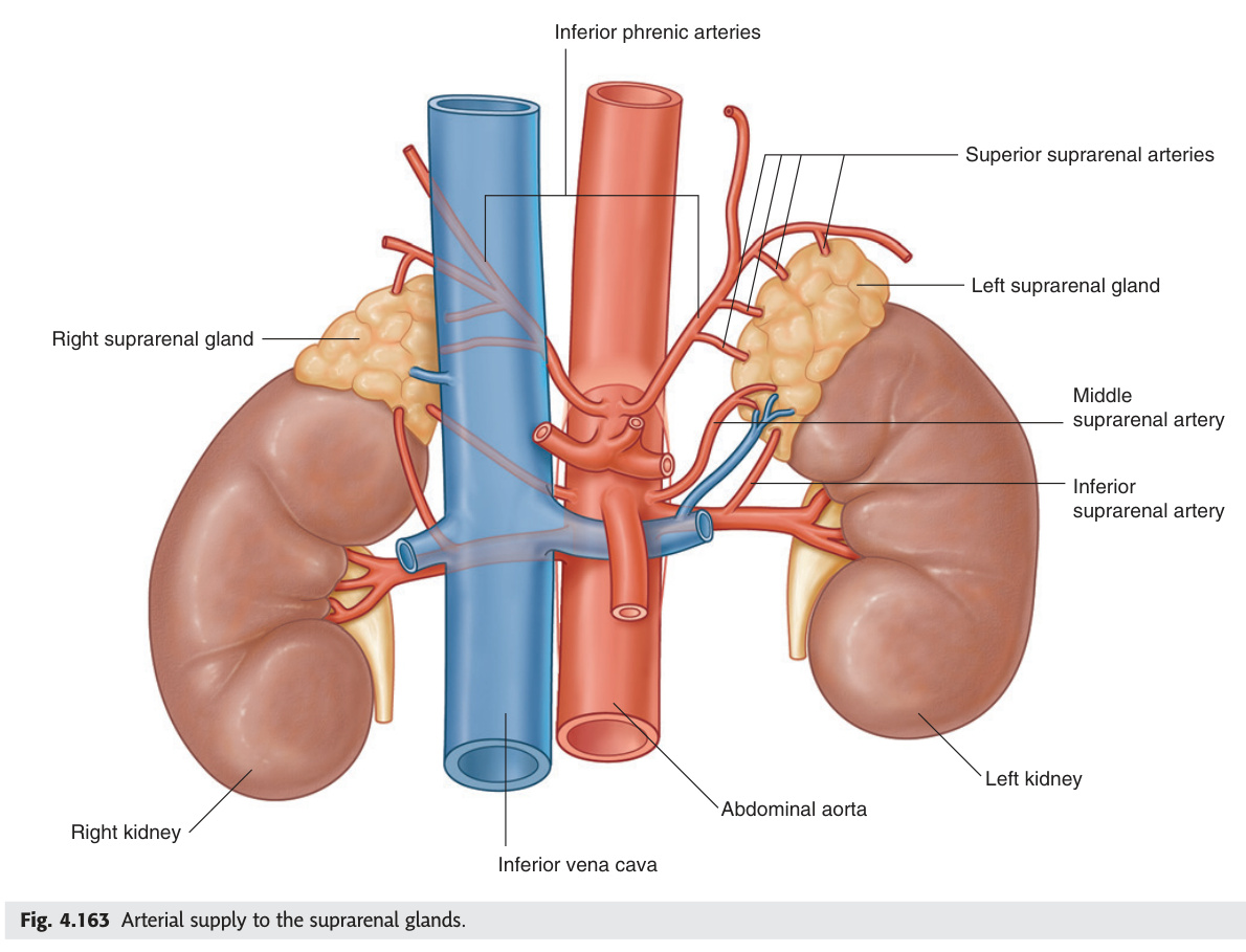
| Right Suprarenal | Left Suprarenal | |
|---|---|---|
| Shape | Pyramidal | Semilunar / crescentic |
| Position | Superomedial to right kidney; posterior to IVC | Superomedial to left kidney; posterior to stomach/pancreas tail |
| Vein drainage | Right suprarenal vein → IVC (directly; very short) | Left suprarenal vein → left renal vein |
| Arteries (3) | Superior suprarenal (from inferior phrenic artery) · Middle suprarenal (from aorta directly) · Inferior suprarenal (from renal artery) | |
Note: The right suprarenal vein drains directly into the IVC — this short vein is easily torn during right adrenalectomy, causing significant haemorrhage. The left vein is longer (drains to left renal vein).
| Layer | Zone (Cortex) | Hormone | Disease of Excess → / Deficiency |
|---|---|---|---|
| Cortex (derived from mesoderm) | Zona glomerulosa (outer) | Mineralocorticoids — Aldosterone | Excess → Conn's syndrome (primary hyperaldosteronism): hypertension + hypokalaemia + low renin. Cause: adrenal adenoma (70%) or bilateral hyperplasia |
| Zona fasciculata (middle, largest) | Glucocorticoids — Cortisol | Excess → Cushing's syndrome: central obesity, moon face, buffalo hump, striae, hypertension, DM, osteoporosis. Deficiency + zona reticularis → Addison's disease | |
| Zona reticularis (inner) | Sex steroids — DHEA, androgens | Congenital adrenal hyperplasia (CAH): 21-hydroxylase deficiency → excess androgens → virilisation in females + salt-wasting crisis in neonates | |
| Medulla (derived from neural crest) | — (chromaffin cells) | Adrenaline (80%) + Noradrenaline (20%) | Phaeochromocytoma: paroxysmal hypertension + headache + sweating + palpitations. "Rule of 10%": 10% bilateral, 10% extra-adrenal, 10% malignant, 10% familial. Diagnose: plasma/urine metanephrines. Treat: alpha-blockade (phenoxybenzamine) FIRST, then beta-blockade, then surgery |
"GFR" (like kidney GFR but for adrenal cortex zones): Glomerulosa (outer — Gluco for salt = mineralocorticoids) · Fasciculata (middle — Fat = glucocorticoids) · Reticularis (inner — Retro = sex steroids / androgens)
"The deeper you go, the sexier it gets" — innermost zone = sex steroids.
Or: "Salt, Sugar, Sex" (outer → inner).
Destruction of adrenal cortex (>90% must be destroyed). In developed world: autoimmune (most common — anti-21-hydroxylase antibodies). Worldwide: TB (most common cause). Features: chronic fatigue, weight loss, postural hypotension, hyponatraemia + hyperkalaemia (aldosterone deficiency), hypoglycaemia (cortisol deficiency), hyperpigmentation (elevated ACTH causes MSH activity — pigments skin creases, buccal mucosa, scars). Treat: hydrocortisone + fludrocortisone. Addisonian crisis (acute): triggered by stress/infection → shock, vomiting → treat with IV hydrocortisone + saline.
- Q: Name the three cortical zones and their hormones (outer → inner). — Glomerulosa → aldosterone (mineralocorticoid); fasciculata → cortisol (glucocorticoid); reticularis → DHEA/sex steroids. Mnemonic: "Salt, Sugar, Sex" or "GFR".
- Q: Right vs left adrenal vein drainage — why does the right matter surgically? — Right drains directly to IVC (short, easily torn = haemorrhage risk); left drains to left renal vein (longer, safer). Always ligate right adrenal vein with care in right adrenalectomy.
- Q: Phaeochromocytoma — rule of 10s and why must you alpha-block BEFORE surgery? — 10% bilateral, 10% extra-adrenal (paraganglioma), 10% malignant, 10% familial (MEN2, NF1, VHL). Alpha-block first (phenoxybenzamine) to prevent hypertensive crisis from tumour handling; add beta-blocker after (never beta-first = unopposed alpha = severe hypertension).
- Q: Conn's syndrome vs Cushing's syndrome — key distinguishing electrolyte finding. — Conn's (hyperaldosteronism): hypertension + hypokalaemia + LOW renin (autonomous aldosterone suppresses renin). Cushing's: hypertension but potassium may be normal unless ectopic ACTH (ACTH-secreting tumours give profound hypokalaemia via cortisol's mineralocorticoid effect).
- Q: Addison's disease — why does hyperpigmentation develop? — Loss of cortisol → unrestrained ACTH secretion (no negative feedback) → ACTH and its precursor POMC are cleaved to produce MSH → melanocyte stimulation → pigmentation of creases, buccal mucosa, scars.
Nephrotic vs Nephritic Syndrome ★★★
Nephrotic and nephritic syndromes both damage the glomerulus but by completely different mechanisms — understanding the mechanism predicts every clinical feature. Nephrotic = loss of the glomerular charge/size barrier → albumin pours into urine → low oncotic pressure → oedema, and the liver compensates by making more lipoproteins → hyperlipidaemia. No haematuria because there is no glomerular inflammation. Nephritic = glomerular inflammation (immune complex deposition or antibody attack on GBM) → haematuria + red cell casts (cells squeezed through inflamed basement membrane), hypertension (reduced GFR → sodium/water retention), reduced renal function. In practice, the two can overlap, but the dominant syndrome guides the differential. Red cell casts on urine microscopy are pathognomonic of glomerulonephritis — their presence essentially proves nephritic syndrome regardless of the protein level. In children, the most common nephrotic syndrome is minimal change disease (podocyte foot process effacement on EM, invisible on light microscopy, steroid-responsive); in adults it is membranous nephropathy (anti-PLA2R antibodies, subepithelial deposits).
| Feature | Nephrotic Syndrome | Nephritic Syndrome |
|---|---|---|
| Core mechanism | Loss of glomerular charge/size barrier → massive protein loss into urine | Glomerular inflammation → haematuria + hypertension + reduced GFR |
| Proteinuria | >3.5 g/day (massive) — diagnostic criterion | Mild–moderate (<3.5 g/day) |
| Haematuria | Absent | Present — RBC casts in urine (pathognomonic of glomerulonephritis) |
| Oedema | Severe (periorbital in AM, ankle/sacral, ascites, pleural effusion) — from low oncotic pressure | Mild |
| Blood pressure | Normal or low | Hypertension (↓ GFR → sodium retention) |
| Serum albumin | ↓↓ (urinary losses) | Normal or mildly ↓ |
| Lipids | Hyperlipidaemia + lipiduria (Maltese-cross fat bodies on microscopy) | Normal |
| GFR / renal function | Usually preserved early | Reduced — azotaemia, oliguria, ↑ creatinine |
| Complement | Normal (except in MPGN, SLE) | ↓ C3 in post-streptococcal GN, MPGN, lupus |
| Urinary casts | Hyaline + fatty casts (oval fat bodies) | Red cell casts (diagnostic) |
| Syndrome | Primary (Idiopathic) | Secondary |
|---|---|---|
| Nephrotic | Minimal change disease (children, MCC in <8 yrs — steroid-responsive, "nil on LM") · FSGS (adults, HIV, heroin, sickle cell) · Membranous nephropathy (MCC in adults — anti-PLA2R antibodies) | Diabetic nephropathy (most common worldwide overall) · Amyloidosis (AL/AA) · SLE (membranous pattern) · Hepatitis B/C (membranous) · Drugs: NSAIDS, gold, penicillamine |
| Nephritic | IgA nephropathy (Berger's — most common GN worldwide; episodic macroscopic haematuria 1–2 days after URTI; IgA deposits in mesangium) · Post-streptococcal GN (children, 2–3 weeks after Group A Strep pharyngitis; "lumpy-bumpy" IgG + C3 deposits on IF; ↓C3; resolves spontaneously) · Anti-GBM disease (Goodpasture's — linear IgG on GBM; pulmonary haemorrhage + rapidly progressive GN; anti-GBM antibody positive; treat: plasmapheresis + steroids) | Lupus nephritis (Class III/IV) · Henoch-Schönlein Purpura (IgA vasculitis; children; palpable purpura + haematuria + arthritis + abdominal pain) · MPGN |
HITE (consequences of lost proteins): Hypercoagulability (↓ antithrombin III, protein C/S lost → DVT/renal vein thrombosis — particularly membranous nephropathy) · Infections (↓ immunoglobulins → pneumococcal peritonitis, sepsis) · Thrombosis (renal vein thrombosis → flank pain + haematuria + pulmonary embolism) · Eodem — see above. Also: hyperlipidaemia (↑ hepatic lipoprotein synthesis to maintain oncotic pressure) → accelerated atherosclerosis.
- Q: What single urine finding is pathognomonic of glomerulonephritis (nephritic)? — Red cell (RBC) casts. Formed when red cells are squeezed through the inflamed glomerular basement membrane into tubules where they become trapped in Tamm-Horsfall protein casts.
- Q: Why do nephrotic patients develop hyperlipidaemia? — Low oncotic pressure (from albumin loss) stimulates hepatic lipoprotein synthesis to try to restore colloid osmotic pressure → hyperlipidaemia + lipiduria (Maltese-cross oval fat bodies on polarised microscopy).
- Q: Minimal change disease — what does the biopsy show on each microscopy type? — Light microscopy: normal. Immunofluorescence: negative. Electron microscopy: diffuse podocyte foot process effacement/fusion. First-line: oral prednisolone (80–90% respond in children).
- Q: Membranous nephropathy — what antibody, what demographics, what EM finding? — Anti-PLA2R antibodies (phospholipase A2 receptor on podocytes). Most common primary nephrotic syndrome in adults. EM: subepithelial "spike and dome" deposits. Associations: hepatitis B/C, SLE, malignancy (lung, colon — always screen adults).
- Q: Name two dangerous complications unique to nephrotic syndrome from the lost proteins. — Hypercoagulability (loss of antithrombin III, protein C/S → DVT, renal vein thrombosis — especially membranous) and increased infection susceptibility (loss of immunoglobulins → pneumococcal peritonitis — vaccinate nephrotic children).
Inguinal Canal & Hernias ★★★
The inguinal canal is an oblique 4 cm passage that exists because the testis had to descend — it is not a design flaw but a developmental necessity, and the oblique direction (not straight through) is the primary mechanical defence against herniation. The key landmark that every exam question hinges on is the inferior epigastric artery: indirect hernias pass lateral to it (through the deep inguinal ring, the path the testis took), while direct hernias bulge medial to it through the weakened posterior wall (Hesselbach's triangle). Femoral hernias are anatomically below the inguinal ligament and lateral to the pubic tubercle — the opposite of inguinal hernias which are above and medial. The clinical stakes differ sharply by type: femoral hernias have the highest strangulation risk because the femoral ring is a rigid inelastic collar (bounded by inguinal ligament anteriorly, pectineal/Cooper's ligament posteriorly, and lacunar ligament medially) — prompt surgical repair is mandatory even if asymptomatic in elderly women.
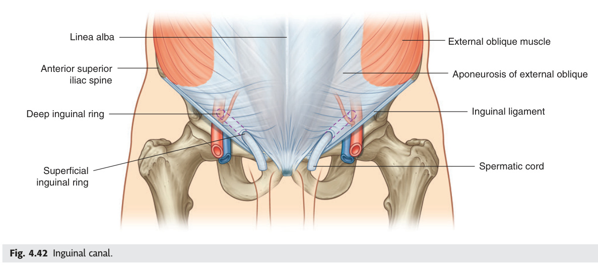
The inguinal canal is an oblique intermuscular passage (~4 cm long in adults) running from the deep (internal) inguinal ring to the superficial (external) inguinal ring, directed superomedially to inferomedially, following the direction of the external oblique fibres.
| Wall | Structure |
|---|---|
| Anterior wall | External oblique aponeurosis (entire length) + internal oblique muscle (lateral 1/3 only) |
| Posterior wall | Transversalis fascia (entire length) + conjoint tendon (medial 1/3 = inguinal falx — reinforces posterior wall where direct hernia protrudes) |
| Roof | Arching fibres of internal oblique + transversus abdominis (these contract during straining, reinforcing the canal — "shutter mechanism") |
| Floor | Inguinal ligament (Poupart's ligament, rolled-under lower edge of external oblique aponeurosis) + lacunar ligament (medially) |
Superficial (external) inguinal ring: a triangular aperture in the external oblique aponeurosis, located just above and lateral to the pubic tubercle. The ilioinguinal nerve exits here. Medial crus and lateral crus form its margins; intercrural fibres prevent lateral spread.
Key rule: the inferior epigastric artery distinguishes hernia types — indirect hernias pass lateral to it (through the deep ring); direct hernias emerge medial to it (through the posterior wall).
| Male | Female | |
|---|---|---|
| Main structure | Spermatic cord (ductus deferens + testicular artery + pampiniform plexus + cremasteric artery + artery to vas + genital branch of genitofemoral nerve + autonomic fibres) | Round ligament of uterus (attaches uterine fundus → labia majora) |
| Nerve | Ilioinguinal nerve (L1) — travels in canal but NOT inside the spermatic cord; exits at superficial ring → scrotum/medial thigh | Same ilioinguinal nerve → labia majora + medial thigh |
| Feature | Indirect Inguinal | Direct Inguinal | Femoral |
|---|---|---|---|
| Path | Through deep inguinal ring → inguinal canal → superficial ring → may enter scrotum | Through posterior wall of canal (Hesselbach's triangle) → superficial ring | Through femoral canal → below inguinal ligament → into femoral triangle |
| Relation to inferior epigastric | Lateral | Medial | Below inguinal ligament |
| Relation to pubic tubercle | Above and medial | Above and medial | Below and lateral |
| Demographics | Any age, M >> F; congenital (patent processus vaginalis); most common overall | Middle-aged/elderly males; acquired (weak transversalis fascia) | F > M (wider pelvis); elderly females; rare in children |
| Strangulation risk | Low–moderate (wide neck) | Low (wide neck) | High (narrow inelastic femoral ring) |
| Reducibility with patient lying | Usually reducible | Usually reducible | Often irreducible |
| Cough impulse (Malgaigne's fossa) | Controlled by finger at deep ring | Not controlled by deep ring occlusion | Present below inguinal ligament |
Boundaries: Medial = lateral border of rectus abdominis · Lateral = inferior epigastric artery · Inferior = inguinal ligament. Direct hernias protrude through the posterior wall (transversalis fascia) within this triangle — medial to the inferior epigastric artery.
Femoral canal (medial compartment of femoral sheath): boundaries — anteriorly inguinal ligament, posteriorly pectineal ligament (Cooper's ligament) + pectineus muscle, medially lacunar ligament, laterally femoral vein. Contains only fat + lymph node of Cloquet. The femoral canal is the path of femoral hernias. The ring is narrow and inelastic → herniated bowel rapidly becomes ischaemic (strangulation) → emergency surgery. Femoral hernias present as a lump below and lateral to the pubic tubercle (unlike inguinal = above and medial). More common in multiparous women (increased intra-abdominal pressure + wide pelvis).
- Q: What is the single most important landmark distinguishing indirect from direct inguinal hernia? — The inferior epigastric artery. Indirect = lateral to it (through deep inguinal ring). Direct = medial to it (through posterior wall/Hesselbach's triangle). Confirmed clinically: occlude deep ring → impulse controlled = indirect; not controlled = direct.
- Q: Name the four walls of the inguinal canal. — Anterior: external oblique aponeurosis (full length) + internal oblique (lateral 1/3). Posterior: transversalis fascia (full) + conjoint tendon (medial 1/3). Roof: arching fibres of internal oblique + transversus. Floor: inguinal ligament + lacunar ligament medially.
- Q: Femoral hernia — where is the lump relative to the pubic tubercle and inguinal ligament? — Below the inguinal ligament and lateral to the pubic tubercle (opposite of inguinal = above + medial to pubic tubercle). More common in multiparous women; highest strangulation risk of all groin hernias.
- Q: What are the boundaries of Hesselbach's triangle? — Medial: lateral border of rectus abdominis; lateral: inferior epigastric artery; inferior: inguinal ligament. Direct hernias protrude through transversalis fascia within this triangle.
- Q: Why does the ilioinguinal nerve NOT provide the cremaster reflex? — The cremaster reflex efferent arc is the genital branch of the genitofemoral nerve (L1–L2), which travels inside the spermatic cord. The ilioinguinal nerve (L1) runs in the canal beside the cord but exits at the superficial ring to provide only cutaneous sensory supply.