Alimentary Glands & Respiratory System
Liver
The liver is the body's metabolic hub — its dual blood supply (75% portal, 25% hepatic artery) reflects its twin roles as a processor of absorbed nutrients and a detoxification centre for everything the portal circulation delivers. Its modern surgical anatomy is the Couinaud system: eight independent segments each with their own portal pedicle and hepatic venous drainage, allowing surgeons to remove individual segments without devascularising the rest. The zone (acinar) model explains injury patterns: Zone 3 (centrilobular) is most vulnerable to hypoxia and drug toxicity because it receives blood last and contains the highest concentration of CYP2E1, the enzyme that converts paracetamol to the hepatotoxin NAPQI. Portal hypertension — the end result of chronic liver disease — opens predictable portosystemic collaterals at every anatomical site where portal and systemic veins run side by side, and knowing those sites lets you explain oesophageal varices, caput medusae, and haemorrhoidal engorgement from first principles.
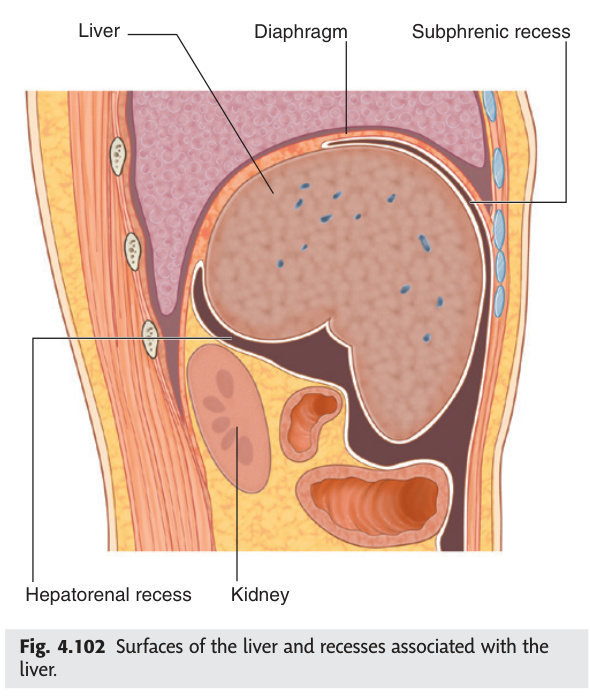
The largest gland in the body (~1.5 kg). Occupies the right hypochondrium and epigastrium, protected by ribs 7–11 on the right. The falciform ligament divides it into right lobe (larger) and left lobe (smaller) — this is the anatomical division.
| Lobe / Structure | Location | Notes |
|---|---|---|
| Right lobe | Right hypochondrium | Largest; inferior surface = gallbladder fossa + right colic flexure + right kidney + suprarenal |
| Left lobe | Epigastrium | Smaller; overlies stomach |
| Caudate lobe | Posterior between IVC (right) and ligamentum venosum (left) | Receives blood from both right + left portal branches and both hepatic arteries; drains directly into IVC → preserved in Budd-Chiari syndrome |
| Quadrate lobe | Inferior surface between gallbladder fossa (right) and round ligament (left) | Functionally part of the left lobe (Couinaud segment IV) |
| Porta hepatis | H-shaped groove on inferior surface | Entry/exit of portal vein, hepatic artery, bile ducts. Left limb = ligamentum teres (obliterated umbilical vein); right limb = gallbladder fossa; crossbar = porta hepatis proper |
Hepatic artery proper (25% of flow, 50% of O2): oxygenated blood from coeliac trunk → common hepatic artery → hepatic artery proper. Divides into right + left hepatic arteries at porta hepatis.
Both converge in the portal tracts and mix in the hepatic sinusoids. Drained by three hepatic veins (right, middle, left) → IVC.
The hepatic artery has the most variable anatomy of any major abdominal vessel (~40% anatomical variants). Most common: replaced right hepatic from SMA (10–15%); replaced left hepatic from left gastric (10%). Critical to identify on pre-operative cross-sectional imaging before liver resection or Whipple's procedure.
The liver is divided into 8 functional segments (Couinaud classification) based on independent blood supply, biliary drainage, and hepatic venous drainage. Each segment can be resected independently. Segments I (caudate) to VIII — separated by the 3 hepatic veins and the main portal fissure (Cantlie's line: IVC to gallbladder fossa).
Liver = most common site of metastatic disease (portal venous drainage from colon, stomach, pancreas). Colorectal metastases: up to 30% of patients at presentation. Resectable if confined to 1–2 segments with adequate functional remnant. Laparoscopic or robotic hepatectomy increasingly used. 5-year survival with resection ~40–50% for colorectal mets.
The hepatic acinus (Rappaport's model) is the functional unit of the liver, arranged around the portal tract. Blood flows from the portal tract (Zone 1) towards the central vein (Zone 3).
| Zone | Location | O₂ / Nutrient Level | Damage Pattern | Clinical Examples |
|---|---|---|---|---|
| Zone 1 (periportal) | Surrounding portal tracts; first to receive blood | Highest O₂ + nutrients | First affected in ischaemia of portal blood (toxic metabolites); periportal necrosis | Eclampsia of pregnancy; phosphorus poisoning; viral hepatitis (periportal inflammation) |
| Zone 2 (midzonal) | Intermediate | Intermediate | Yellow fever (midzonal necrosis — distinctive) | Yellow fever = Councilman bodies in Zone 2 |
| Zone 3 (centrilobular) | Surrounding central vein; last to receive blood | Lowest O₂ | Most vulnerable to hypoxia + toxic metabolites of cytochrome P450; centrilobular necrosis | Paracetamol (acetaminophen) overdose · CCl₄ poisoning · Right heart failure (congestive hepatopathy = "nutmeg liver") · Halothane hepatitis |
Portal hypertension = portal venous pressure >5 mmHg above IVC pressure (normal 5–10 mmHg). Clinical complications arise when portal pressure exceeds 12 mmHg (varices form; HVPG >10 mmHg = decompensation threshold).
| Classification | Site of Obstruction | Common Causes |
|---|---|---|
| Pre-hepatic | Portal or splenic vein (before liver) | Portal vein thrombosis (neonatal sepsis, hypercoagulable states) · Splenic vein thrombosis · Schistosomiasis (worldwide most common cause of portal hypertension) |
| Hepatic — pre-sinusoidal | Portal tracts (before sinusoids) | Primary biliary cholangitis · Sarcoidosis · Congenital hepatic fibrosis · Early schistosomiasis |
| Hepatic — sinusoidal | Sinusoids | Liver cirrhosis (most common cause in developed world): alcoholic, viral (HBV/HCV), NASH, autoimmune hepatitis, Wilson's, haemochromatosis |
| Hepatic — post-sinusoidal | Central veins (within liver) | Veno-occlusive disease (hepatic sinusoidal obstruction syndrome) — HSCT complication, chemotherapy |
| Post-hepatic | Hepatic veins → IVC (beyond liver) | Budd-Chiari syndrome (hepatic vein thrombosis — polycythaemia, JAK2 mutation, OCP use) · Right heart failure · Constrictive pericarditis |
Clinical features: splenomegaly → hypersplenism (pancytopenia); ascites (↓ oncotic pressure + ↑ hydrostatic + aldosterone activation → Na⁺/water retention); portosystemic collaterals (oesophageal varices, caput medusae, rectal varices — see Section 5.8.2); hepatic encephalopathy (ammonia + other toxins bypass liver → cerebral dysfunction); coagulopathy (↓ clotting factor synthesis); jaundice (hepatocellular failure).
Investigation: USS Doppler (portal vein flow); HVPG (hepatic venous pressure gradient — gold standard); OGD (varices grading); LFTs + albumin + INR (synthetic function).
Management: Primary prophylaxis of variceal bleeding — non-selective beta-blockers (propranolol/carvedilol, target HR 55–60). Acute variceal haemorrhage — terlipressin (vasopressin analogue) + IV tazobactam + emergency OGD band ligation; Sengstaken-Blakemore tube if uncontrolled. Secondary prophylaxis — band ligation ± beta-blocker. Refractory — TIPS (transjugular intrahepatic portosystemic shunt) creates a communication between portal and hepatic veins via a stent; reduces portal pressure but risks hepatic encephalopathy. Budd-Chiari — anticoagulation ± TIPS ± liver transplant.
- Q: Dual blood supply of the liver — proportions and sources? — Portal vein 75% of volume (50% of O₂), nutrient-rich from gut; hepatic artery proper 25% of volume (50% of O₂), from coeliac trunk. Both converge in sinusoids.
- Q: Couinaud segments — how many and what divides them? — 8 segments; divided by 3 hepatic veins (left, middle, right) and the main portal fissure (Cantlie's line: IVC to gallbladder fossa). Each resectable independently.
- Q: Which zone is damaged by paracetamol overdose and why? — Zone 3 (centrilobular): highest CYP2E1 concentration (converts paracetamol → NAPQI) + lowest O₂ + least glutathione. Antidote = N-acetylcysteine.
- Q: Three main portosystemic anastomosis sites in portal hypertension? — Oesophagus (left gastric ↔ azygos → varices); umbilicus (paraumbilical ↔ epigastric → caput medusae); rectum (superior rectal ↔ middle/inferior rectal).
- Q: Why is the caudate lobe preserved in Budd-Chiari syndrome? — Caudate lobe drains directly into the IVC via independent hepatic veins, bypassing the obstructed main hepatic veins.
Biliary System
The biliary tree's clinical significance is almost entirely about obstruction — where it occurs and what syndrome it produces. Gallstones cause 95% of biliary pathology, and their anatomical location determines the presentation: cystic duct obstruction gives biliary colic; a stone in the common bile duct gives cholangitis (Charcot's triad) or jaundice; ampullary obstruction gives both jaundice and pancreatitis. Calot's triangle is the most surgically consequential three-centimetre patch in abdominal anatomy — every iatrogenic bile duct injury during laparoscopic cholecystectomy traces back to failure to achieve the "critical view of safety" within it. Courvoisier's law links anatomy to clinical diagnosis: chronic gallstone disease fibroses and contracts the gallbladder wall so it cannot distend, whereas a sudden external compression from pancreatic carcinoma obstructs a previously normal, distensible gallbladder — hence painless jaundice plus palpable gallbladder = malignancy until proven otherwise.
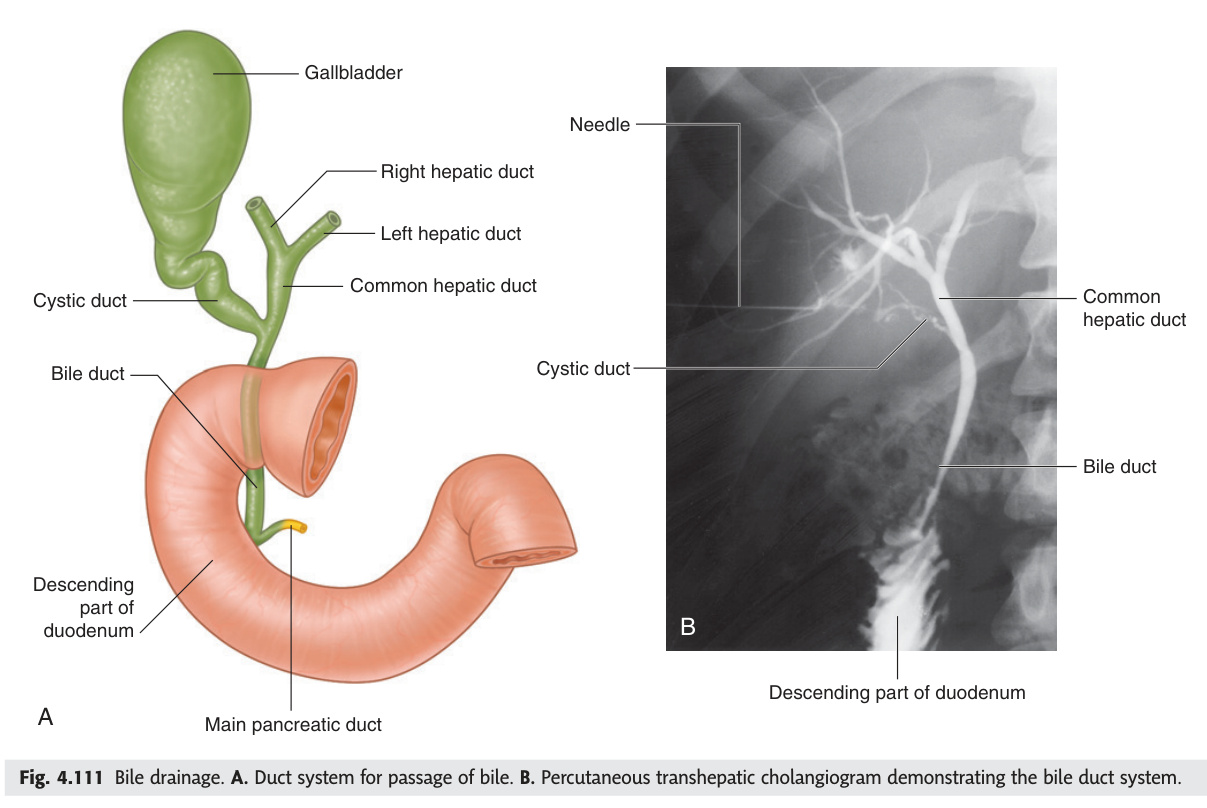
| Structure | Formation/Notes |
|---|---|
| Right + Left hepatic ducts | Emerge from porta hepatis; join immediately to form CHD |
| Common hepatic duct (CHD) | ~3 cm; descends in free edge of lesser omentum, anterior to portal vein |
| Cystic duct | From gallbladder neck; spiral valve of Heister (prevents collapse); joins CHD to form CBD |
| Common bile duct (CBD) | ~8 cm total; 4 parts: supraduodenal (in lesser omentum) → retroduodenal → pancreatic (in pancreatic head) → intraduodenal (joins pancreatic duct). Enters 2nd part of duodenum at ampulla of Vater |
The hepatoduodenal ligament (free right edge of lesser omentum) contains:
From left to right (anterior view): Bile duct (right) · Hepatic artery (left) · Portal vein (posterior)
Mnemonic: "ABP" — Anterior: bile duct + artery; Posterior: portal vein. Pringle's manoeuvre = digital compression of hepatoduodenal ligament between thumb (in epiploic foramen) and index finger to control liver bleeding.
Pear-shaped muscular sac (~10 cm long) on the inferior surface of the right liver lobe in the gallbladder fossa. Parts: fundus (projects below liver edge at tip of 9th costal cartilage — Murphy's sign), body, neck (leads to cystic duct).
Critical in laparoscopic cholecystectomy — the "critical view of safety" requires complete dissection of Calot's triangle to clearly identify both the cystic duct and cystic artery before clipping and division. Failure → bile duct injury.
Biliary colic: stone obstructs cystic duct → RUQ/epigastric pain radiating to right shoulder (phrenic nerve, C4). Cholecystitis = stone + infection. Murphy's sign = inspiratory arrest on deep palpation of RUQ. Courvoisier's law: palpable gallbladder in jaundiced patient is unlikely to be gallstones (chronic stones → fibrosed GB). More likely carcinoma of the pancreatic head. Charcot's triad (ascending cholangitis): RUQ pain + jaundice + fever/rigors.
- Q: Contents of the hepatoduodenal ligament (portal triad) — arrangement? — Bile duct (right, anterior), Hepatic artery (left, anterior), Portal vein (posterior). Pringle's manoeuvre compresses all three between fingers in the epiploic foramen to control haemorrhage.
- Q: Boundaries of Calot's triangle and what must be identified within it? — Superior: inferior liver surface; right: cystic duct; left: common hepatic duct. The cystic artery (usually from right hepatic artery) must be identified before clipping to avoid bile duct injury.
- Q: Courvoisier's law — state it and explain the anatomical basis. — Palpable gallbladder in a jaundiced patient is unlikely due to gallstones. Chronic stones → fibrosis → non-distensible GB; external malignant compression obstructs a normal GB that can still dilate.
- Q: Spiral valve of Heister — what is it and where? — A mucosal fold in the cystic duct that prevents it from collapsing or overdistending; clinically it can trap small stones, causing intermittent obstruction.
- Q: Charcot's triad — components and diagnosis? — RUQ pain + fever/rigors + jaundice = ascending cholangitis (bacterial infection of the biliary tree). Add hypotension + altered mental status → Reynolds' pentad (severe/septic cholangitis).
Pancreas
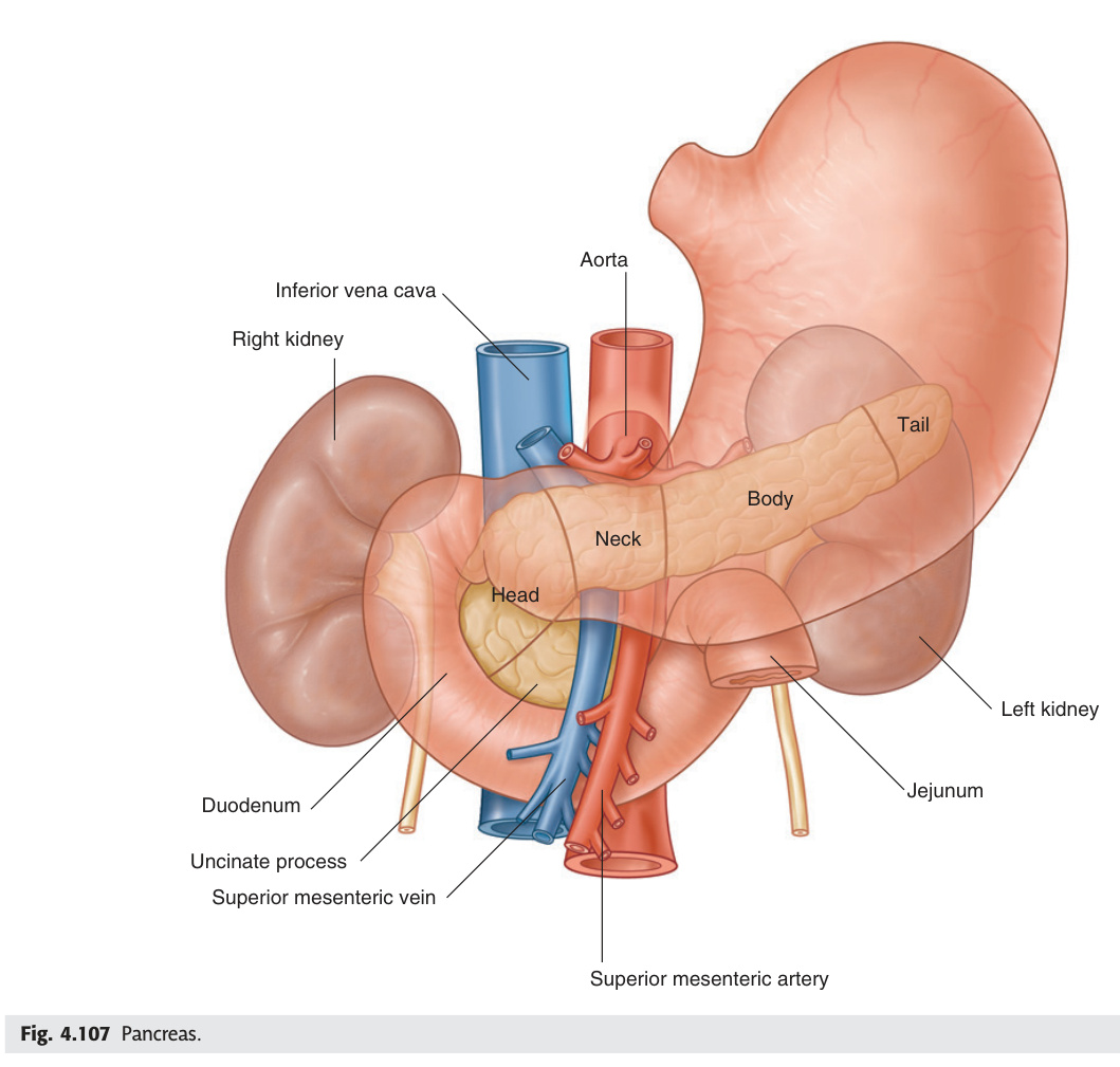
The pancreas is a retroperitoneal gland with dual exocrine (digestive enzyme) and endocrine (insulin, glucagon) roles, and its anatomy maps directly onto its disease patterns. The head sits in the C-loop of the duodenum and shares the common bile duct — carcinoma of the pancreatic head obstructs the CBD early, causing painless progressive jaundice (Courvoisier's gallbladder) and requiring the Whipple's pancreaticoduodenectomy that removes both structures en bloc. The neck lies directly anterior to the SMV–portal vein junction — vascular involvement at this plane is the key surgical boundary for resectability. Acute pancreatitis activates digestive enzymes within the gland itself; the retroperitoneal position explains why haemorrhagic extravasation tracks along fascial planes to the periumbilical skin (Cullen's sign via the falciform ligament) and flanks (Grey Turner's sign via retroperitoneal fat).
| Part | Location / Relations | Notes |
|---|---|---|
| Head | C-loop of duodenum (right of L2). Posterior = CBD + IVC + right renal vessels. Uncinate process wraps behind SMV | Head = most common site of pancreatic carcinoma; obstructs CBD → painless jaundice |
| Neck | Anterior to SMV + portal vein junction (SMV + splenic vein form portal vein here) | Surgical landmark; no capsule → pancreatic juice extravasates freely in pancreatitis |
| Body | Crosses L2 vertebra; posterior = splenic vein (→ portal vein) | Body = common site of pseudocyst formation after pancreatitis |
| Tail | Intraperitoneal; within splenorenal ligament; contacts splenic hilum | Tail = common site of islet cell tumours (insulinoma, glucagonoma) |
Causes: "GET SMASHED" — Gallstones, Ethanol, Trauma, Steroids, Mumps, Autoimmune, Scorpion/snake bite, Hyperlipidaemia/Hypercalcaemia, ERCP, Drugs. Cullen's sign: periumbilical bruising (haemorrhagic pancreatitis tracking along falciform). Grey Turner's sign: flank bruising (retroperitoneal spread). Amylase + lipase elevated. CT scan (Balthazar grading). Severe: APACHE II score, necrosis on CT → ICU.
- Q: Where does the main pancreatic duct of Wirsung open and what guards it? — Joins the CBD at the ampulla of Vater, opening at the major duodenal papilla in the 2nd part of the duodenum; guarded by the sphincter of Oddi.
- Q: Pancreas divisum — what is it and what complication can it cause? — Failure of fusion of the dorsal and ventral pancreatic buds → majority of the pancreas drains through the minor papilla (accessory duct of Santorini) → recurrent pancreatitis from inadequate drainage.
- Q: Mnemonic for causes of acute pancreatitis? — "GET SMASHED": Gallstones, Ethanol, Trauma, Steroids, Mumps, Autoimmune, Scorpion sting, Hyperlipidaemia/Hypercalcaemia, ERCP, Drugs.
- Q: Cullen's vs Grey Turner's sign — mechanism? — Both indicate haemorrhagic pancreatitis. Cullen's = periumbilical bruising (blood tracks along falciform ligament); Grey Turner's = flank bruising (retroperitoneal haematoma dissects through fascial planes).
- Q: Which part of the pancreas is most commonly affected by carcinoma, and what vessel determines resectability? — Head (70%); SMV/portal vein involvement makes it unresectable (encasement = borderline; occlusion = unresectable).
Salivary Glands
Three pairs of salivary glands produce up to 1.5 litres of saliva daily, and their clinical anatomy is governed by two asymmetries. The parotid is the largest but the most surgically dangerous — CN VII does not supply it, but splits into five terminal branches within it, so malignancy or surgery here threatens the whole facial nerve and produces ipsilateral facial palsy. The submandibular duct (Wharton's duct) is the longest and most tortuous, running against gravity from the floor of the mouth upward to the submandibular triangle — the "uphill" course allows saliva to pool and calcium salts to deposit, explaining why 80% of salivary calculi form here. Secretomotor innervation is parasympathetic in both cases but via different routes: parotid via CN IX (glossopharyngeal → lesser petrosal → auriculotemporal), submandibular and sublingual via CN VII (chorda tympani → lingual nerve → submandibular ganglion).
| Gland | Location | Duct | Nerve | Notes |
|---|---|---|---|---|
| Parotid | Preauricular, anterior to ear + masseter | Parotid duct (Stensen's): pierces buccinator, opens at upper 2nd molar | Secretomotor: CN IX → lesser petrosal → auriculotemporal nerve; CN VII passes through (but does not supply) the gland | Largest salivary gland; produces serous (watery) saliva; most common site of salivary gland tumours (pleomorphic adenoma) |
| Submandibular | Submandibular triangle; wraps around posterior edge of mylohyoid | Wharton's duct: opens at sublingual papilla beside frenulum | Secretomotor: CN VII → chorda tympani → lingual nerve → submandibular ganglion | Mixed serous + mucous; most common site of salivary calculi (stones — Wharton's duct longest and most tortuous) |
| Sublingual | Floor of mouth, sublingual fold | 10–20 small ducts of Rivinus (open along sublingual fold) + Bartholin's duct (to Wharton's duct) | Same as submandibular (chorda tympani) | Smallest; mainly mucous; no distinct capsule; rarely forms calculi |
CN VII (facial nerve) passes through the parotid gland (not in its substance but through it), dividing into 5 terminal branches (temporal, zygomatic, buccal, marginal mandibular, cervical). Parotid malignancy or radical parotidectomy risks CN VII injury → facial palsy. Pleomorphic adenoma (benign mixed tumour) = 75% of parotid tumours; treated by superficial parotidectomy with CN VII preservation. Warthin's tumour (cystadenolymphoma) = second most common benign; bilateral in 10%.
- Q: CN VII — does it supply the parotid? What happens if the parotid is removed? — CN VII passes through but does NOT innervate the parotid gland. Radical parotidectomy (malignancy) severs its five branches → complete ipsilateral facial palsy.
- Q: Secretomotor nerve to the parotid — full pathway? — CN IX (glossopharyngeal) → tympanic plexus → lesser petrosal nerve → otic ganglion → auriculotemporal nerve (branch of V3) → parotid.
- Q: Why does the submandibular duct most often form calculi? — Wharton's duct is the longest salivary duct and runs against gravity (uphill from floor of mouth to gland), slowing flow and promoting calcium deposition.
- Q: Most common salivary gland tumour and its treatment? — Pleomorphic adenoma of the parotid (75% of parotid tumours); treated by superficial parotidectomy with CN VII preservation. Risk of recurrence if capsule ruptured.
- Q: Frey's syndrome — what is it and why does it occur? — Gustatory sweating after parotidectomy; aberrant regeneration of auriculotemporal nerve (parasympathetic) fibres into the sympathetic nerve supply to sweat glands of the skin over the parotid.
Trachea
The trachea runs from the cricoid cartilage (C6) to the carina at T4/T5 — the level of the sternal angle, which is simultaneously the most important thoracic surface landmark in anatomy, marking the 2nd rib, the start of the aortic arch, and the boundary between superior and inferior mediastinum. Its 16–20 C-shaped cartilage rings leave the posterior wall membranous (trachealis muscle), which is why posterior tracheal tears are the most common complication of emergency intubation. The carina's normal angle is ~70°; widening signals left atrial enlargement (Samson's sign on CXR) or subcarinal lymphadenopathy. The recurrent laryngeal nerves ascend in the tracheo-oesophageal groove bilaterally, explaining why thyroid surgery, tracheal tumours, and oesophageal carcinoma all present with hoarseness as their first neurological symptom.
| Feature | Detail |
|---|---|
| Length | ~12 cm; from cricoid cartilage (C6) to carina (T4/T5 level = sternal angle) |
| Structure | 16–20 C-shaped hyaline cartilage rings (open posteriorly — posterior wall = trachealis smooth muscle) |
| Relations (cervical) | Anterior: thyroid isthmus (at rings 2–4); posterior: oesophagus. Recurrent laryngeal nerves in tracheo-oesophageal groove bilaterally |
| Relations (thoracic) | Anterior: thymus/great vessels; right side: right brachiocephalic vein + SVC; left side: aortic arch (pushes trachea slightly to the right) |
| Carina | T4/T5 (sternal angle); bifurcation into right and left main bronchi; carina angle ~70°; widening of carina angle = enlarged left atrium or subcarinal lymphadenopathy on CXR |
Tracheostomy levels: elective = through rings 2–3 (avoid ring 1 → subglottic stenosis; avoid ring 4 down → innominate artery in thoracic inlet). Emergency cricothyrotomy: through cricothyroid membrane (below thyroid cartilage, above cricoid) — quickest and safest emergency airway. Recurrent laryngeal nerves at risk in thyroid surgery in the tracheo-oesophageal groove.
- Q: Level of the carina and how to identify it on the surface? — T4/T5 vertebral level = sternal angle (angle of Louis); also marks the 2nd rib, start of aortic arch, and division between superior and inferior mediastinum.
- Q: Why do inhaled foreign bodies preferentially lodge in the right bronchus? — Right main bronchus is shorter (~2.5 cm vs 5 cm), wider, and more vertical (~25° vs ~45° from trachea) — three structural features that make it the preferential path.
- Q: Elective tracheostomy — which rings and what to avoid above and below? — Rings 2–3. Avoid ring 1 above (→ subglottic stenosis); avoid ring 4+ below (→ innominate artery crosses at thoracic inlet → catastrophic haemorrhage).
- Q: Emergency airway — which membrane and anatomical landmarks? — Cricothyroid membrane; lies below the thyroid cartilage and above the cricoid cartilage, palpable in the midline as a soft depression between two firm structures.
- Q: Widening of the carina angle on CXR — two causes? — Left atrial enlargement (pushes left main bronchus superiorly and laterally) and subcarinal lymphadenopathy (e.g. lymphoma, TB, sarcoidosis, lung carcinoma).
Bronchi & Bronchopulmonary Segments
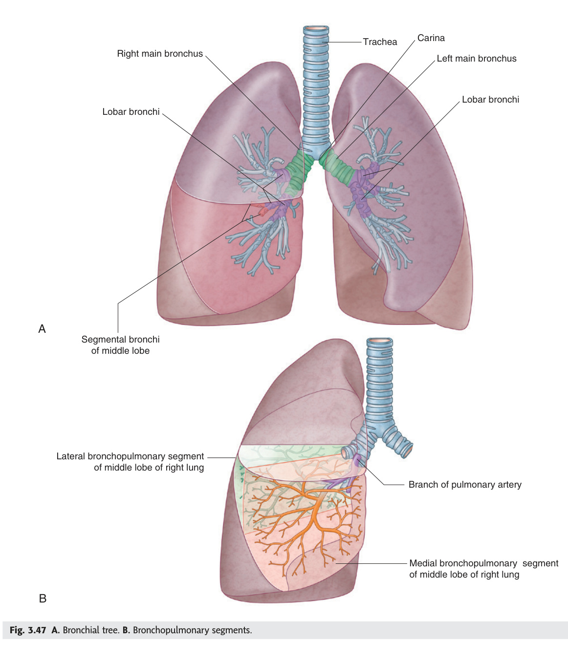
The bronchopulmonary segment is the anatomical unit that underpins modern lung surgery and bronchoscopy — each segment has its own segmental bronchus, artery, and intersegmental venous drainage, making it independently resectable without devascularising adjacent segments. The right upper lobe bronchus is eparterial (the only bronchus in the body that runs above its accompanying pulmonary artery), while all other bronchi are hyparterial — an anatomical distinction that appears in every respiratory anatomy viva. The right lower lobe is the most common site of aspiration pneumonia because the right bronchus is more vertical and gravity directs aspirated material here, especially when supine (specifically into the superior segment of the right lower lobe, the "dependent" segment).
| Right Main Bronchus | Left Main Bronchus | |
|---|---|---|
| Length | ~2.5 cm | ~5 cm |
| Angle from trachea | ~25° (more vertical) | ~45° (more horizontal) |
| Lobar bronchi | 3 (upper, middle, lower) | 2 (upper, lower) |
| Segments total | 10 bronchopulmonary segments | 8–10 segments |
| Relation to pulmonary artery | Eparterial (right upper lobe bronchus above right PA) — "eparterial" = unique to right ULB | All branches hyparterial (below PA) |
Right lung (10 segments): Upper (3): apical, posterior, anterior; Middle (2): lateral, medial; Lower (5): superior, medial basal, anterior basal, lateral basal, posterior basal.
Left lung (8–10 segments): Upper (divided into upper + lingula): apical-posterior, anterior; Lingula (2): superior + inferior; Lower (4–5): superior, anterior-medial basal, lateral basal, posterior basal.
Clinically key: Right lower lobe = most common site of aspiration pneumonia (supine + right bronchus more vertical). Right middle lobe = middle lobe syndrome (lymphadenopathy/tumour compresses middle lobe bronchus → collapse).
- Q: Three features making the right main bronchus preferential for foreign body aspiration? — Shorter (~2.5 cm), wider diameter, more vertical angle (~25° from trachea vs ~45° left).
- Q: What is an eparterial bronchus and which is it? — A bronchus that lies above its accompanying pulmonary artery; the right upper lobe bronchus is the only eparterial bronchus in the body.
- Q: How many bronchopulmonary segments in each lung? — Right = 10; Left = 8–10 (apical + posterior fuse, and medial basal + anterior basal often merge).
- Q: Aspiration pneumonia — which lobe/segment most commonly affected and why? — Right lower lobe, superior segment (when supine) — most dependent segment when lying flat; right bronchus more vertical directs material there.
- Q: Right middle lobe syndrome — cause and mechanism? — Lymphadenopathy (TB, sarcoid) or tumour compresses the long narrow middle lobe bronchus → lobar collapse; the bronchus is particularly vulnerable because it is long with an acute take-off angle.
Lungs
The left lung is smaller than the right — the cardiac notch and the cardiac impression on its mediastinal surface reflect the heart's left-sided displacement — and this structural asymmetry explains why the left has only two lobes and one fissure while the right has three lobes and two fissures. Hilar asymmetry on CXR is physiological: the left hilum is always higher than the right because the left pulmonary artery arches over the left main bronchus, while on the right the bronchus is above the artery in the upper lobe region; a left hilum lower than the right is pathological (left lower lobe collapse). The lung root contains the bronchus, pulmonary artery, and two pulmonary veins (superior and inferior), and their three-dimensional arrangement differs between sides in ways that matter for thoracic surgery and radiological interpretation.
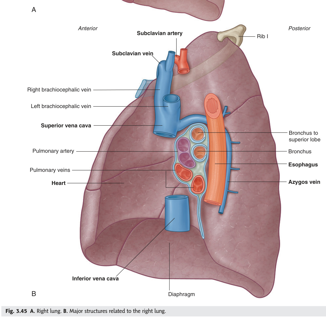
| Right Lung | Left Lung | |
|---|---|---|
| Weight | ~625 g | ~565 g (smaller — cardiac notch) |
| Lobes | 3: upper, middle, lower | 2: upper (+ lingula), lower |
| Fissures | Oblique (major) + horizontal (minor) | Oblique (major) only |
| Horizontal fissure | From right anterior chest (4th costal cartilage) to oblique fissure (5th rib, midaxillary line) | Absent |
| Oblique fissure | From T4 (posteriorly) around to 6th rib (anteriorly) | Same orientation; from T4 to 6th rib |
| Lingula | — | Tongue-like projection of upper lobe (equivalent to right middle lobe) |
Right hilum arrangement (front to back): upper lobe bronchus (eparterial) → pulmonary artery → lower lobe bronchus; (vertical): pulmonary veins anteroinferior.
Left hilum arrangement (front to back): pulmonary artery (arches over left main bronchus) → bronchus → pulmonary veins. Left pulmonary artery is higher than the right.
Pneumonic for right hilum vertical arrangement: RALS = Ringing A Bell Sounds → Right: from above down = Eparterial bronchus, Artery, Bronchus (lower), Veins.
Bilateral hilar lymphadenopathy: sarcoidosis (most common), TB, lymphoma. Unilateral: primary lung carcinoma, TB, lymphoma. On CXR: "double contour" to hilum. Eggshell calcification of hilar nodes = silicosis. Right paratracheal nodes + right hilar = "Pawnbroker's sign" (right-sided prominence) in primary TB.
Pulmonary arteries: deoxygenated blood from right ventricle → lungs for gas exchange. Pulmonary veins (4): oxygenated blood → left atrium. Bronchial arteries: oxygenated blood for lung tissue nutrition. Right bronchial artery = from right 3rd posterior intercostal; left bronchial arteries (2) = from descending thoracic aorta.
- Q: Right vs left lung — lobes, fissures, and weight? — Right: 3 lobes, 2 fissures (oblique + horizontal), ~625 g. Left: 2 lobes + lingula, 1 fissure (oblique only), ~565 g (smaller due to cardiac notch).
- Q: What is the lingula and what is its clinical equivalent? — Tongue-shaped projection of the left upper lobe filling the cardiac notch; functionally equivalent to the right middle lobe (same venous and bronchial drainage pattern).
- Q: Left vs right hilum — which is higher and why? — Left hilum is higher; the left pulmonary artery arches over the left main bronchus, raising the hilum. A lower left hilum signals left lower lobe collapse.
- Q: Contents of the lung root — what does each hilum contain? — Main bronchus + pulmonary artery + 2 pulmonary veins (superior + inferior) + bronchial vessels + lymphatics + autonomic nerves.
- Q: Origin of bronchial arteries — right vs left? — Right = from right 3rd posterior intercostal artery; Left (2) = directly from descending thoracic aorta. Bronchial veins drain to the azygos/hemiazygos system.
Pleura
The pleural space is normally a potential space containing just enough lubricating fluid to allow frictionless respiratory excursion — any pathological accumulation of air, blood, or fluid disrupts this and compresses the lung. The critical anatomical fact is the two-rib gap between the lower lung border and the lower pleural reflection (the costodiaphragmatic recess): the lung ends at rib 6 (MCL), rib 8 (MAL), rib 10 (posterior), while the pleura continues to rib 8, 10, and 12 respectively — this recess is where fluid collects first and from where thoracocentesis is safely performed. Parietal pleura has somatic nerve supply (intercostal + phrenic) and generates sharp localised pleuritic pain; visceral pleura has only autonomic supply and is insensate — inflammation of the lung parenchyma is therefore painless until it reaches the parietal pleura.
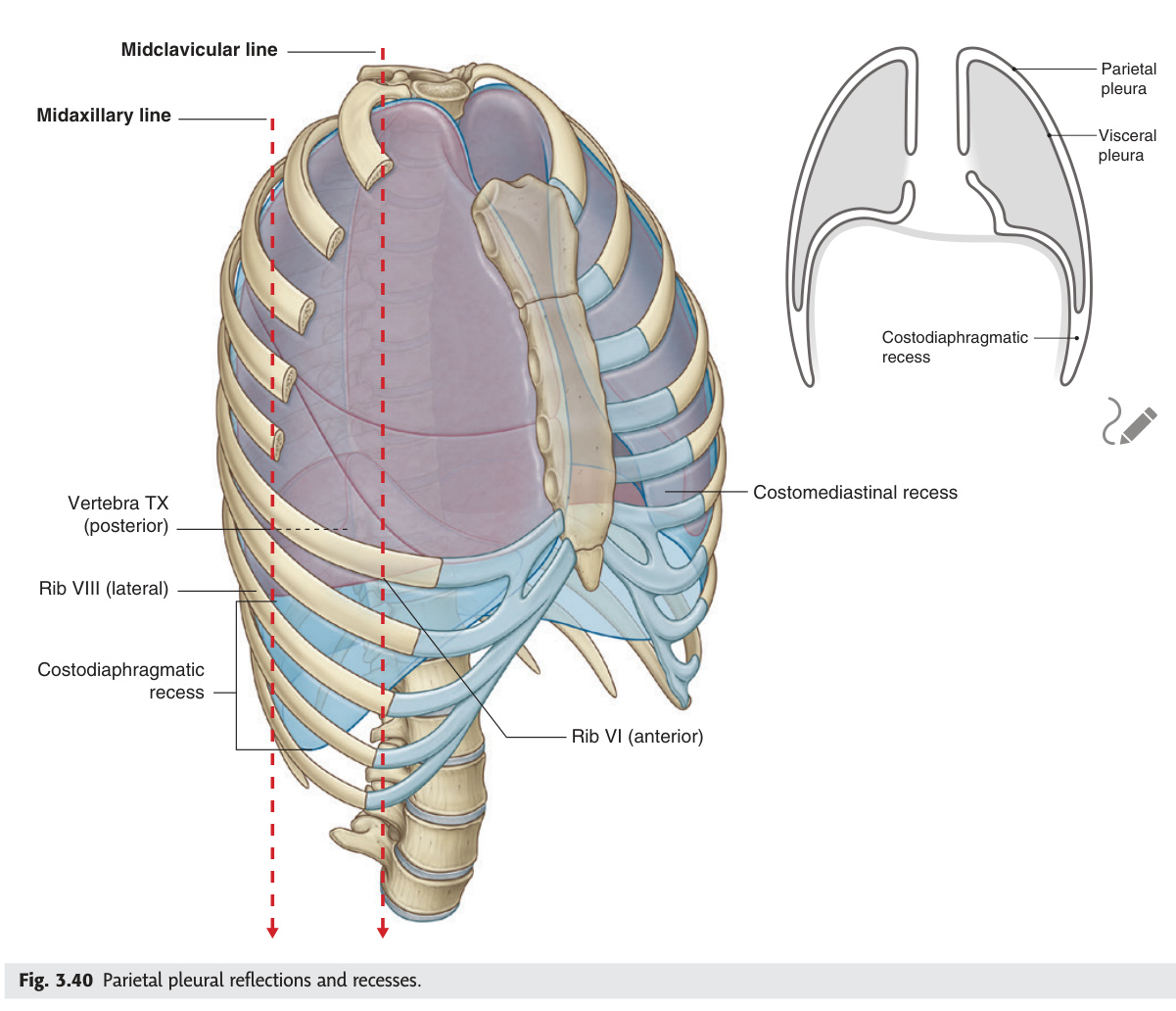
Parietal pleura: lines the thoracic wall, diaphragm, and mediastinum. Has 4 named parts: costal, diaphragmatic, mediastinal, cervical (cupola — extends 2.5 cm above medial clavicle). Innervated by somatic nerves → sharp, well-localised pleural pain. Visceral pleura: covers lung surface; innervated by autonomic → insensitive to pain; cannot be separated from lung.
| Pleural Recess | Location | Clinical Significance |
|---|---|---|
| Costodiaphragmatic recess | Between costal and diaphragmatic pleura. Deepest at midaxillary line (rib 8 → rib 10) | Fluid first collects here on CXR (blunting of costophrenic angle — requires ~200–300 mL). Site for diagnostic pleural aspiration (thoracocentesis) |
| Costomediastinal recess | Between costal and mediastinal pleura anteriorly | Smaller; accomodates anterior lung expansion; relevant in anterior mediastinal procedures |
| Landmark | Right | Left |
|---|---|---|
| Anterior (upper) | Passes behind sternoclavicular joint → midline | Same → diverges at 4th costal cartilage (cardiac notch) |
| Lower border (costal) | Rib 8 (MCL) → Rib 10 (mid-axillary) → Rib 12 (posterior) | Same bilaterally |
| Lung lower border | Rib 6 (MCL) → Rib 8 (MAL) → Rib 10 (posterior) | Same |
| Pleural-lung gap | 2 ribs below lung lower border = costodiaphragmatic recess | Same |
Spontaneous pneumothorax: rupture of apical subpleural blebs; tall young males; dyspnoea + ipsilateral pleuritic chest pain; absent breath sounds + hyper-resonance. Small (<2 cm rim on CXR) = observe; large = needle aspiration 2nd ICS MCL or intercostal drain. Tension pneumothorax = medical emergency: one-way valve → air accumulates → mediastinal shift → trachea deviates to opposite side → raised JVP + hypotension + absent breath sounds. Do NOT wait for CXR. Immediate needle decompression 2nd ICS MCL (large-bore cannula), then chest drain. Chest drain safe triangle: 4th–5th ICS, midaxillary line, insert above upper border of lower rib (avoid NVB).
1. Pleural fluid protein / serum protein > 0.5
2. Pleural fluid LDH / serum LDH > 0.6
3. Pleural fluid LDH > 2/3 upper limit of normal serum LDH
If none met → transudate. Sensitivity 98% / specificity 83% for exudate.
Transudate causes (altered Starling's forces): left heart failure (most common, bilateral), nephrotic syndrome, liver cirrhosis, hypoalbuminaemia, hypothyroidism, Meigs' syndrome (ovarian fibroma + right pleural effusion + ascites).
Exudate causes (inflammation/malignancy/infection): pneumonia (parapneumonic → empyema if pH <7.2), malignancy (lung, breast, mesothelioma — most common), TB (lymphocytic exudate), PE (haemorrhagic exudate), RA/SLE (autoimmune).
Thoracocentesis site: costodiaphragmatic recess posterolaterally — aspirate below scapular tip, above 9th rib (avoid liver on right). Ultrasound-guided.
Ready to test Unit 06?
Liver, gallbladder, lungs, pleura — practice MCQs and essays.
- Q: Lower borders of the lung and pleura — rib levels at MCL, MAL, and posterior? — Lung: rib 6 (MCL), rib 8 (MAL), rib 10 (posterior). Pleura: rib 8 (MCL), rib 10 (MAL), rib 12 (posterior). Two-rib gap = costodiaphragmatic recess.
- Q: Why does parietal pleura hurt and visceral pleura not? — Parietal pleura: somatic innervation (intercostal nerves + phrenic) → sharp, localised pleuritic pain. Visceral pleura: autonomic only → insensate.
- Q: Light's criteria — when is an effusion an exudate? — ≥1 of: pleural protein/serum protein >0.5; pleural LDH/serum LDH >0.6; pleural LDH >2/3 upper limit of normal serum LDH. Transudate if none met.
- Q: Safe triangle for chest drain — boundaries and rib rule? — 4th–5th ICS, mid-axillary line. Insert above the upper border of the lower rib to avoid the neurovascular bundle (nerve-artery-vein runs in the costal groove under the rib above).
- Q: Tension pneumothorax — clinical signs and immediate management? — Trachea deviated away, absent breath sounds ipsilateral, raised JVP, hypotension. Do NOT wait for CXR. Immediate needle decompression at 2nd ICS MCL.
Spleen
The spleen lies obliquely along the 10th rib in the left hypochondrium, protected by the lower ribs — it must double in size before it becomes palpable below the left costal margin, which is why massive splenomegaly (malaria, kala-azar, myelofibrosis) is so striking when you finally feel it. As the body's largest lymphoid organ, the white pulp mounts immune responses to encapsulated bacteria (Streptococcus pneumoniae, Haemophilus influenzae, Neisseria meningitidis), explaining why post-splenectomy patients face overwhelming infection from exactly these organisms and require lifelong vaccination and prophylaxis. The tail of the pancreas physically contacts the splenic hilum within the splenorenal ligament — distal pancreatectomy for tail tumours typically becomes a combined splenopancreatectomy. Kehr's sign (left shoulder pain from splenic haemorrhage) is the classic example of referred pain via the phrenic nerve (C4).
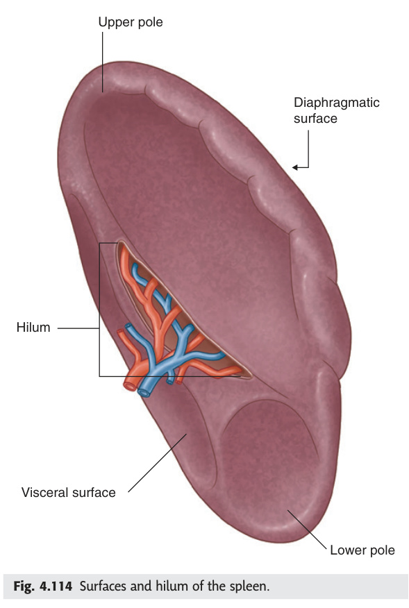
The largest lymphoid organ in the body (~150 g). Intraperitoneal in the left hypochondrium, related to the 9th–11th ribs. It is NOT normally palpable below the left costal margin — if palpable, it is at least twice its normal size.
| Feature | Details |
|---|---|
| Position | Left hypochondrium; long axis along 10th rib; deep to ribs 9–11 |
| Surfaces | Diaphragmatic (smooth, convex) + Visceral (concave): gastric impression (anteromedial), renal impression (posteromedial), colic impression (inferior pole) |
| Hilum | Visceral surface; splenic artery + vein enter/exit; tail of pancreas contacts the hilum |
| Peritoneal ligaments | Splenorenal ligament (to left kidney; contains splenic vessels + tail of pancreas) · Gastrosplenic ligament (to greater curvature; contains short gastric + left gastroepiploic vessels) |
| Blood supply | Splenic artery (largest branch of coeliac trunk; tortuous course along upper border of pancreas). Splenic vein drains to join SMV → portal vein behind neck of pancreas |
| Notch | Superior border has 1–2 notches (remnant of lobulation) — palpable in massive splenomegaly; confirms spleen vs renal mass |
| Function | Details |
|---|---|
| Red pulp | Filtration: removes old/abnormal red cells + Howell-Jolly bodies (nuclear remnants), Heinz bodies, siderocytes. After splenectomy → these appear in blood film |
| White pulp | Lymphoid tissue (B-cell follicles + T-cell periarterial sheaths); immune surveillance; produces IgM; responds to encapsulated organisms |
| Reservoir | Stores 1/3 of platelets; in disease (portal hypertension) → hypersplenism: sequestration of RBCs, WBCs, platelets → pancytopenia |
Most commonly injured solid organ in blunt abdominal trauma (left lower ribs). Kehr's sign: left shoulder pain (haemoperitoneum irritates left diaphragm → referred to C4/left shoulder via phrenic nerve). Haemodynamically stable → CT + non-operative management (splenic angioembolisation). Unstable → emergency splenectomy. Post-splenectomy: vaccinate against encapsulated organisms (pneumococcus, meningococcus, H. influenzae) before elective splenectomy (2 weeks prior) or after emergency (at discharge). Long-term penicillin V prophylaxis in under-16s. Risk of OPSI (overwhelming post-splenectomy infection) — typically Streptococcus pneumoniae sepsis.
Infective: EBV (infectious mononucleosis — most common in young adults), malaria, kala-azar (visceral leishmaniasis — massive), typhoid, bacterial endocarditis.
Congestion: portal hypertension (cirrhosis, Budd-Chiari) → congestive splenomegaly + hypersplenism.
Infiltrative: amyloidosis, sarcoidosis, Gaucher's disease.
Autoimmune: SLE, RA (Felty's syndrome = RA + splenomegaly + neutropenia).
- Q: Why is the spleen not normally palpable? — Lies deep to ribs 9–11 in the left hypochondrium along the 10th rib; must enlarge to at least twice its normal size (~150 g) before it descends below the costal margin.
- Q: Three encapsulated organisms requiring post-splenectomy prophylaxis? — Streptococcus pneumoniae, Haemophilus influenzae type b, Neisseria meningitidis. Vaccine all three before elective splenectomy (2 weeks prior); penicillin V prophylaxis for life in under-16s.
- Q: Kehr's sign — what is it and why does it occur? — Left shoulder tip pain from splenic haemorrhage (haemoperitoneum); blood irritates the undersurface of the left diaphragm → referred via the left phrenic nerve (C4 dermatome = left shoulder tip).
- Q: Contents of the splenorenal ligament? — Splenic vessels (artery + vein) and the tail of the pancreas; these structures reach the splenic hilum via this peritoneal fold.
- Q: How do you distinguish splenomegaly from a large left kidney clinically? — Spleen: superior border has notch(es), cannot ballot, moves towards right iliac fossa on inspiration, dull to percussion. Kidney: ballottable, moves inferiorly on inspiration, resonant anteriorly (overlying colon).
Jaundice ★★★
Jaundice is a symptom with three anatomical addresses: pre-hepatic (too much bilirubin being produced), hepatic (the liver cannot process it), or post-hepatic (the processed bilirubin cannot escape). The bilirubin pathway is the physiological skeleton that decodes all three: unconjugated bilirubin is lipid-soluble and albumin-bound so it cannot reach the urine (no urinary bilirubin in haemolytic jaundice); conjugated bilirubin is water-soluble and filtered by the glomerulus (dark urine in obstructive jaundice); stercobilin colours stools brown — pale stools mean bile is not reaching the gut. Gilbert's syndrome — a UGT1A1 promoter polymorphism causing isolated unconjugated hyperbilirubinaemia with completely normal LFTs in 5–10% of the population — is one of the most commonly missed benign conditions in medicine, and students who know the bilirubin pathway will never confuse it with hepatocellular disease.
Jaundice (icterus) = yellow discolouration of skin, sclera, and mucous membranes due to bilirubin deposition. Clinically detectable when serum bilirubin >35 µmol/L (normal <17 µmol/L). Scleral icterus (earliest sign) detectable at ~35 µmol/L; skin yellowing at >50 µmol/L.
2. Hepatocytes take up unconjugated bilirubin → conjugate with glucuronic acid (enzyme: UGT1A1) → conjugated bilirubin (water-soluble, can be filtered by kidneys).
3. Conjugated bilirubin secreted into bile → reaches small intestine → reduced by gut bacteria (in terminal ileum/colon) to urobilinogen.
4. Most urobilinogen → oxidised to stercobilin → excreted in stool (brown colour). Small fraction reabsorbed → enterohepatic circulation → re-excreted in bile; small amount filtered by kidneys → urinary urobilinogen (trace amount normal).
In obstructive jaundice: no bile reaches gut → no stercobilin → pale/clay stools; conjugated bilirubin accumulates in blood → filtered → dark urine (bilirubinuria); urinary urobilinogen absent (no gut production).
| Feature | Pre-hepatic (Haemolytic) | Hepatic (Hepatocellular) | Post-hepatic (Obstructive / Cholestatic) |
|---|---|---|---|
| Mechanism | Excess haem breakdown → overwhelms hepatic conjugation capacity | Hepatocyte damage → impaired uptake/conjugation/secretion | Obstruction of bile flow → conjugated bilirubin regurgitates into blood |
| Causes | Haemolysis (sickle cell, spherocytosis, G6PD deficiency, malaria, autoimmune) · Neonatal jaundice (physiological) · Gilbert's syndrome (UGT1A1 polymorphism — benign, stress-triggered) | Viral hepatitis (A/B/C/E) · Alcoholic hepatitis · DILI (paracetamol, isoniazid) · Autoimmune hepatitis · Cirrhosis · Sepsis · Wilson's disease · Haemochromatosis | CBD stone (choledocholithiasis, most common) · Pancreatic head carcinoma · Cholangiocarcinoma · Mirizzi syndrome · Primary sclerosing cholangitis · Stricture post-ERCP/cholecystectomy |
| Bilirubin type | Unconjugated ↑↑ (indirect) | Both conjugated + unconjugated ↑ (mixed) | Conjugated ↑↑ (direct) |
| Urine bilirubin | Absent (unconjugated = not filtered) | Present (conjugated bilirubin spillage) | Present ↑↑ (dark urine) |
| Urine urobilinogen | ↑↑ (excess production → excess absorbed) | ↑ or normal | Absent (no bile reaching gut) |
| Stool colour | Normal or dark (excess stercobilin) | Variable (pale if cholestatic component) | Pale/clay-coloured (absent stercobilin) |
| Pruritus | Absent | Variable | Prominent (bile salt deposition in skin) |
| LFTs pattern | AST/ALT mildly ↑ or normal; bilirubin ↑ (unconjugated) | AST/ALT markedly ↑ (hepatocellular pattern); ALP mildly ↑ | ALP + GGT markedly ↑ (cholestatic pattern); AST/ALT mildly ↑ |
| Spleen | Enlarged (haemolysis) | Variable (enlarged in portal hypertension) | Normal (unless underlying cirrhosis) |
- Q: Dark urine + pale stools + jaundice — which type and what does each finding mean? — Post-hepatic (obstructive). Dark urine = conjugated bilirubin filtered by glomerulus. Pale stools = no stercobilin (bile cannot reach gut). Both indicate the same obstruction below the hepatocyte.
- Q: Which type of jaundice has no urinary bilirubin and why? — Pre-hepatic (haemolytic); excess unconjugated bilirubin is lipid-soluble and albumin-bound → not filtered by the glomerulus → urine remains bilirubin-free.
- Q: Light's criteria — state them. — Exudate if ≥1 of: pleural/serum protein >0.5; pleural/serum LDH >0.6; pleural LDH >2/3 upper limit of serum LDH. (Note: also the pleural criteria for completeness.)
- Q: Gilbert's syndrome — gene, mechanism, trigger, and clinical significance? — UGT1A1 promoter polymorphism → reduced UDP-glucuronosyltransferase → impaired conjugation → isolated unconjugated hyperbilirubinaemia. Triggered by fasting, stress, illness. LFTs otherwise normal. Benign; no treatment.
- Q: Painless progressive jaundice + palpable gallbladder — diagnosis and law? — Carcinoma of the head of the pancreas (most likely). Courvoisier's law: palpable gallbladder in jaundiced patient is unlikely due to gallstones (chronic stones fibrosed the GB wall).
Mediastinum ★★★
The mediastinum is divided at the sternal angle (T4/T5) — the single most important surface landmark in thoracic anatomy — into superior and inferior compartments, with the inferior split into anterior (thymic/lymphoid), middle (cardiac/vascular), and posterior (neural/oesophageal). This compartment scheme is a clinical algorithm: the anterior "4 Ts" (thymoma, teratoma, terrible lymphoma, thyroid) account for the great majority of anterior mediastinal masses; posterior masses are almost always neurogenic. The left recurrent laryngeal nerve's intrathoracic loop under the aortic arch at the ligamentum arteriosum is the most clinically exposed cranial nerve branch in the chest — aortic aneurysm, left hilar tumour, and mediastinal lymphadenopathy all cause hoarseness by the same mechanism. The thoracic duct is the drainage route for all lymph below the diaphragm and the entire left side above it; chylothorax (milky fluid, TG >1.1 mmol/L) after oesophagectomy is the classic complication of inadvertent division.
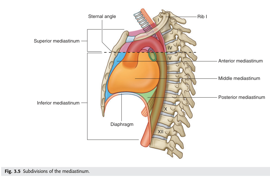
The mediastinum is the central compartment of the thoracic cavity, between the two pleural cavities. Divided by the sternal angle (angle of Louis, T4/T5 level) into superior (above) and inferior (below, further divided into anterior/middle/posterior).
| Compartment | Key Contents |
|---|---|
| Superior mediastinum (above T4/T5) | Thymus (anterior) · Arch of aorta + 3 branches (brachiocephalic trunk → right common carotid + right subclavian; left common carotid; left subclavian) · Brachiocephalic veins + SVC (upper) · Trachea (ends at carina T4/5) · Oesophagus · Thoracic duct (left side) · Vagus nerves (CN X) · Left recurrent laryngeal nerve (loops under arch of aorta at ligamentum arteriosum) · Phrenic nerves (C3, C4, C5) |
| Anterior mediastinum (between sternum and pericardium) | Thymic remnants · Internal thoracic (mammary) vessels · Lymph nodes · Fat · Sternopericardial ligaments |
| Middle mediastinum | Pericardium + heart · Ascending aorta + pulmonary trunk + pulmonary veins · SVC (lower) + IVC · Phrenic nerves (run laterally through middle mediastinum) · Carina + main bronchi · Tracheal bifurcation lymph nodes |
| Posterior mediastinum (between pericardium and vertebral column) | Descending thoracic aorta (T5–T12) · Oesophagus + oesophageal plexus · Azygos + hemiazygos veins · Thoracic duct · Vagus nerves (reforming as oesophageal plexus) · Thoracic sympathetic chain + splanchnic nerves · Thoracic vertebral bodies |
| Mass | Compartment | Key Features |
|---|---|---|
| Thymoma | Anterior | Most common anterior mediastinal mass in adults. Associated with myasthenia gravis (10–15% of myasthenia have thymoma; 30–50% of thymomas have MG). Also associated with pure red cell aplasia, hypogammaglobulinaemia. Surgical thymectomy (improves MG outcomes) |
| Teratoma / Germ cell tumour | Anterior | Mature teratoma (benign, contains teeth/hair/cartilage on imaging). Malignant: non-seminomatous GCT (AFP ↑) or seminoma (LDH/hCG ↑). Treat malignant GCT with BEP chemotherapy |
| Terrible lymphoma | Anterior & middle | Hodgkin's lymphoma (mediastinal involvement in ~60%) or high-grade NHL. Bilateral hilar + anterior mediastinal mass ("bat wing"). Associated with B symptoms (fever, night sweats, weight loss) |
| Thyroid (retrosternal goitre) | Anterior–superior | Descends from neck. Pemberton's sign: raising both arms above head → facial flushing + JVP rise + inspiratory stridor (thoracic inlet compression). Moves on swallowing. Tracheal deviation on CXR. Treat surgically if compressive symptoms |
| Neurogenic tumour | Posterior | Most common posterior mediastinal mass. Children: neuroblastoma/ganglioneuroblastoma. Adults: neurofibroma, schwannoma (Nerve sheath tumour). "Dumb-bell" extension through neural foramina → cord compression. Associated with NF1 |
| Bronchogenic/pericardial cyst | Middle | Smooth, round, well-defined water-density cyst. Usually asymptomatic. Bronchogenic cysts more often paratracheal; pericardial cysts at right cardiophrenic angle |
| Structure | Course / Clinical Relevance |
|---|---|
| Left recurrent laryngeal nerve (RLN) | Leaves vagus at arch of aorta → loops under arch at ligamentum arteriosum (T4) → ascends in left tracheo-oesophageal groove → supplies all intrinsic laryngeal muscles except cricothyroid. Vulnerable in: aortic aneurysm, left hilar lymphadenopathy, oesophageal/lung carcinoma, mediastinal masses, thyroid surgery. Ortner's syndrome: hoarseness from enlarged left atrium compressing the left RLN. Right RLN hooks under right subclavian artery (shorter course — right-sided neck surgery risk) |
| Phrenic nerve (C3, C4, C5) | Descends from neck → enters superior mediastinum → runs anterior to lung root (along pericardium) → reaches diaphragm. Motor to diaphragm; sensory to central diaphragm, pericardium, mediastinal pleura. Clinical: phrenic nerve palsy → ipsilateral diaphragmatic paralysis → elevated hemidiaphragm on CXR. Causes: lung/mediastinal tumour, cardiac surgery (ice-slush hypothermia), aortic surgery, cervical spine injury (C3–C5 → "C3,4,5 keeps the diaphragm alive") |
| Azygos vein | Drains right posterior intercostal veins → arches over right main bronchus at T4 → drains into SVC. Provides collateral venous drainage when SVC is obstructed. Enlarged azygos on CXR (>7 mm in erect position) = SVC obstruction, portal hypertension, IVC anomaly |
| Thoracic duct | Originates from cisterna chyli (L1–L2) → ascends in posterior mediastinum between aorta (left) and azygos (right) → crosses midline at T5 → runs in left posterior mediastinum → arches forward at T1 → drains into junction of left internal jugular + left subclavian veins. Drains ALL lymph from below diaphragm + left side of body above diaphragm. Damage (oesophageal surgery, subclavian line, trauma) → chylothorax (milky fluid; TG >1.1 mmol/L; lymphocytes) |
- Q: "4 Ts" of the anterior mediastinum — list and most common in adults? — Thymoma, Teratoma (germ cell tumour), Terrible lymphoma (Hodgkin's/NHL), Thyroid (retrosternal goitre). Thymoma is the most common primary anterior mediastinal mass in adults.
- Q: Thymoma + myasthenia gravis — what antibody and what treatment? — Anti-acetylcholine receptor (anti-AChR) antibodies. Treatment: thymectomy improves MG outcomes even without thymoma; pyridostigmine (AChE inhibitor) for symptoms; immunosuppression; IVIG/plasmapheresis for crisis.
- Q: Left RLN — where does it loop and what structures can injure it? — Loops under the arch of the aorta at the ligamentum arteriosum (T4). Vulnerable to: aortic aneurysm, left hilar lymphadenopathy/lung carcinoma, mediastinal masses, oesophageal carcinoma, cardiac surgery.
- Q: SVC obstruction syndrome — signs and most common cause? — Facial/upper limb oedema, dilated collateral chest veins, non-pulsatile raised JVP, headache, Pemberton's sign. Most common cause: right-sided lung carcinoma (especially small cell).
- Q: Thoracic duct — route, what it drains, and consequence of injury? — Cisterna chyli (L1–L2) → posterior mediastinum → crosses midline at T5 → left side → drains at left subclavian–IJV junction. Drains all lymph below diaphragm + left side above. Injury → chylothorax (milky fluid, TG >1.1 mmol/L).