The Alimentary Canal
Pharynx ★★★
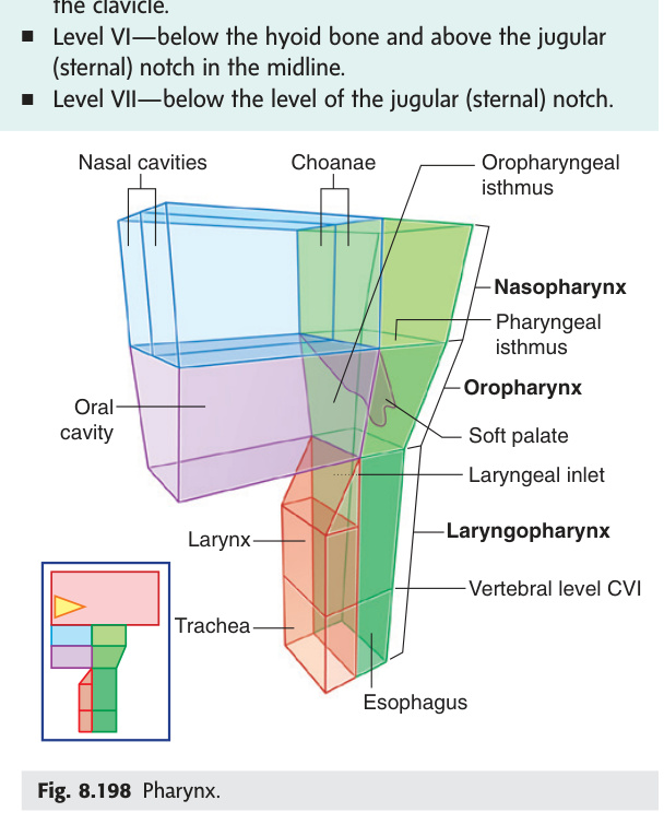
The pharynx is a muscular funnel that serves double duty: it is the shared highway for both the respiratory and alimentary systems. Air and food both travel through it, then split — air into the larynx anteriorly, food into the oesophagus posteriorly. This is why the pharyngeal stage of swallowing is so precisely coordinated: the soft palate must seal the nasopharynx, the epiglottis must tilt over the laryngeal inlet, and the vocal cords must adduct, all within a fraction of a second. The pharynx is divided into three parts by two natural gates: the soft palate (divides naso- from oropharynx) and the upper border of the epiglottis (divides oro- from laryngopharynx). Clinically, the piriform recess of the laryngopharynx is the classic site where swallowed fish bones and other sharp foreign bodies lodge — and where the internal laryngeal nerve runs immediately beneath the mucosa, which is why applying topical anaesthetic to the piriform recess blocks the superior laryngeal nerve.
The pharynx is a fibromuscular tube (~12 cm) extending from the base of the skull to the level of C6, where it becomes continuous with the oesophagus. It is the common passage of both the alimentary and respiratory systems. It has a complete posterior and lateral wall but an incomplete anterior wall that opens into the nasal, oral, and laryngeal cavities. Divided into three parts by the soft palate and the epiglottis.
| Part | Extent | Anterior Communication | Key Structures |
|---|---|---|---|
| Nasopharynx | Base of skull → soft palate | Nasal cavity via choanae (posterior nasal apertures, 2) | Pharyngeal tonsil (adenoid) — roof; tubal tonsil — near auditory tube; pharyngeal opening of auditory tube (1 cm behind inferior nasal concha); pharyngeal recess (fossa of Rosenmüller) — posterior to tubal torus |
| Oropharynx | Soft palate → superior border of epiglottis | Oral cavity via isthmus of fauces | Palatine tonsils in the tonsillar fossa (lateral wall); posterior 1/3 of tongue with lingual tonsil |
| Laryngopharynx | Superior border of epiglottis → C6 (cricopharyngeus) | Larynx via laryngeal inlet (aditus) | Piriform recess (lateral to laryngeal inlet — foreign bodies lodge here; site where internal laryngeal nerve can be damaged) |
- What divides the nasopharynx from the oropharynx, and the oropharynx from the laryngopharynx? Soft palate (naso/oro); upper border of epiglottis (oro/laryngo).
- At what vertebral level does the pharynx become the oesophagus? C6 — at the lower border of cricopharyngeus (the pharyngeal sphincter).
- What is the piriform recess and why is it clinically important? A lateral recess of the laryngopharynx beside the laryngeal inlet — the classic site for foreign body impaction; the internal laryngeal nerve runs just beneath its mucosa.
- What communicates anteriorly with the nasopharynx? The nasal cavity via the choanae (posterior nasal apertures, ×2).
- What is the fossa of Rosenmüller and why does it matter? The pharyngeal recess — a posterior niche of the nasopharynx behind the tubal torus; a common origin site for nasopharyngeal carcinoma.
Waldeyer's ring is the body's first immunological checkpoint — a complete ring of lymphoid tissue encircling the upper pharynx that samples everything entering through the nose and mouth. Think of it as a circular border patrol: the pharyngeal tonsil (adenoid) covers the roof, tubal tonsils guard the Eustachian tube openings, palatine tonsils occupy the lateral walls, and the lingual tonsil covers the tongue base. The palatine tonsils are by far the most clinically significant: they lie in the tonsillar fossa between the palatoglossal and palatopharyngeal arches, immediately lateral to the isthmus of fauces, with only the thin pharyngeal constrictor muscle separating them from the internal carotid artery. This dangerous proximity is why quinsy (peritonsillar abscess) drainage must be shallow — blind deep incisions risk catastrophic arterial haemorrhage.
The isthmus of fauces is the narrow opening that connects the oral cavity proper posteriorly to the oropharynx. Boundaries: superior = uvula + free margin of the soft palate (palatine velum); lateral (each side) = palatoglossal arches (anterior pillars of fauces); inferior = root of the tongue. It is guarded by the anterior pillars.
The tonsillar ring (Waldeyer's lymphatic ring) is a circular arrangement of lymphoid tissue at the pharyngeal inlet that forms a defensive barrier against pathogens entering from the oral and nasal cavities. Members (in order around the ring): (1) Pharyngeal tonsil (adenoid) — roof/posterior wall of nasopharynx; (2) Tubal tonsils (×2) — near auditory tube opening in nasopharynx; (3) Palatine tonsils (×2) — tonsillar fossa, lateral wall of oropharynx; (4) Lingual tonsil — posterior 1/3 of tongue dorsum.
Mnemonic: P-T-P-L = Pharyngeal · Tubal · Palatine · Lingual.
(A) In the roof of the nasopharynx
(B) In the floor of the oropharynx
(C) In the lateral wall of the nasopharynx
(D) In the tonsillar fossa of the oropharynx
(E) On the posterior 1/3 of the tongue
The palatine tonsil lies in the tonsillar fossa on the lateral wall of the oropharynx, bounded anteriorly by the palatoglossal arch and posteriorly by the palatopharyngeal arch.
A = wrong: the pharyngeal tonsil (adenoid) is in the roof of the nasopharynx.
B = wrong: no tonsil occupies the floor of the oropharynx specifically.
C = wrong: the tubal tonsil lies in the lateral wall of the nasopharynx (near the auditory tube opening).
E = wrong: the lingual tonsil lies on the posterior 1/3 of the tongue (also part of Waldeyer's ring).
Peritonsillar abscess (quinsy): pus collects between the tonsillar capsule and the superior constrictor muscle, usually following acute tonsillitis. Features: severe sore throat, dysphagia, hot-potato voice (muffled), trismus (spasm of medial pterygoid), uvular deviation away from the affected side. Treatment: needle aspiration or incision & drainage + IV antibiotics; interval tonsillectomy. Surgical danger: the internal carotid artery lies immediately lateral to the superior constrictor — never penetrate beyond the tonsillar capsule.
Adenoid hypertrophy: enlargement of the pharyngeal tonsil in children → nasal obstruction + mouth breathing + snoring. Blocks the pharyngeal opening of the auditory tube (Eustachian tube) → otitis media with effusion (glue ear) → conductive hearing loss. Treatment: adenoidectomy ± grommet insertion.
- Name the four members of Waldeyer's ring in order around the ring. → Pharyngeal (adenoid) → Tubal (×2) → Palatine (×2) → Lingual; mnemonic P-T-P-L
- Where do sharp foreign bodies (e.g. fish bones) most commonly lodge in the laryngopharynx? → Piriform recess (lateral to the laryngeal inlet)
- In quinsy, the uvula deviates — which direction and why? → Away from the affected side; intact levator veli palatini on healthy side pulls uvula across
- Why does adenoid hypertrophy cause glue ear? → Enlarged adenoid blocks the pharyngeal opening of the Eustachian tube → middle ear effusion → conductive hearing loss
- What structure lies immediately lateral to the palatine tonsil — clinical danger? → Internal carotid artery (through thin superior constrictor) — risk of catastrophic haemorrhage in deep drainage of quinsy
Oesophagus
The oesophagus is a 25 cm muscular tube with one critical anatomical quirk: it has no serosa. Every other part of the GI tract is wrapped in peritoneum or adventitia that slows tumour spread, but the oesophagus is naked — only a fibrous adventitia on its outside, meaning oesophageal carcinoma invades mediastinal structures early and silently. Its muscle composition changes along its length: the upper third is skeletal (voluntary, under somatic control for the initiation of swallowing), the lower third is smooth (autonomic, vagal), and the middle is a mixed zone. This transition matters clinically — achalasia affects the smooth muscle lower oesophagus (loss of myenteric plexus ganglion cells → failure of LOS relaxation), whereas pharyngeal dysphagia (e.g. motor neuron disease) affects the striated upper portion. The three constrictions are the most exam-tested oesophageal facts: remember "15-25-40" for the distances from upper incisors on endoscopy — cricopharyngeus (C6), arch of aorta/left bronchus (T4), and diaphragmatic hiatus (T10).
| Feature | Detail |
|---|---|
| Length | ~25 cm (from cricoid/C6 to T11) |
| Start | Cricopharyngeus (inferior pharyngeal constrictor) at C6 |
| Entry to stomach | Passes through diaphragm at T10, joins stomach at gastro-oesophageal junction (T11) |
| Layers | No serosa (outer layer = adventitia only — enables tumour spread). Mucosa, submucosa, muscularis. Upper 1/3 = skeletal muscle; middle 1/3 = mixed; lower 1/3 = smooth muscle |
| Nerve supply | Sympathetic: T5–T9 (via thoracic splanchnic); Parasympathetic: vagus nerve (CN X) throughout; lower 2/3 = oesophageal plexus of vagus |
2. Aortic/bronchial constriction — T4/T5, where the aortic arch crosses anteriorly then left main bronchus crosses.
3. Diaphragmatic constriction (lower oesophageal sphincter) — T10, where it passes through the oesophageal hiatus in the diaphragm.
"15–25–40" (cm from upper incisors on endoscopy / nasogastric tube):
15 cm = cricopharyngeal constriction (upper oesophageal sphincter, C6)
25 cm = aortic/bronchial constriction (T4/T5)
40 cm = diaphragmatic constriction / lower oesophageal sphincter (T10)
Clinically vital for: NGT insertion (tip at ~50 cm = stomach), OGD landmarks, foreign body localisation, oesophageal cancer level reporting. Oesophageal length total ~25 cm (C6 to T11); stomach begins at T11 (~45 cm from teeth).
Oesophageal carcinoma: squamous cell carcinoma in upper 2/3; adenocarcinoma in lower 1/3 (from Barrett's). The absence of serosa allows early spread to mediastinal structures. Dysphagia progresses from solids → liquids. Barrett's oesophagus: metaplastic replacement of squamous by columnar (intestinal-type) epithelium in lower oesophagus from chronic GORD. Pre-malignant. Pharyngeal (Zenker's) diverticulum: herniation through Killian's dehiscence (gap between thyropharyngeus and cricopharyngeus) → upper dysphagia, regurgitation of undigested food.
| Region | Artery | Vein | Lymph |
|---|---|---|---|
| Cervical oesophagus | Inferior thyroid artery | Inferior thyroid vein | Deep cervical nodes |
| Thoracic oesophagus | Oesophageal branches of descending aorta + bronchial arteries | Azygos + hemiazygos veins | Posterior mediastinal nodes |
| Abdominal oesophagus | Left gastric artery (branch of coeliac) | Left gastric vein → portal system | Left gastric nodes |
The abdominal oesophageal veins drain into the left gastric vein (portal) and also anastomose with the azygos system (systemic). In portal hypertension → backflow into oesophageal submucosal veins → oesophageal varices → massive haematemesis. Management: endoscopic banding or TIPS procedure.
- Why does oesophageal cancer spread early to mediastinal structures? → No serosa — only adventitia; tumour extends directly into adjacent mediastinum without peritoneal barrier
- Three constrictions at endoscopy — distances from upper incisors? → 15 cm = cricopharyngeus (C6); 25 cm = aortic arch / left main bronchus (T4); 40 cm = diaphragmatic hiatus (T10)
- Which part of the oesophagus drains to the portal system? → Abdominal oesophagus via left gastric vein → portal; anastomoses with azygos (systemic) → varices in portal hypertension
- Muscle type in upper vs lower oesophagus? → Upper 1/3 = skeletal (voluntary); middle = mixed; lower 1/3 = smooth muscle (autonomic, vagal)
- Barrett's oesophagus — metaplastic change and risk? → Squamous → columnar (intestinal-type) epithelium from chronic GORD; pre-malignant → adenocarcinoma
Swallowing looks simple but is one of the most complex motor acts: it requires the precise sequential firing of over 25 pairs of muscles within about one second. The oral (voluntary) stage is initiated consciously — the tongue presses the bolus against the hard palate and tips it posteriorly. Once the bolus touches the posterior pharyngeal wall, the reflex takes over entirely: the brainstem swallowing centre (nucleus tractus solitarius receives afferent, nucleus ambiguus sends motor output) coordinates the pharyngeal and oesophageal stages automatically. The key airway protection manoeuvres all happen simultaneously — soft palate elevation seals the nasopharynx, laryngeal elevation tilts the epiglottis, and vocal cord adduction closes the glottis. Any disruption — by stroke (CN X palsy), bulbar palsy (MND), or myopathy — causes aspiration because these simultaneous gates fail in sequence. The test of choice is a videofluoroscopic swallow study (modified barium swallow) to catch silent aspiration.
| Stage | Control | Description | CN Involved |
|---|---|---|---|
| Oral (Stage 1) | Voluntary | Food bolus formed by tongue and teeth; pushed posteriorly into oropharynx by tongue pressing against hard palate | CN V (chewing) · CN XII (tongue) · CN VII (lips) |
| Pharyngeal (Stage 2) | Involuntary (reflex) | Soft palate elevates (closes nasopharynx); larynx elevates + epiglottis tilts back; vocal cords adduct (closes airway); pharyngeal constrictors propel bolus; upper oesophageal sphincter (cricopharyngeus) relaxes | CN IX (afferent) · CN X (efferent — motor to pharynx, larynx) · CN XI (pharyngeal constrictors) |
| Oesophageal (Stage 3) | Involuntary (peristalsis) | Primary peristalsis propels bolus; secondary peristalsis clears residue; lower oesophageal sphincter relaxes before bolus arrives | CN X (vagus) — oesophageal plexus |
Dysphagia for solids only: mechanical obstruction (stricture, carcinoma, external compression). Dysphagia for solids + liquids simultaneously: motility disorder (achalasia, diffuse oesophageal spasm). Achalasia: failure of LOS relaxation due to loss of Auerbach's (myenteric) plexus ganglion cells → bird-beak appearance on barium swallow; treat with pneumatic dilatation or Heller myotomy. Odynophagia (painful swallowing): oesophagitis (candida, herpes, reflux, corrosive).
Herniation of the stomach (or other abdominal viscus) through the oesophageal hiatus of the diaphragm (T10) into the posterior mediastinum.
| Type | Anatomy | Epidemiology | Clinical / Management |
|---|---|---|---|
| Type I — Sliding (95%) | GEJ + cardia slide superiorly through the hiatus → GEJ lies above the diaphragm. LOS mechanism lost → GORD | F > M; obesity, pregnancy, advanced age, raised intra-abdominal pressure | Heartburn, regurgitation, worse lying flat. Diagnosis: barium swallow (shows B-ring / Schatzki ring at squamocolumnar junction). Management: weight loss, PPI, head-of-bed elevation. Laparoscopic Nissen fundoplication for refractory GORD or when complications arise (Barrett's, stricture). |
| Type II — Rolling / Para-oesophageal (5%) | GEJ remains below diaphragm but fundus/greater curvature herniates alongside the oesophagus through the hiatus. GEJ competence preserved (no GORD initially) | Less common; older patients; can enlarge to involve most of the stomach | Post-prandial epigastric/chest discomfort; dysphagia. Risk: gastric volvulus (organoaxial rotation → ischaemia/strangulation = emergency). Elective surgical repair recommended even if asymptomatic (risk of incarceration). |
| Type III — Mixed | Both GEJ + fundus herniate through the hiatus | — | Combine features of Types I + II; highest risk of complications. |
- Sliding vs rolling hiatus hernia — which has GORD and which risks volvulus? → Sliding (Type I, 95%) → GEJ above diaphragm → GORD. Rolling (Type II) → GEJ normal but fundus herniates → gastric volvulus risk
- Borchardt's triad — what three features suggest gastric volvulus? → Sudden chest pain + retching without vomiting + inability to pass NGT
- Which stage of swallowing is voluntary? → Oral stage only; pharyngeal + oesophageal are involuntary brainstem reflexes
- Dysphagia for solids only vs solids + liquids — what does each imply? → Solids only = mechanical obstruction (stricture, cancer). Solids + liquids simultaneously = motility disorder (achalasia, diffuse spasm)
- Achalasia — what is lost and what is the barium appearance? → Auerbach's (myenteric plexus) ganglion cells → LOS fails to relax → "bird-beak" (rat-tail) tapering on barium swallow
Stomach
The stomach is a J-shaped muscular bag that does much more than store food — it is a chemical and mechanical processor. Its three-layered muscle coat (outer longitudinal, middle circular, inner oblique — unique to the stomach) allows the churning that breaks solids into chyme. The most critical clinical feature is its blood supply: the stomach is the most richly vascularised organ in the abdomen, with two complete arterial arcades (lesser and greater curvature) all originating from the coeliac trunk. This is why the stomach survives radical surgery (such as oesophagogastric resection preserving only a single arcade) but also why posterior gastric ulcers are so dangerous — erosion of a posterior ulcer hits the gastroduodenal artery posteriorly, causing massive retroperitoneal haemorrhage that is hidden from the peritoneal cavity. The gastric mucosal cell types are exam gold: parietal cells (HCl + intrinsic factor), chief cells (pepsinogen), G cells in antrum (gastrin), and the ECL cells that histamine uses to amplify parietal cell stimulation.
| Part | Location / Description |
|---|---|
| Cardia | Surrounds the gastro-oesophageal junction (GOJ). Lower oesophageal sphincter (physiological — no discrete muscle) |
| Fundus | Superior dome above the cardia; contains swallowed air. Contacts the left dome of the diaphragm |
| Body | Largest part. Has greater curvature (left border) and lesser curvature (right border) |
| Pyloric antrum | Funnel-shaped part before the pylorus; gastrin-secreting G-cells here |
| Pylorus | Thick pyloric sphincter (thickened circular muscle) → pyloric canal → pyloric orifice. Marks transition to duodenum. At L1 level |
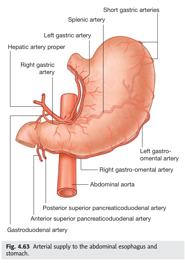
| Artery | Origin | Region |
|---|---|---|
| Left gastric | Coeliac trunk (directly) | Lesser curvature + lower oesophagus |
| Right gastric | Hepatic artery proper | Lesser curvature (right part) |
| Left gastro-omental (gastroepiploic) | Splenic artery | Greater curvature (left part) |
| Right gastro-omental | Gastroduodenal artery (from hepatic) | Greater curvature (right part) |
| Short gastric arteries (5–7) | Splenic artery | Fundus |
| Posterior gastric artery | Splenic artery | Posterior body/fundus |
The stomach has two arterial arcades:
Lesser curvature arcade: Left gastric + Right gastric
Greater curvature arcade: Left + Right gastro-omental
All supply ultimately from the coeliac trunk (first branch of abdominal aorta at T12).
Sympathetic: T6–T10 via greater splanchnic → coeliac plexus. Inhibits motility, causes vasoconstriction.
Parasympathetic: Left vagus (anterior gastric nerve) and Right vagus (posterior gastric nerve) from oesophageal plexus. Stimulates acid secretion + motility.
Lesser omentum: connects lesser curvature to liver (hepatoduodenal ligament + hepatogastric ligament). Contains portal triad (portal vein, hepatic artery, bile duct) in the free edge.
Greater omentum: hangs from greater curvature like apron; 4 layers; "policeman of the abdomen" — walls off infection.
| Cell Type | Location | Secretion | Clinical Relevance |
|---|---|---|---|
| Surface mucous cells | Entire gastric surface | Mucus + bicarbonate (HCO₃⁻) — protective layer | NSAIDs inhibit prostaglandins → reduce mucus → ulceration |
| Parietal (oxyntic) cells | Fundus + body (gastric glands) | HCl (via H⁺/K⁺-ATPase) + Intrinsic factor (IF) | PPIs block H⁺/K⁺-ATPase → reduce acid. Autoimmune destruction (pernicious anaemia) → no IF → B12 deficiency. Carcinoid tumours stimulate → massive acid |
| Chief (zymogenic) cells | Fundus + body | Pepsinogen (→ pepsin at pH <2); gastric lipase | Pepsin begins protein digestion. Low pH needed — reduced in achlorhydria |
| G cells | Pyloric antrum | Gastrin → stimulates parietal cells (via CCK-B receptor) | Zollinger-Ellison syndrome: gastrinoma (G cell tumour, often in duodenum/pancreas) → massively elevated gastrin → severe refractory PUD + diarrhoea |
| D cells | Antrum + duodenum | Somatostatin → inhibits gastrin (paracrine) | Negative feedback loop on acid secretion |
| Enterochromaffin-like (ECL) cells | Fundus + body | Histamine → stimulates parietal cells (H₂ receptor) | H₂ blockers (ranitidine) reduce acid by blocking ECL→parietal pathway |
Hypertrophy of the pyloric circular muscle in neonates (2–6 weeks). M > F (4:1); first-born males. Projectile non-bilious vomiting after feeds (no bile = obstruction ABOVE ampulla of Vater). "Olive" = palpable pyloric mass in the epigastrium. Hypochloraemic hypokalaemic metabolic alkalosis (losing HCl in vomit → kidney retains H⁺, excretes K⁺ → hypokalaemia + alkalosis; paradoxical aciduria late). Ultrasound confirms (pyloric muscle width >3 mm, length >16 mm). Treat: Ramstedt's pyloromyotomy.
Anterior duodenal ulcer (1st part, superior surface): perforates into peritoneal cavity → sudden onset peritonitis (board-like abdomen) → air under diaphragm on erect CXR. Posterior duodenal ulcer: erodes into gastroduodenal artery → massive haemorrhage (haematemesis/melaena). Gastric ulcer on lesser curvature: erodes left gastric artery. H. pylori is the cause in >70%; NSAIDs second most common. Treat H. pylori with triple therapy.
- Which gastric cell secretes intrinsic factor — and why does this matter? → Parietal (oxyntic) cells; autoimmune destruction → no IF → vitamin B12 malabsorption → pernicious anaemia + SACD of the cord
- G cells are in the antrum — what do they secrete and what disease results from a G-cell tumour? → Gastrin; gastrinoma (Zollinger-Ellison) → massively elevated gastrin → severe refractory peptic ulcers + diarrhoea
- Posterior duodenal ulcer erodes which artery? Clinical presentation? → Gastroduodenal artery → massive haematemesis/melaena (retroperitoneal bleeding, not peritonitis)
- Hypertrophic pyloric stenosis — metabolic disturbance and why? → Hypochloraemic, hypokalaemic metabolic alkalosis; vomiting HCl → kidney retains H⁺, excretes K⁺
- Left vs right vagus on stomach surface? → Left vagus → anterior gastric nerve; right vagus → posterior gastric nerve (mnemonic: LARP — Left Anterior, Right Posterior)
Duodenum
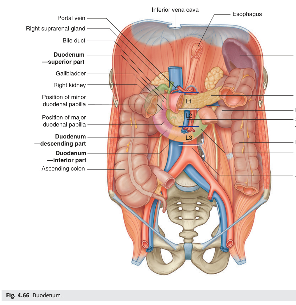
The duodenum is the most anatomically complex segment of the GI tract — a C-shaped retroperitoneal frame (except for the first 2.5 cm "cap") that wraps around the head of the pancreas and receives bile and pancreatic juice via the major duodenal papilla (ampulla of Vater) on the posteromedial wall of its second part. It is the site of the most important surgical watershed in the abdomen: everything supplied by the coeliac trunk meets everything supplied by the SMA at the level of the major papilla. Above the papilla = coeliac (gastroduodenal artery); below = SMA (inferior pancreaticoduodenal artery). This dual supply makes Whipple's pancreaticoduodenectomy technically demanding but also means that neither the coeliac nor SMA alone can be sacrificed without risk. The third part of the duodenum is pinched between the SMA anteriorly and the aorta posteriorly — in cachectic patients who lose the mesenteric fat cushion, this produces SMA syndrome (high GI obstruction). The duodenojejunal flexure is held in place by the ligament of Treitz — the surgical landmark that defines the start of the small bowel for counting purposes.
| Part | Level | Description / Relations | Notes |
|---|---|---|---|
| 1st (superior) | L1 | Cap = intraperitoneal, mobile; rest = retroperitoneal. Anterior = gallbladder. Posterior = gastroduodenal artery, portal vein, bile duct | Most common site of peptic ulcers. "Duodenal cap" on barium meal |
| 2nd (descending) | L1–L3 | Retroperitoneal. Major duodenal papilla (of Vater) at medial wall — opening of bile duct + main pancreatic duct. Minor papilla 2 cm above — accessory pancreatic duct | Head of pancreas intimately related medially. SMA crosses anterior to 3rd part |
| 3rd (horizontal) | L3 | Retroperitoneal, crosses midline. Anterior = SMA + SMV. Posterior = aorta + IVC | SMA syndrome: compression of 3rd part between SMA and aorta in thin/cachectic patients → high GI obstruction |
| 4th (ascending) | L2–L3 | Ascends to duodenojejunal (DJ) flexure at L2 level. Supported by ligament of Treitz (suspensory ligament of duodenum) | DJ flexure = surgical landmark for small bowel; ligament of Treitz = fibromuscular band from right crus to DJ junction |
The major duodenal papilla (papilla of Vater / bile papilla) is an elevation on the posteromedial wall of the descending (2nd) part of the duodenum at approximately L2. It is the site where the common bile duct and the main pancreatic duct (of Wirsung) unite to form the hepatopancreatic ampulla (ampulla of Vater), which opens at the summit of the papilla. The opening is controlled by the sphincter of Oddi (hepatopancreatic sphincter). The minor duodenal papilla lies ~2 cm above (anterosuperiorly) and carries the opening of the accessory pancreatic duct (of Santorini).
Clinical significance:
• Obstructive jaundice: carcinoma of the head of the pancreas or an impacted gallstone in the distal CBD obstructs flow here → bile backs up → jaundice + pale stools + dark urine + steatorrhoea.
• ERCP (Endoscopic Retrograde Cholangiopancreatography): endoscope cannulates the major duodenal papilla → inject contrast or perform therapeutic manoeuvres (stone extraction, sphincterotomy, stent insertion).
• Non-bilious vomiting in pyloric stenosis: obstruction is at the pylorus, above the major papilla → bile and pancreatic juice cannot reflux into the stomach → vomit is non-bilious (distinguishes high GI obstruction above the papilla from obstruction below it).
- Where is the major duodenal papilla and what opens there? → Posteromedial wall of 2nd (descending) duodenum; CBD + main pancreatic duct → hepatopancreatic ampulla of Vater; sphincter of Oddi controls opening
- Which part of the duodenum is intraperitoneal (mobile)? → First 2.5 cm (duodenal cap) only; rest is retroperitoneal
- SMA syndrome — which part compressed and why? → 3rd (horizontal) part pinched between SMA and aorta; loss of mesenteric fat pad in cachectic patients removes the cushion
- Why is vomiting non-bilious in pyloric stenosis? → Obstruction at pylorus is above the major papilla — bile cannot reflux into stomach
- Ligament of Treitz — what and why it matters? → Fibromuscular band from right crus to DJ flexure (L2); surgical landmark defining start of small bowel
Jejunum & Ileum
The jejunum and ileum together form about 6 metres of small bowel, and their differences reflect their different jobs. The jejunum is optimised for rapid absorption — thick walls, tall plicae circulares, and long finger-like villi maximise surface area in the segment that receives freshly digested chyme from the duodenum. By the ileum, most nutrients are already absorbed, so the wall is thinner and the folds are fewer; instead, the ileum is packed with Peyer's patches (submucosal lymphoid aggregates) as it approaches the immune challenge of the colon. The terminal ileum has two unique functions that no other segment can substitute: active absorption of vitamin B12 (only here, bound to intrinsic factor) and active reabsorption of bile salts (enterohepatic circulation). Lose the terminal ileum to Crohn's or resection, and you lose both — B12 deficiency and bile salt diarrhoea are the predictable consequences. Meckel's diverticulum (rule of 2s) is the commonest congenital anomaly of the GI tract, sitting 60 cm from the ileocaecal valve; its gastric ectopic mucosa secretes acid, ulcerating the adjacent ileal wall and causing painless rectal bleeding in children.
| Feature | Jejunum | Ileum |
|---|---|---|
| Length | Upper 2/5 (~2.5 m) | Lower 3/5 (~3.5 m) |
| Location | Left upper quadrant, umbilical region | Right iliac fossa and pelvis |
| Wall thickness | Thick (prominent folds) | Thin (fewer folds) |
| Plicae circulares | Tall + numerous (circular folds; increase surface area) | Few and low |
| Villi | Tall; finger-like; maximum absorption | Shorter |
| Peyer's patches | Few/absent | Numerous (lymphoid aggregates on anti-mesenteric border) |
| Mesentery fat | Thin; arcades fewer (1–2 long vasa recta) | More fat; 3–5 arcades (short vasa recta) |
| Blood supply | SMA branches (jejunal arteries) | SMA branches (ileal arteries + ileocolic) |
Meckel's diverticulum: remnant of the vitello-intestinal (omphalomesenteric) duct. Rule of 2s: 2% of population, 2 feet (60 cm) from ileocaecal valve, 2 inches long, 2× more common in males, 2 types of ectopic mucosa (gastric > pancreatic). Gastric mucosa secretes acid → ulceration → painless rectal bleeding in children. Technetium-99m pertechnetate scan (Meckel's scan) detects ectopic gastric mucosa. Complications: bleeding, obstruction (intussusception), diverticulitis mimicking appendicitis.
| Nutrient | Primary Site | Mechanism / Clinical Note |
|---|---|---|
| Iron (Fe²⁺) | Duodenum + proximal jejunum | Reduced to Fe²⁺ by vitamin C; DMT1 transporter. Coeliac (villous atrophy) → iron deficiency anaemia |
| Calcium | Duodenum (vitamin D-dependent) + jejunum (passive) | Vitamin D induces calbindin. Fat-soluble vitamin D malabsorption → osteomalacia |
| Fat + fat-soluble vitamins (A, D, E, K) | Jejunum → via lacteals as chylomicrons | Requires bile salt micelles. Steatorrhoea if bile salt / lipase deficit. Vitamin K deficiency → prolonged PT |
| Vitamin B12 | Terminal ileum (with intrinsic factor) | Crohn's / terminal ileal resection / pernicious anaemia → B12 deficiency → megaloblastic anaemia + SACD of cord |
| Bile salts | Terminal ileum (active; enterohepatic circulation) | Terminal ileal disease → bile salt diarrhoea + depleted pool → cholesterol gallstones |
| Folate (B9) | Proximal jejunum | Coeliac disease (proximal gut damage) → folate deficiency → megaloblastic anaemia |
- Two unique functions of the terminal ileum nothing else substitutes? → Active B12 absorption (with intrinsic factor) + active bile salt reabsorption (enterohepatic circulation)
- Meckel's rule of 2s — list them. → 2% population; 2 feet (60 cm) from ileocaecal valve; 2 inches long; 2× more common in males; 2 types ectopic mucosa (gastric > pancreatic)
- Where is iron absorbed and what transporter? → Duodenum + proximal jejunum; DMT1 transporter (Fe²⁺); vitamin C maintains reduced state
- Why does fat travel via lymph rather than portal blood? → Chylomicrons too large for portal capillaries → lacteals → cisterna chyli → thoracic duct → subclavian vein
- Jejunum vs ileum on barium — key macroscopic difference? → Jejunum: thick wall, prominent plicae circulares (feathery pattern); ileum: thin wall, smooth, multiple short arcades, fat-laden mesentery
Caecum & Appendix
Blind-ended pouch in the right iliac fossa; connects to the ascending colon superiorly and receives the ileum at the ileocaecal valve. Usually intraperitoneal (mobile). Taeniae coli converge on the appendix base — useful intraoperative landmark.
The appendix is a blind-ended tube of lymphoid tissue hanging off the caecum, and its position is the single most important reason appendicitis is clinically treacherous. In 65% of people it is retrocaecal — lying behind the caecum — where it does not irritate the anterior peritoneum. This means the classical McBurney's point tenderness can be absent, the abdomen may remain soft, and the diagnosis is made late. When the appendix hangs into the pelvis (30%), it can mimic ovarian pathology in women or produce urinary symptoms if it lies against the bladder. The blood supply is an end artery (appendicular artery, from the ileocolic branch of SMA) — there are no collaterals. When the lumen obstructs (most commonly by a faecolith), pressure builds, the end artery thromboses, and the tip becomes gangrenous within 24–72 hours, well before the body can mount an adequate inflammatory response. This is why appendicitis is a time-critical surgical emergency.
| Feature | Detail |
|---|---|
| Position | Variable: retrocaecal (most common, ~65%); pelvic (30%); others rare |
| Length | 2–20 cm (average 9 cm) |
| Surface marking | McBurney's point: junction of lateral 1/3 and medial 2/3 of line from ASIS to umbilicus = site of maximum tenderness in appendicitis |
| Blood supply | Appendicular artery (branch of ileocolic artery from SMA). End artery — no collateral → thrombosis → gangrene |
| Nerve supply | T10 → referred pain to umbilical region early in appendicitis |
Classic progression: periumbilical (T10 visceral) pain → anorexia/nausea → pain migrates to RIF (McBurney's point somatic) → fever. Signs: Rovsing's sign (LIF palpation causes RIF pain), Psoas sign (hip extension pain = retrocaecal appendix), Obturator sign (hip IR pain = pelvic appendix). Alvarado score. Rovsing's + McBurney's = most reliable. Perforation risk after 24–72h if untreated.
- McBurney's point — exact surface marking? → Junction of lateral 1/3 and medial 2/3 of line from ASIS to umbilicus
- Why does appendicitis pain start periumbilical then shift to RIF? → Early = visceral pain (T10 dermatome, poorly localised to umbilical region); late = somatic parietal peritoneum irritation (localised to RIF)
- Psoas sign vs Obturator sign — which position suggests which appendix? → Psoas sign (pain on hip extension) = retrocaecal appendix; Obturator sign (pain on hip IR) = pelvic appendix
- Why does appendicitis progress to gangrene within 24–72 h? → Appendicular artery is an end artery (no collaterals); luminal obstruction → pressure → thrombosis → ischaemia
- How do taeniae coli help find the appendix intraoperatively? → All three taeniae converge at the appendix base on the caecum — follow any taenia to the appendix
Colon
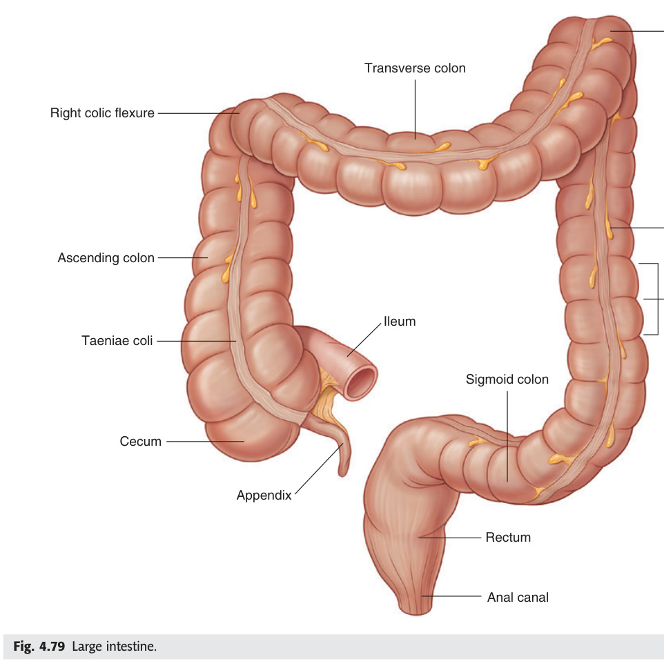
The colon's job is water reclamation — it converts liquid ileal effluent (~1.5 L/day) into solid faeces by absorbing water and electrolytes. Its three unique macroscopic features (taeniae coli, haustra, appendices epiploicae) are the radiologist's and surgeon's key identifiers. The taeniae coli are three condensed bands of the outer longitudinal muscle layer that run the colon's full length; because they are shorter than the colon wall they cause puckering into haustra. This haustral pattern disappears in ulcerative colitis ("lead-pipe colon") as the muscle hypertrophies and shortens. The most important vascular watershed in the colon is the splenic flexure — the point where SMA territory ends and IMA territory begins. The marginal artery of Drummond is the anastomotic arcade that runs along the colon's mesenteric border connecting these two systems, but it is weakest at the splenic flexure. After abdominal aortic aneurysm repair where the IMA is ligated, splenic flexure ischaemic colitis is the feared complication if the Drummond anastomosis is inadequate.
Three features unique to the colon:
1. Taeniae coli: 3 narrow bands of longitudinal smooth muscle running along the outer colon surface (shortened → haustra formation)
2. Haustra: sacculations of the colon wall between taeniae (seen on colonoscopy and CT)
3. Appendices epiploicae: small fat-filled peritoneal pouches on outer surface
| Part | Position | Artery |
|---|---|---|
| Ascending colon | Retroperitoneal, right side; ends at hepatic flexure | Right colic artery + ileocolic (SMA) |
| Hepatic (right colic) flexure | Right hypochondrium; deep to liver. More acute angle than splenic | Right colic (SMA) |
| Transverse colon | Intraperitoneal (transverse mesocolon); most mobile | Middle colic artery (SMA) |
| Splenic (left colic) flexure | Left hypochondrium. Higher + more acute than hepatic | Left colic artery (IMA) |
| Descending colon | Retroperitoneal, left side | Left colic artery (IMA) |
| Sigmoid colon | Intraperitoneal; highly mobile; sigmoid mesocolon. Ends at S3 | Sigmoid arteries (IMA) |
| Condition | Key Anatomy / Details |
|---|---|
| Diverticular disease | Pulsion diverticula (false — mucosa/submucosa only, no muscle) herniate through weak points where the vasa recta penetrate the colonic wall. Sigmoid colon most commonly affected (high intraluminal pressure). Diverticulosis (asymptomatic) → Diverticulitis (LIF pain, fever, leucocytosis) → complications: perforation, abscess, fistula (colovesical = pneumaturia), stricture, haemorrhage. CT abdomen is investigation of choice. Hinchey classification grades severity. |
| Sigmoid volvulus | Axial rotation of sigmoid on its mesentery → closed-loop obstruction. Predisposed by long, redundant sigmoid mesocolon (elderly, chronic constipation, institutionalised). X-ray: "coffee bean" / "omega loop" sign. Decompress with flexible sigmoidoscopy; elective sigmoid resection to prevent recurrence. |
| Caecal volvulus | Mobile caecum twists (requires a caecal mesentery). Right-sided obstruction. CT confirms. Surgical: right hemicolectomy. |
| Intussusception | Telescoping of one bowel segment (intussusceptum) into adjacent (intussuscipiens). Commonest in children 6–18 months; ileocaecal junction (ileum into caecum). Triad: episodic colicky abdominal pain + redcurrant jelly stool (blood + mucus) + right-sided mass ("sausage-shaped" on palpation). Adults: usually has pathological lead point (polyp, Meckel's, carcinoma). Ultrasound shows target/doughnut sign. Treat with air enema (pneumatic reduction) in children; surgery if failed. |
| Hirschsprung's disease | Congenital absence of myenteric (Auerbach's) and submucosal (Meissner's) ganglion cells in the rectum and variable length of colon, due to failure of neural crest cell migration (5th–12th weeks). Always involves the rectum; extends proximally for a variable length (short-segment = rectosigmoid, 80%; long-segment = to splenic flexure; total colonic aganglionosis rare). The aganglionic segment fails to relax → functional obstruction. Presentation: neonate — delayed passage of meconium (>48 h); bilious vomiting; abdominal distension; ribbon-like stools; explosive release of stool on DRE. Radiology: barium enema shows narrow aganglionic segment with transition zone and dilated proximal colon. Diagnosis: suction rectal biopsy (absence of ganglion cells + hypertrophied nerve trunks + ↑ acetylcholinesterase staining). Treatment: pull-through procedure (Swenson/Soave/Duhamel) — removes aganglionic segment and pulls innervated bowel to the anus. Associated with Down syndrome (trisomy 21) and RET proto-oncogene mutations. |
- Three macroscopic features unique to the colon (not in small bowel)? → Taeniae coli (3 longitudinal muscle bands) + haustra (sacculations) + appendices epiploicae (fat tags)
- Splenic flexure watershed — which two arteries meet there and clinical significance? → SMA (middle colic) meets IMA (left colic) at splenic flexure; weakest point of marginal artery → ischaemic colitis after IMA ligation in AAA repair
- Sigmoid volvulus — X-ray sign and treatment? → "Coffee bean" / omega loop sign; decompress with flexible sigmoidoscopy then elective sigmoid resection
- Hirschsprung's — what is absent, always involves which segment, and how is it diagnosed? → Absent myenteric + submucosal ganglion cells; always involves rectum; diagnosis = suction rectal biopsy (no ganglion cells + ↑ acetylcholinesterase)
- Intussusception classic triad and investigation? → Episodic colicky pain + redcurrant jelly stool + sausage-shaped mass; ultrasound = target/doughnut sign; air enema for reduction in children
Rectum & Anal Canal
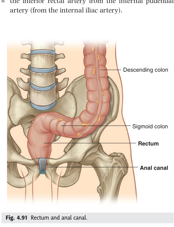
The rectum and anal canal are the most examined centimetres in anatomy — every boundary, curve, nerve, and blood supply difference above and below the pectinate line is fair game. The rectum begins at S3 (where the sigmoid mesocolon ends and the peritoneal covering progressively disappears) and ends at the anorectal junction where puborectalis creates the anorectal angle. This angle — maintained at about 90° at rest by the tonic U-shaped puborectalis sling — is the primary mechanism of faecal continence. Increased intra-abdominal pressure actually reinforces continence by compressing the anorectal junction, not forcing it open. The peritoneal coverage decreases as you descend: upper third has anterior + lateral cover, middle third has anterior only, lower third has none at all. This is clinically significant in anterior resection — the peritoneal reflection defines the plane of dissection, and below it the surgeon must perform total mesorectal excision (TME) in the "holy plane" between the mesorectal fascia and pelvic sidewall to avoid the autonomic nerves controlling bladder and sexual function.
Rectum (~12 cm): starts at S3, ends at anorectal junction (puborectalis). Divided into an upper narrow part and a lower dilated ampulla of the rectum.
| Feature | Detail |
|---|---|
| Two curves in the sagittal plane ★★★ | (1) Sacral flexure: convex posteriorly; follows the concavity of the sacrum and coccyx (upper rectum bends posteriorly to match sacral curve). (2) Perineal flexure (anorectal flexure): at the anorectal junction, a sharp forward bend (~90°) created by the puborectalis sling of the levator ani; convex anteriorly. Maintained by puborectalis contraction at rest → maintains faecal continence. Relaxes during defaecation. |
| Three lateral curves in the coronal plane | Upper and lower curves convex to the right; middle curve convex to the left. Represented internally as the 3 transverse folds of the rectum (valves of Houston) — 2 on the left, 1 on the right (middle). |
| Peritoneal cover | Upper 1/3: front and sides (intraperitoneal). Middle 1/3: front only. Lower 1/3: no peritoneum (extraperitoneal). The rectovesical (male) / rectouterine (female, pouch of Douglas) peritoneal reflection is clinically important. |
| Blood supply | Superior rectal artery (terminal branch of IMA); middle rectal arteries (internal iliac); inferior rectal arteries (internal pudendal). Venous: superior rectal vein → IMV → portal system (portosystemic anastomosis site). |
(2) Perineal flexure (anorectal flexure): at the anorectal junction, the rectum bends sharply anteriorly (~80–90°) due to the puborectalis sling (part of levator ani, origin: pubis, loops around the anorectal junction, insertion: pubis — forms a U-shaped sling). Clinical: (a) The puborectalis is the most important muscle of faecal continence — its tonic contraction maintains the anorectal angle; when it relaxes during defaecation the angle straightens and defaecation proceeds. (b) Injury to puborectalis (childbirth, surgery) → anorectal angle lost → faecal incontinence. (c) Internal sphincter relaxes (rectoanal inhibitory reflex); external sphincter is voluntary control (pudendal nerve).
• Epithelium: columnar (transitional zone just above)
• Blood supply: superior rectal artery (IMA) / superior rectal vein → IMV → portal
• Lymph: internal iliac nodes
• Nerve: autonomic (visceral — no pain sensation, only stretch)
• Internal haemorrhoids — painless
Below the pectinate line (lower anal canal, ectodermal origin):
• Epithelium: stratified squamous (keratinised)
• Blood supply: inferior rectal artery (pudendal) / inferior rectal vein → pudendal → internal iliac → IVC
• Lymph: superficial inguinal nodes
• Nerve: somatic (inferior rectal nerve — exquisitely pain-sensitive)
• External haemorrhoids — painful (somatic nerve)
Internal haemorrhoids (above pectinate line): painless bright red bleeding per rectum; classified by degree of prolapse (Grades 1–4). Anal fistula/fissure: fissure = tear in anoderm usually posteriorly (ischaemic watershed). Fistula = anal gland infection tracking to skin. Goodsall's rule: external openings anterior to transverse anal line track directly; posterior openings track to posterior midline. Rectal carcinoma: most common presentation = altered bowel habit + rectal bleeding. Digital rectal exam + rigid sigmoidoscopy. Total mesorectal excision (TME) is the standard surgery.
- Pectinate line — epithelium above vs below? → Above = columnar (transitional zone); below = stratified squamous (keratinised); ectodermal below, endodermal above
- Internal vs external haemorrhoids — why is one painful and one not? → Above pectinate line = autonomic innervation (no somatic pain) → internal haemorrhoids painless. Below = somatic inferior rectal nerve → external haemorrhoids exquisitely painful
- Lymph drainage above vs below pectinate line? → Above = internal iliac nodes; below = superficial inguinal nodes
- Two curves of the rectum in the sagittal plane? → Sacral flexure (convex posteriorly, follows sacrum) + perineal/anorectal flexure (convex anteriorly, created by puborectalis sling at ~90°)
- Pringle's manoeuvre — what is compressed and what does it achieve? → Index finger through epiploic foramen compresses hepatoduodenal ligament (portal vein + hepatic artery) between finger and thumb → controls hepatic inflow haemorrhage
Blood Supply Summary & Portosystemic Anastomoses
The GI tract's blood supply maps directly onto embryological gut divisions: coeliac trunk for foregut, SMA for midgut, IMA for hindgut. The boundaries are not arbitrary — they reflect the embryological rotation and fixation of the gut. The coeliac/SMA watershed sits at the major duodenal papilla (ampulla of Vater); the SMA/IMA watershed sits at the splenic flexure of the colon. These two watersheds are the most vulnerable points in ischaemia — the papilla watershed is relevant in Whipple's resections, and the splenic flexure watershed is the first region to die in low-flow states (post-cardiac surgery, after AAA repair). The portosystemic anastomoses are where portal and systemic venous drainage overlap; these communications are normally trivial, but in portal hypertension they dilate dangerously at four sites — lower oesophagus (varices, lethal haemorrhage), anterior abdominal wall (caput medusae), rectum (rectal varices), and retroperitoneum (veins of Retzius).
| Artery | Origin | Structures Supplied |
|---|---|---|
| Coeliac trunk | Abdominal aorta T12 | Foregut: lower oesophagus → upper duodenum (to ampulla of Vater); liver, spleen, gallbladder, pancreas (head) |
| SMA | Abdominal aorta L1 | Midgut: lower duodenum → 2/3 transverse colon |
| IMA | Abdominal aorta L3 | Hindgut: splenic flexure → upper rectum |
In portal hypertension, blood is redirected through portosystemic communications:
1. Lower oesophagus: left gastric vein (portal) ↔ azygos vein (systemic) → oesophageal varices (lethal haemorrhage)
2. Anterior abdominal wall: paraumbilical veins (portal) ↔ epigastric veins (systemic) → caput medusae (radiating dilated veins from umbilicus)
3. Rectum: superior rectal vein (IMV/portal) ↔ inferior/middle rectal veins (systemic) → rectal varices
4. Retroperitoneum: veins of Retzius (less clinically visible)
Also: splenomegaly, ascites, hepatic encephalopathy.
- Three arterial territories — boundaries (two key watershed points)? → Coeliac (foregut) ends at ampulla of Vater; SMA (midgut) ends at splenic flexure; IMA (hindgut) supplies to upper rectum
- Four portosystemic anastomosis sites in portal hypertension? → Lower oesophagus (varices) + anterior abdominal wall (caput medusae) + rectum (rectal varices) + retroperitoneum (veins of Retzius)
- Why is the splenic flexure the most vulnerable colon segment in ischaemia? → Watershed between SMA and IMA; marginal artery of Drummond is weakest here; first to die when IMA ligated
- Oesophageal varices — portal vein drains into which vessel that anastomoses with systemic? → Left gastric vein (portal) ↔ azygos/hemiazygos veins (systemic) at lower oesophagus
- Caput medusae — which veins are involved? → Paraumbilical veins (portal, run in falciform ligament) ↔ superior/inferior epigastric veins (systemic) → dilated veins radiate from umbilicus
Test your knowledge of Unit 05
Alimentary canal MCQs, TF and essays in timed exam format.
Inflammatory Bowel Disease: Crohn's vs UC ★★★
Crohn's disease and ulcerative colitis are the two chronic inflammatory bowel diseases, and distinguishing them is one of the highest-yield tasks in both anatomy and medicine exams. The key conceptual difference is depth: UC is a mucosal disease (inflammation stays in the mucosa and submucosa, starts at the rectum, spreads continuously proximally), while Crohn's is transmural (goes through all layers, causing fistulae and strictures, and can affect any segment from mouth to anus with skip lesions between affected areas). This transmural nature explains all of Crohn's complications — fistulae form because inflammation burns through the full wall into adjacent structures; strictures form because transmural fibrosis contracts the lumen. Histologically, non-caseating granulomas are pathognomonic for Crohn's (present in ~60%); UC's hallmark is crypt abscesses. The rule of thumb for surgery: UC is curable by total proctocolectomy (remove all the diseased mucosa); Crohn's is not curable by any surgery because it can recur at any new anastomosis.
| Feature | Crohn's Disease | Ulcerative Colitis |
|---|---|---|
| Location | Any part of GI tract (mouth to anus). Most common: terminal ileum ± proximal colon | Colon only. Always starts at rectum and extends proximally (continuous) |
| Pattern | Skip lesions (normal bowel between affected segments) | Continuous, diffuse from rectum |
| Rectal involvement | Spared in ~50% of cases | Always involved (100%) |
| Wall involvement | Transmural (all layers) → fistulae, abscesses, strictures | Mucosal + submucosal only |
| Histology (key) | Non-caseating granulomas; transmural inflammation; lymphoid aggregates; fissuring ulcers | Crypt abscesses (hallmark); goblet cell depletion; mucosal/submucosal only; no granulomas |
| Gross appearance | Cobblestone mucosa; rose-thorn fissures; creeping fat; strictures; string sign (ileum) on barium | Pseudopolyps; lead-pipe colon (loss of haustra); continuous granular mucosa |
| Bloody diarrhoea | Less common (unless colonic involvement) | Hallmark — frequent bloody, mucoid diarrhoea |
| Perianal disease | Common: fistulae, skin tags, abscesses, fissures | Rare |
| Smoking | Worsens (smoking is a risk factor) | Protective (non-smokers/ex-smokers predominantly affected) |
| Surgery | Not curative (recurrence at anastomosis); bowel-sparing (stricturoplasty) | Total proctocolectomy is curative |
| Cancer risk | Small bowel adenocarcinoma (↑); colorectal if extensive colonic disease | Colorectal carcinoma (↑ with extent + duration; surveillance colonoscopy every 1–3 years from 8–10 years of extensive colitis) |
| System | Manifestation | Notes |
|---|---|---|
| Eyes | Episcleritis (parallels gut activity) · Uveitis (independent of gut activity) | Uveitis can progress to blindness; needs ophthalmology |
| Skin | Erythema nodosum (parallels activity) · Pyoderma gangrenosum (independent) | Pyoderma = deep painful ulcers on lower limbs; treat with steroids |
| Joints | Peripheral arthropathy (parallels activity) · Ankylosing spondylitis / sacroiliitis (independent) | HLA-B27 associated; AS does not improve with colectomy |
| Liver/biliary | Primary sclerosing cholangitis (PSC) — mainly UC (70% of PSC patients have UC) | PSC → stricturing of bile ducts → cholangiocarcinoma risk; MRCP diagnostic; liver transplant only cure |
| Haematological | Anaemia (chronic disease / iron deficiency / B12 in Crohn's terminal ileum) · Thromboembolism (IBD = prothrombotic state) | DVT/PE risk ↑ in active IBD flares |
- Crohn's vs UC — which always involves the rectum? → UC (100%); Crohn's spares rectum in ~50%
- Histological hallmark of Crohn's vs UC? → Crohn's = non-caseating granulomas (pathognomonic, ~60%); UC = crypt abscesses + goblet cell depletion
- Why is Crohn's not curable by surgery but UC is? → UC is a mucosal disease confined to the colon — proctocolectomy removes all diseased tissue. Crohn's can recur at any new anastomosis and can affect any GI segment
- Which IBD type has skip lesions and which is continuous? → Crohn's = skip lesions; UC = continuous from rectum proximally
- PSC — which IBD it's associated with, MRCP appearance, and only cure? → UC (70% of PSC patients); "beads-on-a-string" bile duct strictures on MRCP; liver transplantation
Peritoneum ★★★
The peritoneum is the abdominal cavity's lining and the body's most elegant infection-fighting membrane — its visceral layer drapes organs with a frictionless surface, while the parietal layer forms the cavity wall. The distinction between intraperitoneal and retroperitoneal organs is not just anatomical trivia; it determines how pathology presents and how surgeons approach organs. An intraperitoneal organ surrounded by peritoneum will cause peritonitis when it perforates (the parietal peritoneum's somatic innervation produces exquisite localised pain). A retroperitoneal organ — kidney, pancreas, aorta, duodenum — sits behind the peritoneum and can bleed or leak massively into the retroperitoneal space before causing any peritoneal signs at all. The "SAD PUCKER" mnemonic captures the primary retroperitoneal organs. The peritoneal folds (lesser omentum, greater omentum, mesenteries) are not passive drapes — the greater omentum actively migrates to wall off infection (hence "policeman of the abdomen"), and the lesser omentum's free right edge contains the portal triad, making it the site of Pringle's manoeuvre in liver haemorrhage control.
The peritoneum is a double-layered serous membrane: parietal (lines abdominal/pelvic walls — somatic innervation, localised pain) and visceral (covers organs — autonomic innervation, poorly localised pain). The potential space between them is the peritoneal cavity.
| Category | Organs |
|---|---|
| Intraperitoneal (covered by visceral peritoneum, suspended by mesentery) | Stomach · First 2.5 cm of duodenum (cap) · Jejunum · Ileum · Caecum (usually) · Appendix · Transverse colon · Sigmoid colon · Upper third of rectum (anterior surface only) · Spleen · Liver · Ovaries · Uterus (body) |
| Primary retroperitoneal (never had a mesentery) | Kidneys + adrenals · Aorta + IVC · Ureter · Duodenum (2nd–4th parts) · Pancreas (except tail) |
| Secondary retroperitoneal (fused against posterior wall during development) | Ascending colon · Descending colon · Middle and lower thirds of rectum |
"SAD PUCKER": Suprarenal glands · Aorta & IVC · Duodenum (2nd–4th) · Pancreas (except tail) · Ureters · Colon (ascending + descending) · Kidneys · Esophagus (thoracic) · Rectum (lower 2/3)
| Structure | Connects | Contents / Notes |
|---|---|---|
| Falciform ligament | Anterior abdominal wall → anterior liver surface | Contains round ligament of liver (ligamentum teres = obliterated left umbilical vein). In portal hypertension → paraumbilical veins reopen → caput medusae |
| Lesser omentum | Lesser curvature of stomach + upper duodenum → porta hepatis/liver | Hepatogastric ligament + hepatoduodenal ligament (free right edge). Contains portal triad in free edge: bile duct (right), hepatic artery (left), portal vein (posterior). Pringle's manoeuvre = compress here |
| Greater omentum | Greater curvature → hangs as 4-layered apron over bowel | "Policeman of abdomen" — migrates to wall off infection. Contains right + left gastro-omental vessels. 4 layers of peritoneum fused (with omental bursa obliterated). Site of omental metastases in peritoneal carcinomatosis ("omental cake") |
| Gastrosplenic ligament | Greater curvature (fundus) → spleen | Short gastric arteries + left gastro-omental vessels |
| Splenorenal (lienorenal) ligament | Spleen → left kidney | Splenic vessels + tail of pancreas |
| Mesentery proper | DJ flexure (L2) → RIF (along oblique line) | SMA + SMV + lymphatics. Root is 15 cm; gut border is ~6 m. Contains mesenteric fat (increased in Crohn's = "creeping fat") |
| Transverse mesocolon | Transverse colon → posterior abdominal wall | Middle colic vessels. Divides peritoneal cavity into supramesocolic + inframesocolic compartments. Pancreatic pseudocysts → inflate lesser sac → present as mass felt through transverse mesocolon |
| Sigmoid mesocolon | Sigmoid colon → posterior pelvic wall | Sigmoid vessels. V-shaped root (apex at bifurcation of left common iliac artery). Long/mobile mesocolon → predisposes to sigmoid volvulus |
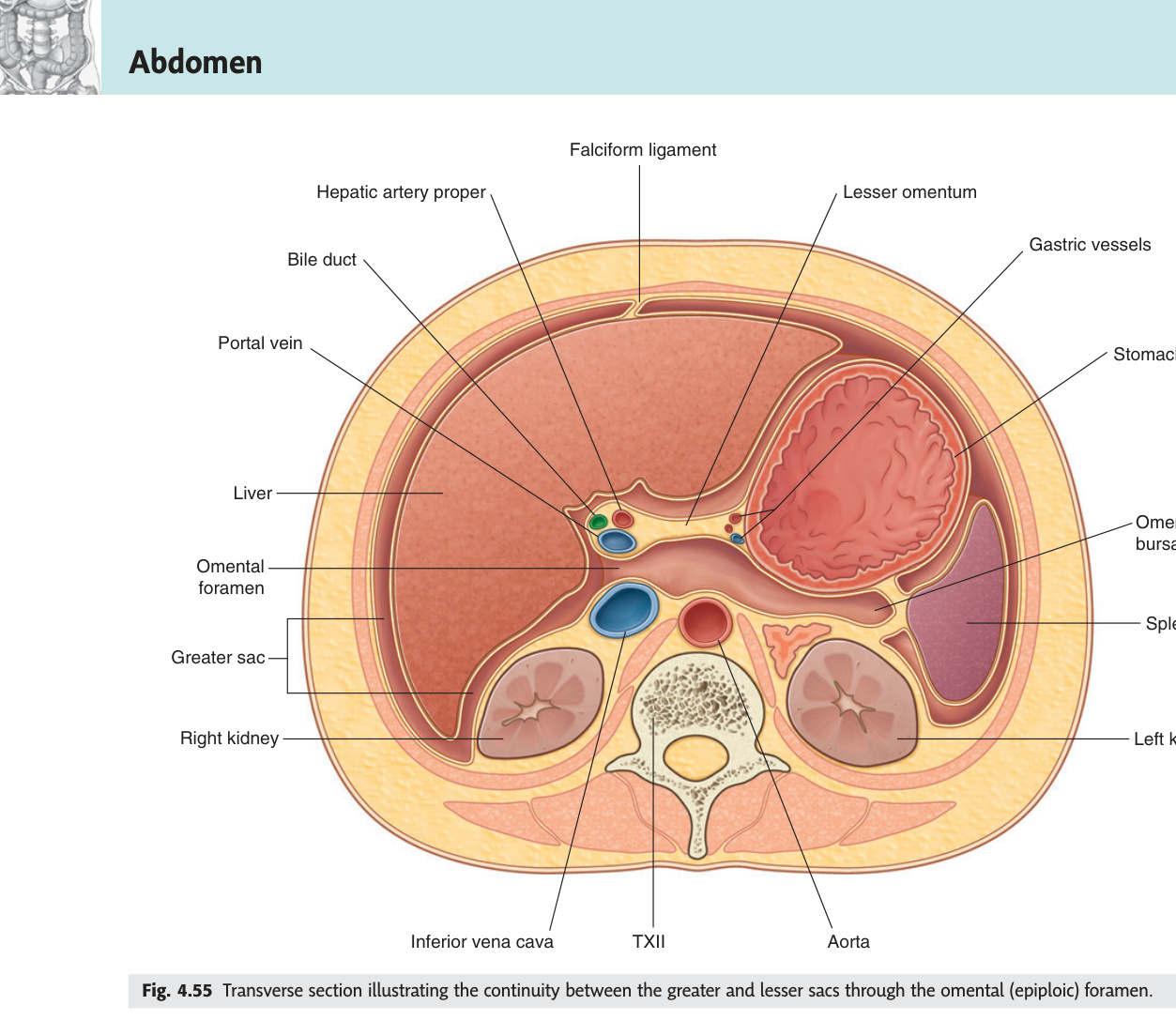
Lesser sac (omental bursa): a potential space posterior to the stomach + lesser omentum and anterior to the pancreas/posterior abdominal wall. Communicates with the greater peritoneal sac through the epiploic foramen of Winslow.
| Boundary of Epiploic Foramen | Structure |
|---|---|
| Anterior | Hepatoduodenal ligament (free edge of lesser omentum) — contains portal vein, hepatic artery, bile duct |
| Posterior | IVC (covered by parietal peritoneum) |
| Superior | Caudate lobe of liver |
| Inferior | 1st part of duodenum + hepatoduodenal ligament |
Morrison's pouch (hepatorenal recess): most dependent part of the peritoneal cavity in the supine position, between the right lobe of the liver and the right kidney. Free fluid (blood, bile, pus) collects here first. Detected by FAST (Focused Assessment with Sonography in Trauma) ultrasound — free fluid appears as black crescent. Pouch of Douglas (rectouterine pouch in females; rectovesical in males): most dependent point of the pelvic peritoneal cavity. Fluid/pus/blood pools here — can palpate fullness/tenderness on per rectal/vaginal exam. Drained by colpotomy (posterior vaginal fornix) or rectal drainage. Lesser sac: pancreatic pseudocysts (post-pancreatitis) rupture into lesser sac → epigastric mass; can drain endoscopically (cystogastrostomy). Lesser sac abscesses from posterior gastric ulcer perforation.
- SAD PUCKER — list all primary retroperitoneal organs. → Suprarenal glands · Aorta & IVC · Duodenum (2nd–4th) · Pancreas (except tail) · Ureters · Colon (ascending + descending) · Kidneys · Oesophagus (thoracic) · Rectum (lower 2/3)
- Contents of the free right edge of the lesser omentum (hepatoduodenal ligament)? → Portal vein (posterior) + hepatic artery proper (left) + common bile duct (right) — portal triad
- Morrison's pouch — where is it and why does fluid collect there? → Hepatorenal recess between right liver lobe and right kidney; most dependent part of peritoneal cavity in supine position — free fluid collects here first; detected on FAST USS
- Lesser sac — bounded by what anteriorly and posteriorly, and accessed how? → Anterior = stomach + lesser omentum; posterior = pancreas + posterior abdominal wall; communicates with greater sac via epiploic foramen of Winslow
- Why does retroperitoneal organ rupture cause less acute peritonism than intraperitoneal? → Retroperitoneal bleeding/leak stays behind the peritoneum — parietal peritoneum not directly irritated → no guarding or rigidity; patient can be haemodynamically compromised with a soft abdomen