Muscles
Back Muscles
These five muscles have nothing to do with moving the spine — they all act on the upper limb and are called “extrinsic” because they migrated onto the back from limb-bud territory during development. That embryological origin explains their nerve supply: trapezius is innervated by CN XI (a cranial nerve!) because it derives from branchial arch mesenchyme, not from spinal muscle. Latissimus dorsi is the great climbing and swimming muscle — it pulls the arm forcefully downward and backward, which is exactly what you do when hauling yourself out of a swimming pool. The clinical analogy that unifies this group: think of each muscle as a rope connecting one body part to another; when the rope is cut (nerve injury), the structure it was holding drifts away.
| Muscle | Origin | Insertion | Action | Nerve |
|---|---|---|---|---|
| Trapezius | Occipital bone, nuchal lig, C7–T12 SPs | Lateral clavicle, acromion, scapular spine | Upper: elevates scapula; Middle: retracts; Lower: depresses; Upper+lower: rotates glenoid up | CN XI (spinal accessory) + C3–C4 |
| Latissimus dorsi | T7–T12 SPs, thoracolumbar fascia, iliac crest, ribs 9–12 | Intertubercular groove of humerus | Extension, adduction, medial rotation of arm; depression of shoulder | Thoracodorsal nerve (C6–C8) |
| Rhomboid major/minor | T2–T5 / C7–T1 spinous processes | Medial border of scapula | Retraction + elevation of scapula; rotates glenoid downward | Dorsal scapular nerve (C4–C5) |
| Serratus anterior | Outer surfaces ribs 1–8 | Costal surface of medial scapular border | Protracts + rotates scapula; keeps scapula against thorax | Long thoracic nerve (C5–C7) |
| Levator scapulae | C1–C4 transverse processes | Superior angle of scapula | Elevates + downwardly rotates scapula | C3–C4 + dorsal scapular (C5) |
Serratus anterior palsy (long thoracic nerve injury — mastectomy, stab wound, sport): medial border of scapula lifts off thoracic wall on pushing against resistance = medial winging. Trapezius palsy (CN XI injury — radical neck dissection): drooped shoulder + lateral winging. Distinguish by direction of winging and action causing it.
• Latissimus dorsi nerve and its action used to climb a rope? → Thoracodorsal (C6–C8); extends + adducts + medially rotates arm (pulling body upward)
• Long thoracic nerve injury: which muscle and what sign? → Serratus anterior; medial scapular winging on pushing against wall
• Rhomboids: action and nerve? → Retract + elevate scapula; dorsal scapular nerve (C4–C5)
The intrinsic back muscles are the true movers and stabilisers of the vertebral column. Think of the erector spinae as three parallel columns of cables running the entire length of the spine: when they shorten symmetrically they pull the column into extension (straightening up from a forward bend); when they shorten on one side they produce lateral flexion. The multifidus sits even deeper, spanning only 2–4 vertebral levels, and acts as a fine-tuning stabiliser rather than a gross mover — it is the muscle that rehabilitation programmes target in chronic low back pain, because it is selectively inhibited and atrophies after a back injury while the superficial erectors compensate. Both groups are supplied by the posterior rami of spinal nerves, which is why they are anatomically and embryologically separate from the anterior abdominal musculature.
Erector spinae (lateral to medial): Iliocostalis (ribs), Longissimus (transverse processes), Spinalis (spinous processes). Supplied by posterior rami of spinal nerves. Actions: extension and lateral flexion of the vertebral column; maintain upright posture.
Multifidus: deep to erector spinae; spans 2–4 vertebrae; most powerful rotator/stabiliser of the spine; important in low back pain rehabilitation.
- Name the three erector spinae columns, lateral to medial. Iliocostalis (ribs), Longissimus (transverse processes), Spinalis (spinous processes).
- What nerve supply do ALL intrinsic back muscles share? Posterior (dorsal) rami of the corresponding spinal nerves — anatomically distinct from anterior trunk muscles.
- Which deep back muscle is selectively targeted in chronic low back pain rehabilitation? Multifidus — it atrophies after back injury while the superficial erectors compensate.
- What does the suboccipital triangle contain? The vertebral artery (V3 segment), suboccipital nerve (dorsal ramus C1), and suboccipital venous plexus.
- What is the unique action of obliquus capitis inferior? Rotates the atlas on the axis (C2), turning the head — it is the only suboccipital muscle that does NOT attach to the skull.
Thorax Muscles & Diaphragm
You take roughly 20,000 breaths a day, and for each quiet breath the diaphragm does virtually all the work. Think of it as a piston: when its muscle fibres contract, the central tendon descends like the bottom of a syringe being pulled, expanding the thoracic volume and drawing air in. It then relaxes passively and recoils upward as the lungs' elastic recoil drives expiration. The genius of the design is the three holes in it — each at a different level, each transmitting different structures. Remembering these three levels is non-negotiable for any anatomy or surgical exam: T8 IVC, T10 oesophagus, T12 aorta (“I 8 ten eggs at 12”). The phrenic nerve originates from C3–C5, which is why diaphragm irritation (blood under the diaphragm, sub-phrenic abscess) refers pain to the tip of the shoulder — both are C4 dermatomal territory.
The dome-shaped musculotendinous partition between the thorax and abdomen. The primary muscle of inspiration — its contraction descends the dome, increasing thoracic vertical diameter.
| Part | Origin | Notes |
|---|---|---|
| Sternal part | Back of xiphoid process | Smallest part; gap with costal part (Morgagni hernia site) |
| Costal part | Inner surfaces of lower 6 costal cartilages + ribs | Largest part; interdigitates with transversus abdominis |
| Lumbar part (crura) | Right crus: L1–L3; Left crus: L1–L2 vertebral bodies | Right crus larger; forms oesophageal sphincter |
| Central tendon | — | Insertion; fused with inferior pericardium |
T8 = I (IVC) — Inferior Vena Cava + right phrenic nerve (at caval hiatus = most anterior, right of midline)
T10 = E (Oesophagus) — Oesophagus + left + right vagus nerves (through muscle of right crus = most muscular)
T12 = A (Aorta) — Aorta + thoracic duct + azygos vein (posterior, between crura)
Mnemonic: "I 8 (ate) 10 Eggs At 12" — T8 IVC, T10 oesophagus, T12 aorta
Nerve supply: Phrenic nerve C3, C4, C5 — "C3, 4, 5 keeps the diaphragm alive". Peripheral: lower 5 intercostal nerves (sensory only). Referred pain from diaphragm: C4 dermatome = tip of shoulder.
Herniation of an abdominal structure through the oesophageal hiatus (T10). Sliding hiatus hernia (Type I, 95%): gastro-oesophageal junction (GOJ) and upper stomach slide superiorly into the posterior mediastinum. Lower oesophageal sphincter (LOS) incompetence → GORD. Symptoms: heartburn worse on lying/bending, acid regurgitation, waterbrash. Diagnosed by barium swallow or endoscopy. Managed medically (PPI + lifestyle); Nissen fundoplication for refractory cases. Rolling (Para-oesophageal) hernia (Type II, 5%): GOJ remains in position, but gastric fundus herniates through a separate peritoneal defect alongside the oesophagus. LOS competent → less acid reflux but risk of gastric volvulus and strangulation. Symptoms: epigastric pain after meals, post-prandial breathlessness, dysphagia from extrinsic compression. Elective surgery recommended even if asymptomatic due to volvulus risk. Type III = combined; Type IV = other abdominal organs (colon, spleen) herniate too.
• Nerve supply to diaphragm and why shoulder pain refers here? → Phrenic C3–C5; referred pain to C4 dermatome (tip of shoulder) from sub-phrenic irritation
• Bochdalek vs Morgagni hernia: which is more common and which side? → Bochdalek 90%, left-sided; Morgagni anterior, usually right-sided
• Which crus is larger and what does it surround? → Right crus (L1–L3); forms the muscular oesophageal sphincter at T10
The three layers of intercostal muscle mimic the three layers of the abdominal wall — external, internal, innermost — and their fibre directions alternate in the same way. The external intercostals run like your fingers in your pockets (“hands in pockets” = inferomedial): they pull the ribs up and apart during inspiration. The internal layer runs the opposite way and is active during forced expiration. The crucial practical point is where the neurovascular bundle runs: it is tucked under the rib in the costal groove, and it runs vein–artery–nerve from top to bottom (“VAN”). Insert a needle or drain just above the upper border of the rib below to stay clear of it.
| Muscle | Fibre Direction | Action | Nerve |
|---|---|---|---|
| External intercostal | Inferomedial (“hands in pockets”) | Elevates ribs during inspiration; active in quiet breathing | Intercostal nerve of same space |
| Internal intercostal | Inferolateral (perpendicular to external) | Depresses ribs during forced expiration | Intercostal nerve |
| Innermost intercostal | Same as internal | Forced expiration | Intercostal nerve |
• Order of VAN in costal groove (top to bottom)? → Vein → Artery → Nerve (most inferior, most at risk)
• Which intercostals are active in forced expiration? → Internal + innermost intercostals (depress ribs)
• Safe triangle for chest drain insertion? → 4th–5th ICS, mid-axillary line, above upper border of lower rib
Anterior Abdominal Wall
The anterior abdominal wall is a four-layer structure that works like a corset: three flat muscles with fibres running in different directions (external oblique inferomedial, internal oblique superomedial, transversus abdominis horizontal) create a grid that resists forces in all directions, much like a sheet of plywood with alternating grain. The fourth muscle, rectus abdominis, runs vertically in the midline enclosed in a sheath formed by the aponeuroses of the three flat muscles. The layered design has one weak point: below the arcuate line (halfway between umbilicus and pubis) the posterior rectus sheath disappears entirely, leaving only thin transversalis fascia on the back of the muscle — this is why direct inguinal hernias form in the lower abdomen. The same nerves that supply the abdominal muscles (T7–T12) also carry sensation from the overlying skin, which is why a high cord lesion at T10 produces periumbilical anaesthesia along with abdominal muscle weakness.
| Muscle | Origin | Insertion | Fibre Direction | Nerve |
|---|---|---|---|---|
| External oblique | Ribs 5–12 (outer surfaces) | Linea alba, ASIS, inguinal ligament, pubic tubercle | Inferomedial (“hands in pockets”) | T7–T11 intercostal + iliohypogastric + ilioinguinal |
| Internal oblique | Thoracolumbar fascia, iliac crest, lateral inguinal lig | Linea alba, pubic crest, ribs 10–12 | Superomedial (perpendicular to EO) | T8–T12 + iliohypogastric + ilioinguinal |
| Transversus abdominis | Thoracolumbar fascia, iliac crest, inguinal lig, lower 6 costal cartilages | Linea alba, pubic crest | Horizontal | T7–T12 + iliohypogastric + ilioinguinal |
| Rectus abdominis | Pubic crest and pubic symphysis | 5th–7th costal cartilages + xiphoid | Vertical; enclosed in rectus sheath | T7–T12 intercostal nerves |
Above arcuate line: Anterior = EO aponeurosis + anterior layer of IO; Posterior = posterior layer of IO + TA aponeurosis.
Below arcuate line: Anterior = ALL three aponeuroses (EO + IO + TA); Posterior = absent (only transversalis fascia). This weakness explains inguinal hernias below the arcuate line.
- Name the three flat muscles and their fibre directions. External oblique (inferomedial), internal oblique (superomedial), transversus abdominis (horizontal) — a three-ply grid resisting forces in all directions.
- What is the arcuate line and what changes below it? Curved line ~1/3 above pubis where the posterior rectus sheath ends; below it all three aponeuroses pass anterior to rectus, leaving only transversalis fascia posteriorly.
- What nerves supply the anterior abdominal wall and where do they travel? T7–T12 (thoracoabdominal nerves) + L1 (iliohypogastric, ilioinguinal); they run in the neurovascular plane between internal oblique and transversus abdominis.
- Why does a cord lesion at T10 cause periumbilical anaesthesia? T10 dermatome maps to the umbilicus — the same segmental nerves supply both the abdominal muscles and the overlying skin.
- Where is the linea semilunaris and what is its surgical significance? Lateral edge of rectus abdominis where the flat-muscle aponeuroses merge; incisions here risk severing T10–T12 segmental nerves, denervating a strip of rectus.
The inguinal canal is an engineering problem: the abdominal wall needs to be airtight to resist the pressure surges of coughing, straining, and lifting, yet it must also allow the spermatic cord (in males) or the round ligament (in females) to pass through. The solution is an oblique tunnel, not a straight hole. By running diagonally through the wall for ~4 cm, the canal creates a natural flap-valve mechanism — when intra-abdominal pressure rises, the canal walls are compressed together rather than blown open. Where this mechanism fails, a hernia forms. The key to distinguishing hernia types is the relationship to the inferior epigastric vessels: indirect hernias pass lateral to these vessels (through the deep ring), while direct hernias bulge medially through the posterior wall of the canal. Think “MD — Medial Direct.” The clinical examination test (press over the deep ring and ask the patient to cough) exploits this anatomy.
An oblique passage through the lower abdominal wall, ~4 cm long, running superomedially from the deep inguinal ring to the superficial inguinal ring. It is formed by the walls of all three flat abdominal muscles.
| Wall | Structure |
|---|---|
| Anterior | External oblique aponeurosis (whole length) + internal oblique (lateral 1/3) |
| Posterior | Transversalis fascia (whole length) + conjoint tendon (medial 1/3) |
| Floor (inferior) | Inguinal ligament (in-folded lower border of EO aponeurosis; ASIS → pubic tubercle) |
| Roof (superior) | Arching fibres of internal oblique + transversus abdominis (form conjoint tendon medially) |
| Deep inguinal ring | Defect in transversalis fascia; lies lateral to inferior epigastric vessels; midpoint of inguinal ligament |
| Superficial inguinal ring | Triangular defect in EO aponeurosis; above + medial to pubic tubercle |
| Male | Female | |
|---|---|---|
| Main structure | Spermatic cord: vas deferens + testicular artery + pampiniform venous plexus + lymphatics + cremasteric artery + genital branch of genitofemoral nerve (+ nerve to cremaster) | Round ligament of uterus |
| Both sexes | Ilioinguinal nerve (runs in canal but outside the spermatic cord / round ligament) | |
| Hernia Type | Path | Relation to Inferior Epigastric Vessels | Key Features |
|---|---|---|---|
| Indirect inguinal | Through deep ring → canal → superficial ring → may enter scrotum (congenital, patent processus vaginalis) | Lateral to inferior epigastric vessels | More common; young males; covered by all 3 spermatic fascial layers; reducible, controlled by pressure over deep ring |
| Direct inguinal | Through posterior wall (Hesselbach's triangle) — bulges anteromedially | Medial to inferior epigastric vessels | Acquired (weakness in elderly men); does NOT descend into scrotum; only covered by transversalis fascia |
| Femoral | Through femoral ring into femoral canal (below inguinal ligament, medial to femoral vein) | Below + lateral to inguinal ligament | More common in females (wider pelvis); high risk of strangulation; NOT an inguinal hernia |
Hesselbach's triangle boundaries: Inguinal ligament (inferior) · Inferior epigastric vessels (lateral) · Rectus abdominis lateral border (medial). Direct inguinal hernias pass through this triangle.
• Indirect: hernia controlled (doesn't reappear) — the deep ring plug blocks it.
• Direct: hernia bulges forward despite pressure — it exits medially through the posterior wall, not through the deep ring.
Also: indirect descends into scrotum; direct does not.
• Indirect vs direct inguinal hernia: relationship to inferior epigastric vessels? → Indirect = lateral (through deep ring); Direct = medial (through posterior wall, Hesselbach's triangle)
• Contents of inguinal canal in both sexes? → Ilioinguinal nerve (both); spermatic cord (male) or round ligament of uterus (female)
• Which hernia has highest risk of strangulation: indirect, direct, or femoral? → Femoral (narrow femoral ring, rigid boundaries — inguinal lig + lacunar lig + femoral vein)
Shoulder & Arm Muscles
The shoulder is the most mobile joint in the body, which means it is also the least stable. The rotator cuff (supraspinatus, infraspinatus, teres minor, subscapularis — mnemonic SITS) compensates for the shallow glenoid socket by acting as a dynamic ring of compressors that press the humeral head into the glenoid, preventing it from riding upward during deltoid-driven abduction. Think of the deltoid as a powerful elevator pulling the arm up, and the rotator cuff as the rope that keeps the humeral head from popping out of the socket as it does so. When supraspinatus tears (the most common rotator cuff injury, typically at its avascular “critical zone” near the greater tubercle), the deltoid can still elevate the arm but only after it gets above the impingement arc — producing the classic painful arc at 60–120°. The brachial plexus roots C5–T1 supply this entire region, and knowing which root each muscle uses is the only way to localise an injury to the correct level.
| Muscle | Origin | Insertion | Action | Nerve |
|---|---|---|---|---|
| Pectoralis major | Medial clavicle, sternum, costal cartilages 1–6 | Lateral lip bicipital groove | Adduction, medial rotation, flexion of arm | Medial + lateral pectoral (C5–T1) |
| Pectoralis minor | Ribs 3–5 (anterior) | Coracoid process | Depresses + protracts scapula | Medial pectoral nerve (C8–T1) |
| Deltoid | Lateral 1/3 clavicle, acromion, scapular spine | Deltoid tuberosity | Middle: abduction; Anterior: flexion + MR; Posterior: extension + LR | Axillary nerve (C5–C6) |
| Supraspinatus | Supraspinous fossa | Greater tubercle (top) | Initiates abduction 0–15° | Suprascapular nerve (C5–C6) |
| Infraspinatus | Infraspinous fossa | Greater tubercle (middle) | External rotation of arm | Suprascapular nerve (C5–C6) |
| Teres minor | Lateral border scapula | Greater tubercle (bottom) | External rotation of arm | Axillary nerve (C5–C6) |
| Subscapularis | Subscapular fossa | Lesser tubercle | Internal rotation of arm | Upper + lower subscapular (C5–C7) |
| Biceps brachii | Short: coracoid; Long: supraglenoid tubercle | Radial tuberosity + bicipital aponeurosis | Flexion elbow; supination (most powerful with elbow flexed) | Musculocutaneous (C5–C6) |
| Brachialis | Lower anterior humerus shaft | Coronoid process of ulna | Pure flexor of elbow | Musculocutaneous (C5–C6) |
| Triceps brachii | Long: infraglenoid; Lateral: posterior humerus; Medial: posterior humerus | Olecranon process | Extension of elbow; long head extends + adducts arm | Radial nerve (C6–C8) |
• Empty can test (Jobe's): arm at 90° abduction, 30° forward flexion, thumb down (empty can position). Examiner resists elevation — pain/weakness = supraspinatus tear/impingement.
• Drop arm test: arm passively abducted to 90°, patient slowly lowers — arm suddenly drops to side = complete supraspinatus tear.
• Painful arc: pain at 60–120° of active abduction (supraspinatus passes under coracoacromial arch) — classic for impingement.
• External rotation resistance (Resisted ER): tests infraspinatus/teres minor. Lift-off test (Gerber's): hand behind back, lift off = subscapularis intact.
Impingement tests: Neer (passive forward flexion — subacromial pain); Hawkins (passive IR in 90° flexion — supraspinatus compressed under acromion).
MRI arthrogram = gold standard for full-thickness tears. Ultrasound = dynamic assessment. Conservative (physio, subacromial injection) → arthroscopic decompression/repair.
Erb's palsy (C5–C6 upper trunk): arm hangs adducted + internally rotated + pronated = "waiter's tip." Loss of deltoid (abduction), biceps (flexion/supination), brachialis. Cause: traction on neck, shoulder dystocia.
Klumpke's palsy (C8–T1 lower trunk): intrinsic hand muscles lost + Horner's syndrome (if T1 ramus). "Claw hand." Cause: upward traction of arm.
Long head biceps tendinopathy: pain in bicipital groove; positive Speed's test; may rupture → "Popeye sign" (muscle belly recoils distally).
• Supraspinatus nerve + most common tear location? → Suprascapular nerve (C5–C6); avascular critical zone near greater tubercle
• Erb's palsy: roots involved and classic posture? → C5–C6 upper trunk; arm adducted + internally rotated + pronated ("waiter's tip")
• Which muscle is the pure elbow flexor regardless of forearm position? → Brachialis (musculocutaneous C5–C6); biceps is strongest when forearm supinated
Forearm & Hand Muscles
The forearm is divided into two compartments by the interosseous membrane: the anterior (flexor) compartment bends the wrist and fingers and pronates the forearm; the posterior (extensor) compartment does the opposite. The nerve that controls each compartment matches its name: the median nerve (with its deep branch, the anterior interosseous nerve) runs anteriorly and supplies most flexors, while the radial nerve (specifically its posterior interosseous nerve) supplies the extensor compartment. The single important exception to this clean divide is the flexor carpi ulnaris and the medial half of flexor digitorum profundus — these anterior muscles are supplied by the ulnar nerve, not the median. This exception matters enormously in nerve injury: in a high ulnar nerve lesion, the patient cannot flex the DIP joints of the ring and little fingers, which paradoxically reduces the severity of the ulnar claw (the “ulnar paradox”).
| Compartment | Key Muscles | Main Actions | Nerve |
|---|---|---|---|
| Anterior (flexor) — superficial | FCR, FCU, Palmaris longus, Pronator teres, FDS | Wrist/finger flexion, pronation | Median (except FCU = ulnar) |
| Anterior (flexor) — deep | FDP, FPL, Pronator quadratus | DIP flexion, thumb IP flexion, pronation | AIN (branch of median) except FDP ring/little = ulnar |
| Posterior (extensor) | ECRL, ECRB, ECU, EDC, EPL, EPB, APL | Wrist/finger/thumb extension | Radial nerve / PIN (posterior interosseous nerve) |
- Which anterior forearm muscles are NOT supplied by the median nerve? Flexor carpi ulnaris and the medial half of flexor digitorum profundus — both supplied by the ulnar nerve.
- What is the "ulnar paradox"? A high ulnar nerve lesion (above elbow) produces LESS clawing than a low lesion because FDP to ring/little fingers is also paralysed, reducing the IP flexion component of the claw.
- What nerve supplies the posterior (extensor) compartment of the forearm? The posterior interosseous nerve (PIN) — the deep branch of the radial nerve after it passes through the radial tunnel.
- Name the three muscles of the deep anterior forearm compartment. Flexor digitorum profundus (FDP), flexor pollicis longus (FPL), pronator quadratus (PQ).
- Where do the radial and ulnar arteries originate? Both branch from the brachial artery in the antecubital fossa; radial runs laterally, ulnar medially, meeting as the superficial and deep palmar arches in the hand.
The intrinsic hand muscles are what make the human hand unique — they allow the precision grip and fine manipulation that no other primate can replicate. Almost all of them are supplied by the ulnar nerve, with the critical exception of the LOAF muscles (supplied by the median nerve's recurrent branch). The lumbricals have a peculiar and beautiful action: they flex the MCP joint while simultaneously extending the PIP and DIP joints — the exact position needed to hold a pen. They can do this because they originate from the flexor digitorum profundus tendons (so they are flexors of MCP) and insert into the extensor expansion (so they extend the IPs). When the ulnar nerve is cut, the ring and little finger lumbricals (L3 and L4) are lost, producing clawing at those fingers: MCP hyperextends because the lumbricals no longer resist it, while the IPs flex because FDP is intact but unopposed by the extensor expansion mechanism.
| Group | Muscles | Action | Nerve |
|---|---|---|---|
| Thenar | Abductor pollicis brevis (APB), Flexor pollicis brevis (FPB), Opponens pollicis | Abduction, flexion, opposition of thumb | Recurrent branch of median nerve |
| Adductor pollicis | Adductor pollicis (transverse + oblique heads) | Adduction of thumb | Deep branch ulnar nerve |
| Hypothenar | AbDM, FDM, Opponens digiti minimi | Abduction, flexion, opposition of little finger | Deep branch ulnar nerve |
| Lumbricals | 1–4 (arise from FDP tendons) | Flex MCP + extend PIP/DIP joints | 1st + 2nd = median; 3rd + 4th = ulnar |
| Palmar interossei (3) | Lateral sides of metacarpals 2, 4, 5 | PAD = Palmar interossei ADduct fingers toward middle finger | Ulnar (deep branch) |
| Dorsal interossei (4) | Adjacent sides of all metacarpals | DAB = Dorsal interossei ABduct fingers from middle finger | Ulnar (deep branch) |
"All intrinsic hand muscles = ulnar nerve, EXCEPT LOAF"
Lumbricals 1 + 2 · Opponens pollicis · Abductor pollicis brevis · Flexor pollicis brevis (superficial head) = median nerve
Everything else in the hand (including adductor pollicis, all interossei, hypothenar, deep FPB) = ulnar nerve
Ulnar claw hand (ulnar nerve palsy): clawing of ring + little fingers (lumbricals 3+4 lost — cannot extend PIP/DIP). More obvious distally at rest. Ulnar paradox: high ulnar lesion claws LESS (FDP ring/little also paralysed, so fingers stay straighter). Median nerve palsy: "ape hand" — flattened thenar eminence, thumb cannot oppose.
• Lumbrical action: what is unique about it? → Flexes MCP + extends PIP/DIP simultaneously (origin on FDP → inserts into extensor expansion)
• PAD and DAB for interossei? → Palmar ADduct (3 palmar); Dorsal ABduct (4 dorsal)
• Ulnar paradox: why does a high ulnar lesion claw less? → FDP to ring/little also paralysed → no flexion force at IPs → fingers hang straighter despite lumbrical loss
Carpal tunnel syndrome is the most common peripheral nerve entrapment, and its presentation follows directly from the anatomy: the median nerve is compressed in a rigid fibro-osseous channel and has nowhere to go. The classic symptom of nocturnal tingling is explained by fluid redistribution when the hand hangs dependently during sleep — venous engorgement within the tunnel raises the pressure further. The first muscle that wastes is abductor pollicis brevis (LOAF mnemonic, recurrent branch of median nerve), which is why you examine the thenar eminence in every patient with hand symptoms. The clinical tests work by reproducing the compression: Phalen's test maximally flexes the wrist (closes the tunnel), while Durkan's test applies direct pressure. Importantly, ulnar nerve sensation is preserved in CTS — if the little finger is numb, look for a more proximal lesion or ulnar nerve pathology at Guyon's canal.
Compression of the median nerve within the carpal tunnel — the fibro-osseous channel bounded by the flexor retinaculum (roof) and the carpal bones (floor + walls). The carpal tunnel transmits: median nerve + 4 tendons of FDS + 4 tendons of FDP + tendon of FPL (9 tendons total). The flexor carpi radialis tendon runs in a separate groove in the retinaculum.
| Feature | Detail |
|---|---|
| Anatomical boundaries | Roof: flexor retinaculum (transverse carpal ligament) — attaches from scaphoid + trapezium (radial side) to pisiform + hook of hamate (ulnar side). Floor + walls: concave carpal bones forming arch |
| What passes OUTSIDE the tunnel | Ulnar nerve + ulnar artery (pass through Guyon's canal, not carpal tunnel); FCR (own groove in retinaculum); palmaris longus (superficial to retinaculum) |
| Symptoms | Pain + paraesthesia (pins and needles) in median nerve distribution: thumb, index, middle, radial half of ring finger. Worse at night (fluid redistribution); relieved by shaking/hanging hand out of bed |
| Signs | Weak + wasted thenar eminence (abductor pollicis brevis — first muscle to waste); weakness of opposition; diminished 2-point discrimination index/middle finger |
| Risk factors | Female sex; pregnancy; hypothyroidism; rheumatoid arthritis; diabetes; renal failure; acromegaly; obesity; repetitive wrist use |
| Clinical tests | Tinel's sign: tap over flexor retinaculum → tingling in median nerve distribution (low sensitivity but specific if positive). Phalen's test: wrists held in full flexion for 60 seconds → symptoms reproduced (more sensitive). Durkan's compression test: sustained direct compression over retinaculum for 30 seconds → symptoms reproduced (most sensitive + specific) |
| Investigations | Nerve conduction studies (NCS) — confirms diagnosis; shows prolonged motor/sensory latency across the wrist. USS shows enlarged median nerve cross-sectional area >10 mm² |
| Treatment | Conservative: wrist splint at night in neutral position + treating underlying cause (thyroid/DM/pregnancy). Corticosteroid injection into tunnel (temporary relief). Surgical: carpal tunnel decompression — division of flexor retinaculum (open or endoscopic) — definitive; relieves pressure instantly |
• First muscle to waste in CTS and why? → Abductor pollicis brevis (recurrent branch of median nerve is most superficial and thus most compressed)
• Phalen's vs Durkan's: which is more sensitive? → Durkan's compression test (most sensitive + specific); Phalen's = sustained wrist flexion 60 s
• Why are CTS symptoms worse at night? → Fluid redistributes to the hand when lying down → venous engorgement increases tunnel pressure
Volkmann's contracture is compartment syndrome’s most feared legacy in the forearm. The anterior compartment contains the deep flexors (FDP, FPL) which are the muscles most vulnerable to ischaemia because they are farthest from the fascial surface and therefore the last to receive blood when pressure rises. When these muscles die and are replaced by fibrous scar, they shorten — pulling the wrist into flexion, the fingers into an IP-flexed, MCP-extended claw. The pathognomonic clinical test is passive wrist extension: extending the wrist takes up slack in the long flexors and forces the fingers into tighter flexion, confirming that the contracture is in the muscle/tendon, not intrinsic to the finger joints. The lesson is always prevention: any swelling of the forearm after a supracondylar fracture is a compartment syndrome until proved otherwise, and a rising pressure >30 mmHg or within 30 mmHg of diastolic mandates immediate fasciotomy.
Definition: a late sequela of forearm compartment syndrome — ischaemic necrosis of flexor muscles (flexor digitorum profundus, flexor digitorum superficialis, flexor pollicis longus — deep anterior compartment most vulnerable) → fibrosis + contracture. Classic setting: supracondylar fracture of humerus in a child with swelling or tight cast → anterior forearm compartment syndrome.
Pathophysiology: forearm compartment pressure rises (oedema/haematoma/tight cast) → compartment pressure approaches diastolic pressure → arteriolar flow ceases → muscle ischaemia in hours (muscle tolerates ~6 hours) → necrosis → fibrous replacement → shortening of FDP/FDS/FPL → characteristic deformity.
Classic deformity: wrist flexed + pronated, MCP extended, IP joints flexed (flexion contracture). Key sign: passively extending the wrist forces the fingers into MORE flexion (passive tenodesis effect — confirms the ischaemic tendon/muscle is the cause, not intrinsic joint). The intrinsic muscles of the hand may also be involved if severe.
Prevention: urgent fasciotomy when compartment syndrome suspected — do NOT wait for all 5 P's (pain on passive stretch + pressure = fasciotomy NOW). Normal compartment pressure <30 mmHg; fasciotomy if >30 mmHg or if within 30 mmHg of diastolic.
Treatment (established contracture): physiotherapy + dynamic splinting for mild; surgical muscle slide (forearm flexor origin slide — Scaglietti procedure) for moderate; segmental excision of necrotic muscle + tendon lengthening for severe. Nerve decompression if median/anterior interosseous nerve compressed.
- What is Volkmann's ischaemic contracture? Late sequela of forearm compartment syndrome — ischaemic necrosis of flexor muscles (FDP and FPL most vulnerable) → fibrous replacement → fixed flexor contracture.
- Classic setting and typical patient? Supracondylar fracture of the humerus in a child with swelling or a tight cast → anterior forearm compartment syndrome.
- Describe the classic deformity. Wrist flexed + pronated, MCPs extended, IP joints flexed — the posture of a long flexor contracture.
- What is the pathognomonic test for Volkmann's contracture? Passive wrist extension forces the fingers into MORE flexion (passive tenodesis) — confirms the contracture is in muscle/tendon, not the finger joints.
- At what compartment pressure is fasciotomy indicated? >30 mmHg or within 30 mmHg of diastolic blood pressure — do NOT wait for all 5 Ps.
Gluteal & Thigh Muscles
Gluteus maximus is the most powerful extensor in the body — it is what drives you up a staircase, out of a deep chair, or off the starting block. Gluteus medius and minimus are the abductors, and they do something even more essential during walking: every time you lift one foot off the ground, the gluteus medius of the stance leg must fire to stop the pelvis from dropping toward the unsupported side. This is the Trendelenburg mechanism, and it explains one of the most clinically important signs in musculoskeletal medicine. The piriformis is important for a different reason: it passes directly over the sciatic nerve, and in a small percentage of people the nerve actually passes through the muscle — this is piriformis syndrome, a cause of sciatica with a normal MRI spine. The gluteal region is also where the superior gluteal nerve (to gluteus medius/minimus) must not be injured during intramuscular injections, which is why the upper outer quadrant of the buttock is used.
| Muscle | Origin | Insertion | Action | Nerve |
|---|---|---|---|---|
| Gluteus maximus | Gluteal surface ilium, sacrum, coccyx, sacrotuberous lig | IT band + gluteal tuberosity femur | Hip extension + lateral rotation; strongest hip extensor | Inferior gluteal (L5–S2) |
| Gluteus medius | Gluteal surface ilium (between anterior + posterior gluteal lines) | Lateral greater trochanter | Hip abduction; medial rotation; prevents pelvic drop in stance | Superior gluteal (L4–S1) |
| Gluteus minimus | Gluteal surface ilium (between anterior + inferior lines) | Anterior greater trochanter | Hip abduction + medial rotation | Superior gluteal (L4–S1) |
| Piriformis | Anterior surface sacrum (S2–S4) | Apex of greater trochanter | Lateral rotation + abduction of hip (flexed >60° = medial rotation) | Nerve to piriformis (S1–S2) |
Patient stands on one leg. Positive Trendelenburg = pelvis drops on the non-weight-bearing (lifted) side = ipsilateral gluteus medius weakness (or hip pathology, short neck of femur, high-riding greater trochanter). During normal walking, gluteus medius on the stance side contracts to keep the pelvis level. Paralysis (superior gluteal nerve injury, e.g. posterior hip dislocation) → waddling Trendelenburg gait.
• Positive Trendelenburg sign: which side drops and which muscle is weak? → Pelvis drops on the non-weight-bearing (lifted) side; ipsilateral gluteus medius is weak
• Piriformis relation to sciatic nerve and clinical relevance? → Sciatic nerve usually exits below piriformis; piriformis syndrome = sciatic pain from piriformis spasm/hypertrophy
• Safe quadrant for IM injection in buttock and why? → Upper outer quadrant — avoids sciatic nerve (central/lower) and superior gluteal nerve
The thigh has three compartments with completely different roles. The anterior compartment (quadriceps + iliopsoas) is the stair-climbing, kicking, standing-from-sitting engine of the lower limb — supplied by the femoral nerve. The medial compartment (adductors) pulls the limb toward the midline during running and provides pelvic stability; it is largely supplied by the obturator nerve (L2–L4). The posterior compartment (hamstrings) is unique because all three muscles cross both the hip and knee: they extend the hip in activities like sprinting and running upstairs, and they flex the knee. The hamstrings are the most commonly torn muscles in sport, usually at or near their origin at the ischial tuberosity during explosive sprinting (the “pop” felt in the back of the thigh). Knowing that the short head of biceps femoris is not a true hamstring (it does not cross the hip and is supplied by the common peroneal division, not the tibial division) is a favourite exam distinction.
| Compartment | Muscles | Main Action | Nerve |
|---|---|---|---|
| Anterior | Iliopsoas (iliacus + psoas major), Rectus femoris, Vastus lateralis, Vastus medialis, Vastus intermedius, Sartorius | Hip flexion (iliopsoas) + knee extension (quadriceps); sartorius = "tailor's" = flexes/abducts/ER hip + flexes knee | Femoral nerve (L2–L4); iliopsoas = L1–L3 (psoas directly) |
| Medial (adductors) | Adductor longus, Adductor brevis, Adductor magnus, Gracilis, Pectineus, Obturator externus | Hip adduction; gracilis + hamstring part of adductor magnus = hip extension | Obturator nerve (L2–L4); pectineus + adductor magnus distal part = femoral/sciatic |
| Posterior (hamstrings) | Biceps femoris (long + short heads), Semitendinosus, Semimembranosus | Hip extension + knee flexion; BF = lateral rotation; ST/SM = medial rotation | Sciatic nerve: long head BF + ST + SM = tibial division; short head BF = common peroneal division |
• All hamstrings except one arise from the ischial tuberosity — which is the exception? → Short head of biceps femoris (arises from linea aspera of femur)
• Femoral nerve supplies which thigh compartment? → Anterior (quadriceps + sartorius); also iliopsoas (directly from lumbar plexus L1–L3)
• Pes anserinus: muscles and their nerve supply? → Sartorius (femoral) + Gracilis (obturator) + Semitendinosus (tibial division of sciatic)
Leg & Foot Muscles
The leg has four tightly enclosed compartments, and this tight enclosure is the reason compartment syndrome occurs here more than anywhere else. The anterior compartment is the most commonly affected: it contains tibialis anterior (the foot drop muscle), EHL, and EDL, and it is bounded by the rigid tibia, fibula, and interosseous membrane with no room to expand. When it swells — from a tibial fracture, a tight cast, or reperfusion after ischaemia — pressure rises quickly and the deep peroneal nerve is the first casualty, producing sensory loss in the first web space and weakness of dorsiflexion. The posterior compartment is divided into superficial (gastrocnemius + soleus = “triceps surae”, inserting via Achilles tendon) and deep (tibialis posterior, FHL, FDL). Tibialis posterior is the key inverter and the muscle whose tendon, when it fails, produces adult flat foot. The lateral compartment (peroneus longus + brevis, supplied by the superficial peroneal nerve) everts the foot, and the peroneus longus tendon crosses the plantar surface diagonally to support the transverse arch — a uniquely human adaptation for bipedal walking.
| Compartment | Key Muscles | Actions | Nerve |
|---|---|---|---|
| Anterior | Tibialis anterior, Extensor hallucis longus (EHL), Extensor digitorum longus (EDL), Peroneus tertius | Ankle dorsiflexion + foot inversion (TA); toe extension (EHL, EDL) | Deep peroneal (fibular) nerve |
| Lateral | Peroneus (fibularis) longus, Peroneus brevis | Ankle eversion + plantarflexion; PL crosses plantar foot to support transverse arch | Superficial peroneal (fibular) nerve |
| Posterior — superficial | Gastrocnemius (medial + lateral heads), Soleus, Plantaris | Plantarflexion + knee flexion (gastroc only). Triceps surae = gastroc + soleus → Achilles tendon | Tibial nerve (S1–S2) |
| Posterior — deep | Tibialis posterior, Flexor hallucis longus (FHL), Flexor digitorum longus (FDL), Popliteus | TP = inversion + plantarflexion (main invertor); FHL/FDL = toe flexion; Popliteus = unlocks knee from extension (medial rotation tibia) | Tibial nerve |
Tibialis anterior: injury or anterior compartment syndrome → foot drop + loss of inversion. Tibialis posterior tendon dysfunction: most common cause of acquired adult flat foot (pes planus) — tendon thickening/tears, swelling behind medial malleolus, too-many-toes sign. Achilles tendon rupture: Thompson's test (squeeze calf, no plantarflexion = positive = ruptured). Compartment syndrome: 5 P's = Pain (especially on passive stretch), Pallor, Paraesthesia, Paralysis, Pulselessness — urgent fasciotomy.
• Thompson's test: positive result means what? → Squeezing the calf produces no plantarflexion → complete Achilles tendon rupture
• Tibialis posterior function and clinical test for its tendon failure? → Inversion + plantarflexion; single heel-rise test fails; "too-many-toes" sign from foot abduction + flat arch
• Peroneus longus crosses the plantar surface to do what? → Supports the transverse arch; its unique diagonal course across the plantar foot is the key to maintaining the arch
Head & Neck Muscles
All muscles of facial expression share two characteristics: they are inserted into skin (not bone-to-bone), and they are all innervated by the facial nerve (CN VII). They develop from the 2nd pharyngeal arch, which is why CN VII — the 2nd arch nerve — supplies them all. The high-yield clinical principle is the UMN vs LMN distinction: the frontalis (forehead) muscle receives bilateral cortical input, so a unilateral stroke (UMN lesion) spares it — the patient can still wrinkle their forehead even though the lower face is weak. In Bell’s palsy (LMN lesion of CN VII itself), the entire ipsilateral face fails including the forehead. This single observation — can the patient wrinkle their brow? — immediately tells you whether the lesion is above or below the level of the facial nerve nucleus in the pons.
| Muscle | Action | Clinical Note |
|---|---|---|
| Orbicularis oculi | Closes eyelid (orbital part = tight; palpebral = gentle) | Bell's palsy → lagophthalmos (incomplete closure) → exposure keratitis |
| Frontalis | Raises eyebrow, wrinkles forehead | Upper motor neuron lesion (stroke) spares frontalis (bilateral cortical representation) — helps localise CN VII lesion |
| Orbicularis oris | Closes and purses lips | Whistling, kissing. Weak in Bell's palsy |
| Buccinator | Compresses cheeks against teeth; keeps food between molars | Pierced by Stensen's duct (parotid duct). "Trumpeter's muscle." Weak = food pouches in cheek |
| Platysma | Draws lower lip + corners of mouth downward; tenses neck skin | Superficial to neck; wrinkles neck skin on exertion. Surgical neck incisions must go through platysma |
- Why are all facial expression muscles innervated by CN VII? → They all derive from the 2nd pharyngeal arch, whose nerve is CN VII
- UMN vs LMN: which can still wrinkle the forehead? → UMN (stroke) — frontalis has bilateral cortical supply, so one hemisphere lesion spares it
- Bell's palsy: which virus reactivates, and where? → Herpes simplex virus type 1; reactivates in the geniculate ganglion of CN VII
- Buccinator: what duct pierces it? → Stensen's duct (parotid duct)
- Incomplete eye closure in Bell's palsy → what complication? → Lagophthalmos → exposure keratitis (corneal ulceration)
The four muscles of mastication all move the mandible and are all supplied by the mandibular division of the trigeminal nerve (CN V3) — because they develop from the 1st pharyngeal arch. Think of them in two functional pairs: jaw closers (masseter + temporalis + medial pterygoid) and one jaw opener (lateral pterygoid — its inferior head depresses the mandible by pulling the condyle and disc anteriorly). The masseter is the most powerful jaw-closer by cross-sectional area; you can feel it bulk up when you clench your teeth. The lateral pterygoid is the only muscle that opens the jaw actively — gravity and digastric assist — which is why a unilateral lateral pterygoid contraction deflects the chin to the opposite side. In trigeminal neuralgia, intense lancinating pain is triggered by chewing, making these muscles clinically important even when structurally intact.
| Muscle | Origin | Insertion | Action |
|---|---|---|---|
| Masseter | Zygomatic arch | Lateral ramus + angle of mandible | Elevation of mandible (closes jaw) — most powerful |
| Temporalis | Temporal fossa + temporal fascia | Coronoid process + anterior ramus | Elevation + retraction of mandible |
| Medial pterygoid | Medial surface lateral pterygoid plate | Medial surface of ramus/angle | Elevation + protrusion + contralateral movement (grinding) |
| Lateral pterygoid | Greater wing sphenoid + lateral pterygoid plate | TMJ disc + condylar neck | Opens jaw (bilateral) + protrusion; side-to-side (unilateral) |
- Why are all muscles of mastication supplied by CN V3? All are derivatives of the 1st pharyngeal arch, which is innervated by the mandibular division of the trigeminal nerve.
- Which is the only muscle that actively OPENS the jaw? Lateral pterygoid — its inferior head pulls the condyle and disc anteriorly, depressing the mandible.
- A unilateral V3 lesion: which way does the jaw deviate on opening? Toward the paralysed side — the intact contralateral lateral pterygoid pushes the jaw across.
- Which jaw-closer is most powerful by cross-sectional area? Masseter — it can be felt hardening under the skin when the teeth are clenched.
- What unique action does temporalis perform that masseter cannot? Temporalis retracts the mandible (via its posterior fibres), in addition to elevation — masseter only elevates.
The sternocleidomastoid (SCM) is the dominant strap of the anterior neck. When both contract together they flex the cervical spine and extend the head at the atlanto-occipital joint (chin protrudes forward, not down — an important distinction). When one contracts alone it tilts the ear toward the ipsilateral shoulder and rotates the chin to the opposite side — the clinical rule is: the muscle turns the face away from itself. SCM receives a dual nerve supply from CN XI (spinal accessory) for movement and C2–C3 for proprioception; this dual supply means that testing SCM with resistance tests CN XI function, making it a simple bedside test for accessory nerve integrity after posterior triangle neck surgery. The scalene muscles are more than just neck flexors: the gap between anterior and middle scalenes (the interscalene triangle) is the exit route for the brachial plexus roots and subclavian artery, making scalene hypertrophy or a cervical rib a cause of thoracic outlet syndrome.
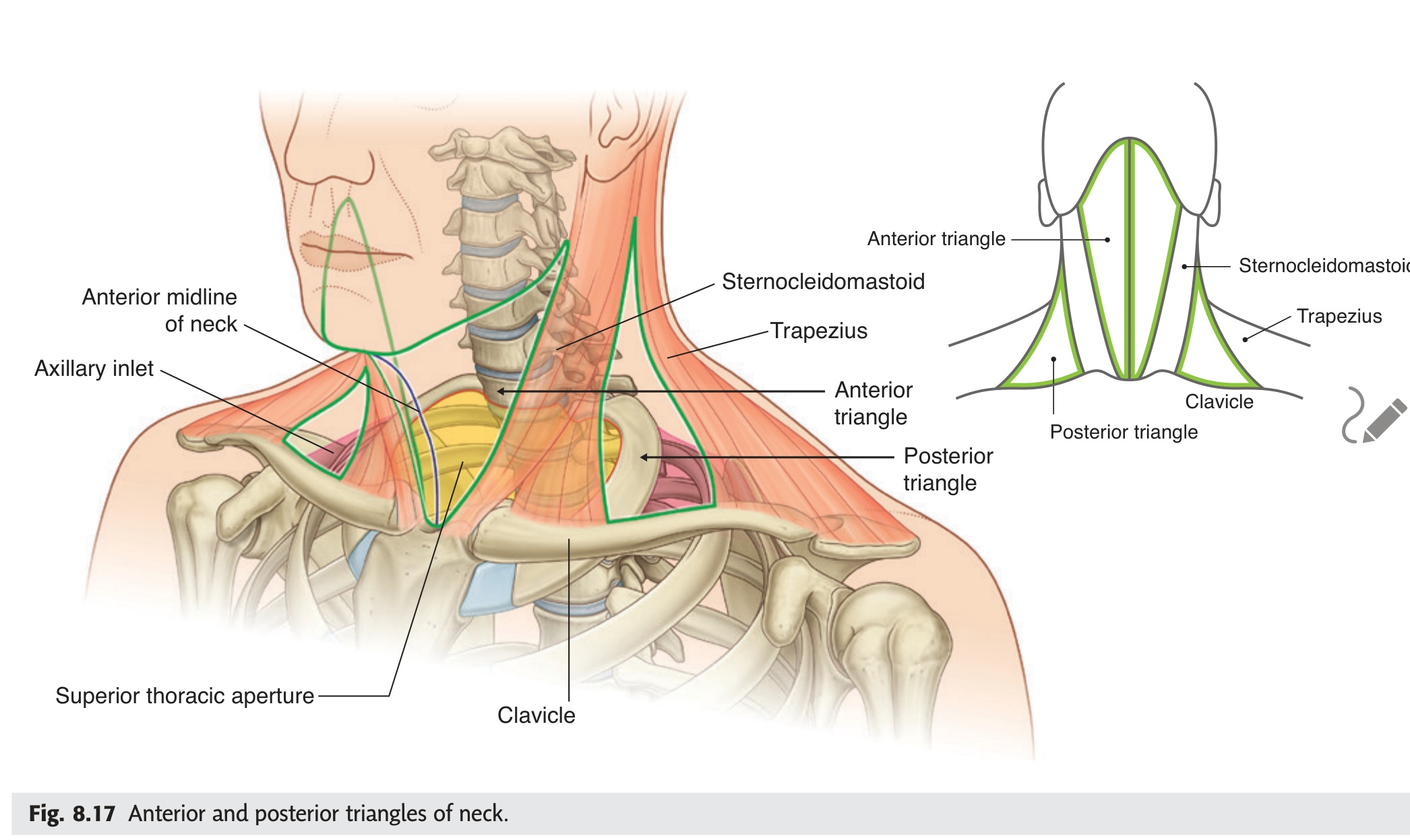
| Muscle | Origin → Insertion | Action | Nerve |
|---|---|---|---|
| Sternocleidomastoid (SCM) | Manubrium + medial clavicle → mastoid process + superior nuchal line | Bilateral: flex neck + extend head; Unilateral: ipsilateral lateral flexion + contralateral rotation | CN XI + C2–C3 |
| Anterior scalene | C3–C6 transverse processes → scalene tubercle rib 1 | Neck flexion; elevates rib 1 (accessory inspiration); brachial plexus exits between anterior + middle scalenes | Cervical nerve roots C4–C6 |
| Sternohyoid / Sternothyroid / Thyrohyoid | Sternum/thyroid → hyoid/thyroid cartilage | Depress hyoid and larynx (swallowing, phonation) | Ansa cervicalis (C1–C3) |
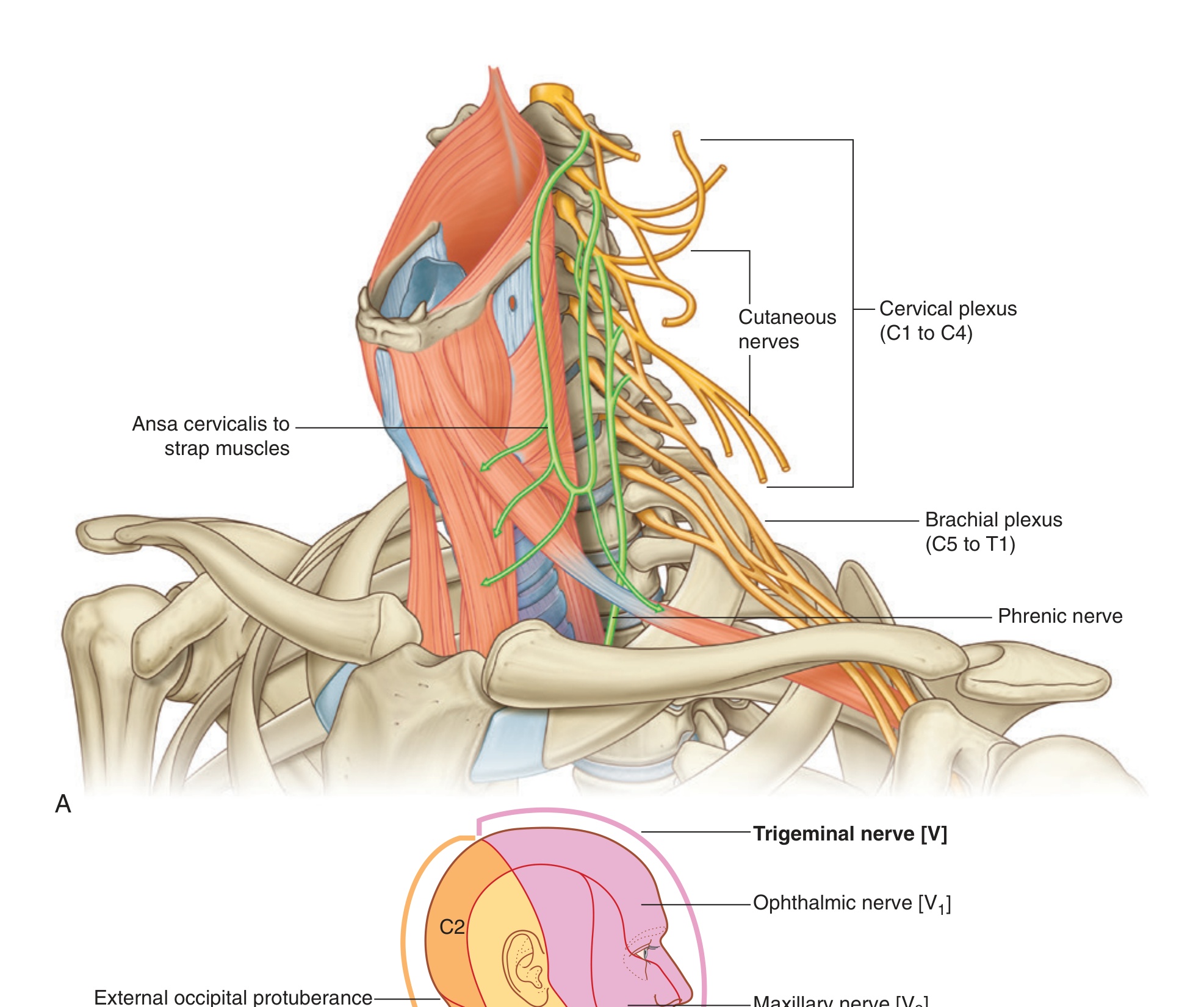
Congenital muscular torticollis: unilateral SCM fibrosis/shortening → head tilts ipsilaterally + rotates contralaterally. Possible cause: SCM compartment syndrome during traumatic delivery (haematoma → fibrosis). Treatment: physiotherapy stretching; surgical release if persistent. The "tumour" felt at birth in SCM is the fibrotic mass.
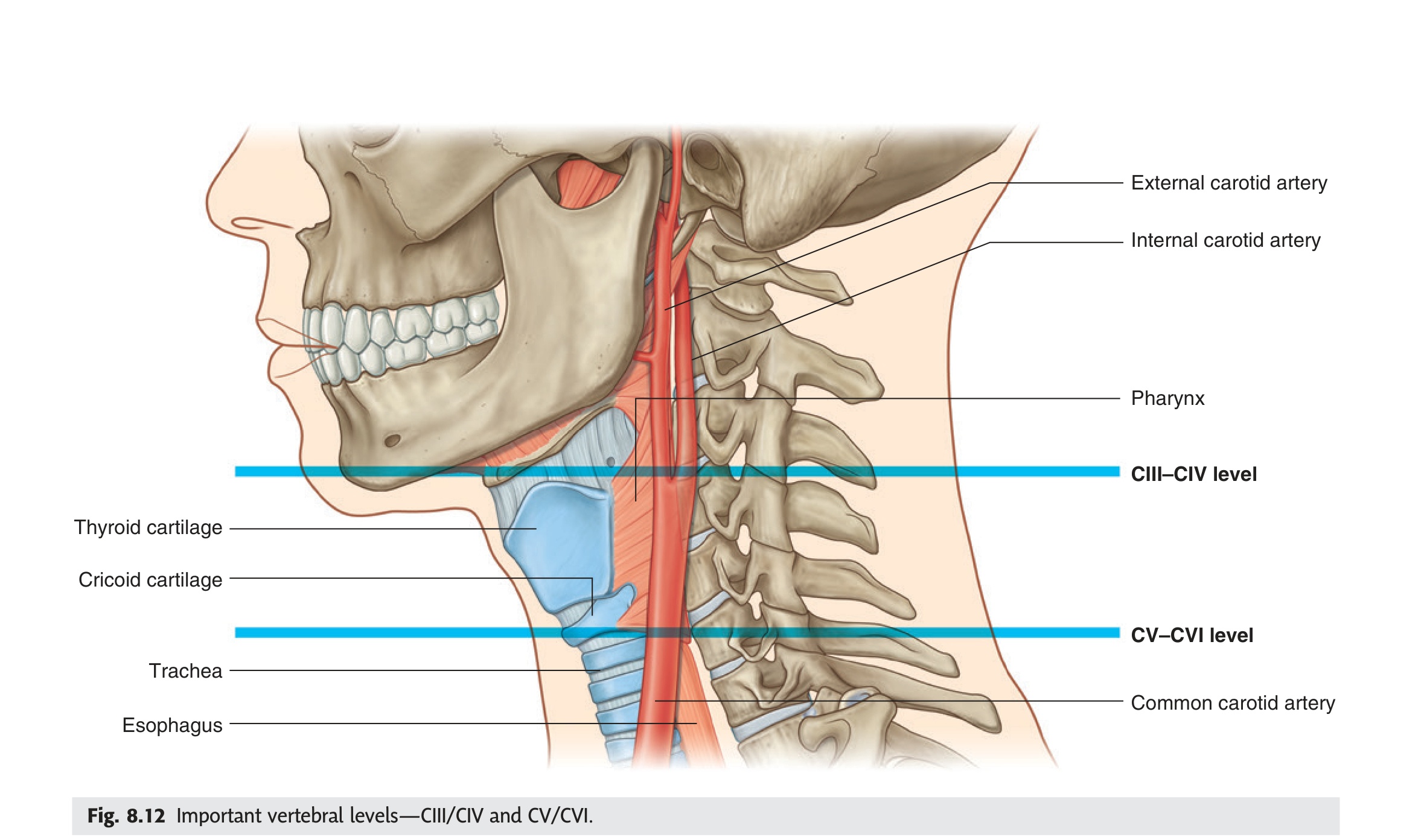
- What is the action of bilateral SCM contraction? Flexes the cervical spine AND extends the head at the atlanto-occipital joint — chin protrudes forward, not simply downward.
- Unilateral SCM contraction: describe the movement. Ipsilateral lateral flexion + contralateral rotation — the face turns AWAY from the contracting muscle.
- What is the dual nerve supply of SCM? CN XI (spinal accessory) for motor power + C2–C3 for proprioception.
- What structures pass through the interscalene triangle and what syndrome results from compression? Brachial plexus roots and subclavian artery — compression causes thoracic outlet syndrome (pain, paraesthesia in arm + vascular symptoms).
- What is the nerve supply to the infrahyoid strap muscles, with exception? Ansa cervicalis (C1–C3) for all strap muscles except thyrohyoid, which receives C1 fibres travelling with CN XII.
The suprahyoid muscles sit above the hyoid bone and form the muscular floor of the mouth. Their collective job is to move the hyoid and tongue during swallowing: when the hyoid is fixed by the infrahyoid muscles, the suprahyoids open the jaw; when the jaw is fixed, they elevate the hyoid and larynx — the upward jerk you can feel under your chin when you swallow is these muscles contracting. The mylohyoid forms a true diaphragm (the oral diaphragm), its right and left halves meeting at a midline raphe; abscesses in the floor of the mouth can spread either above or below it, changing both the clinical presentation and the drainage approach. The digastric has a fascinating dual nerve supply: its anterior belly is a 1st arch muscle (V3) and its posterior belly is a 2nd arch muscle (CN VII) — the intermediate tendon connecting them passes through a fibrous loop anchored to the hyoid, making it the only muscle with a pulley system in the neck.
| Muscle | Origin → Insertion | Action | Nerve |
|---|---|---|---|
| Mylohyoid | Mylohyoid line (body of mandible) → hyoid bone + median raphe | Elevates hyoid + tongue (swallowing); depresses mandible (when hyoid fixed); forms muscular floor of mouth | Mylohyoid nerve (branch of V3 — inferior alveolar) |
| Geniohyoid | Genial tubercle (inner mandible) → body of hyoid | Protrudes + elevates hyoid; widens pharynx for swallowing | C1 fibres via hypoglossal nerve (CN XII) |
| Digastric | Anterior belly: digastric fossa (mandible) → hyoid (intermediate tendon); Posterior belly: mastoid notch → hyoid (intermediate tendon) | Depresses mandible (opens jaw); elevates + retracts hyoid in swallowing | Anterior belly = V3 (mylohyoid nerve); Posterior belly = CN VII (facial) |
| Stylohyoid | Styloid process → body of hyoid (perforated by digastric tendon) | Elevates + retracts hyoid; elongates floor of mouth | CN VII (facial nerve) |
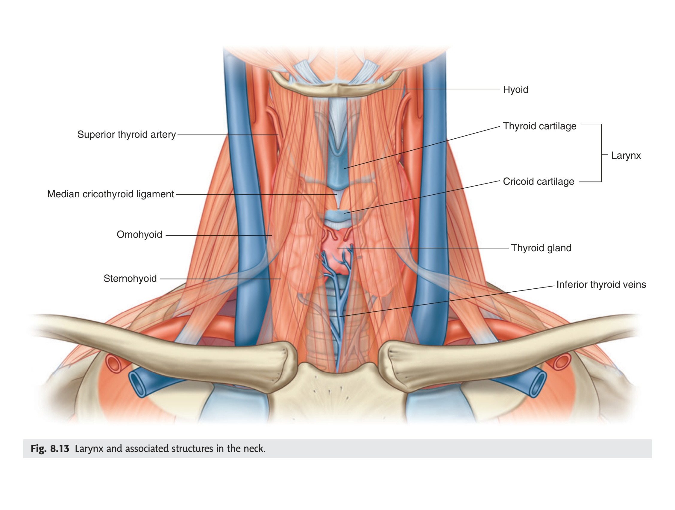
- Why does the digastric have two different nerve supplies? Anterior belly = 1st pharyngeal arch → V3 (mylohyoid nerve); posterior belly = 2nd arch → CN VII (facial nerve).
- What unique anatomical feature does the digastric's intermediate tendon have? It passes through a fibrous pulley loop anchored to the hyoid bone — the only neck muscle with a pulley system.
- What is the anatomical role of the mylohyoid? It forms the muscular floor (oral diaphragm) of the mouth; right and left halves meet at a midline raphe, separating the sublingual space above from the submandibular space below.
- Why is the mylohyoid clinically critical in floor-of-mouth infections? Infection spreading below it causes Ludwig's angina — a rapidly spreading submandibular cellulitis with life-threatening airway compromise.
- What is the nerve supply to geniohyoid, and what is the exam trap? C1 fibres carried by CN XII — geniohyoid is NOT a branch of XII itself; the hypoglossal nerve merely conveys the C1 fibres piggyback.
The soft palate separates the oral cavity from the nasopharynx. During swallowing it must rise and seal against the posterior pharyngeal wall to prevent food entering the nose — a failure here causes nasal regurgitation. Its five muscles are controlled almost entirely by the vagus nerve (CN X) via the pharyngeal plexus, with one critical exception: tensor veli palatini is innervated by V3 (medial pterygoid branch). Tensor has a unique job beyond palate stiffening — its tendon hooks around the pterygoid hamulus and fans out to open the cartilaginous Eustachian tube with each swallow and yawn. This is why children with cleft palate have chronic middle ear effusions ("glue ear"): the abnormal tensor cannot open the Eustachian tube properly. The palatoglossus is the one muscle in this group not innervated by CN XII despite having "glossus" in its name — it too is CN X, a classic exam trap.
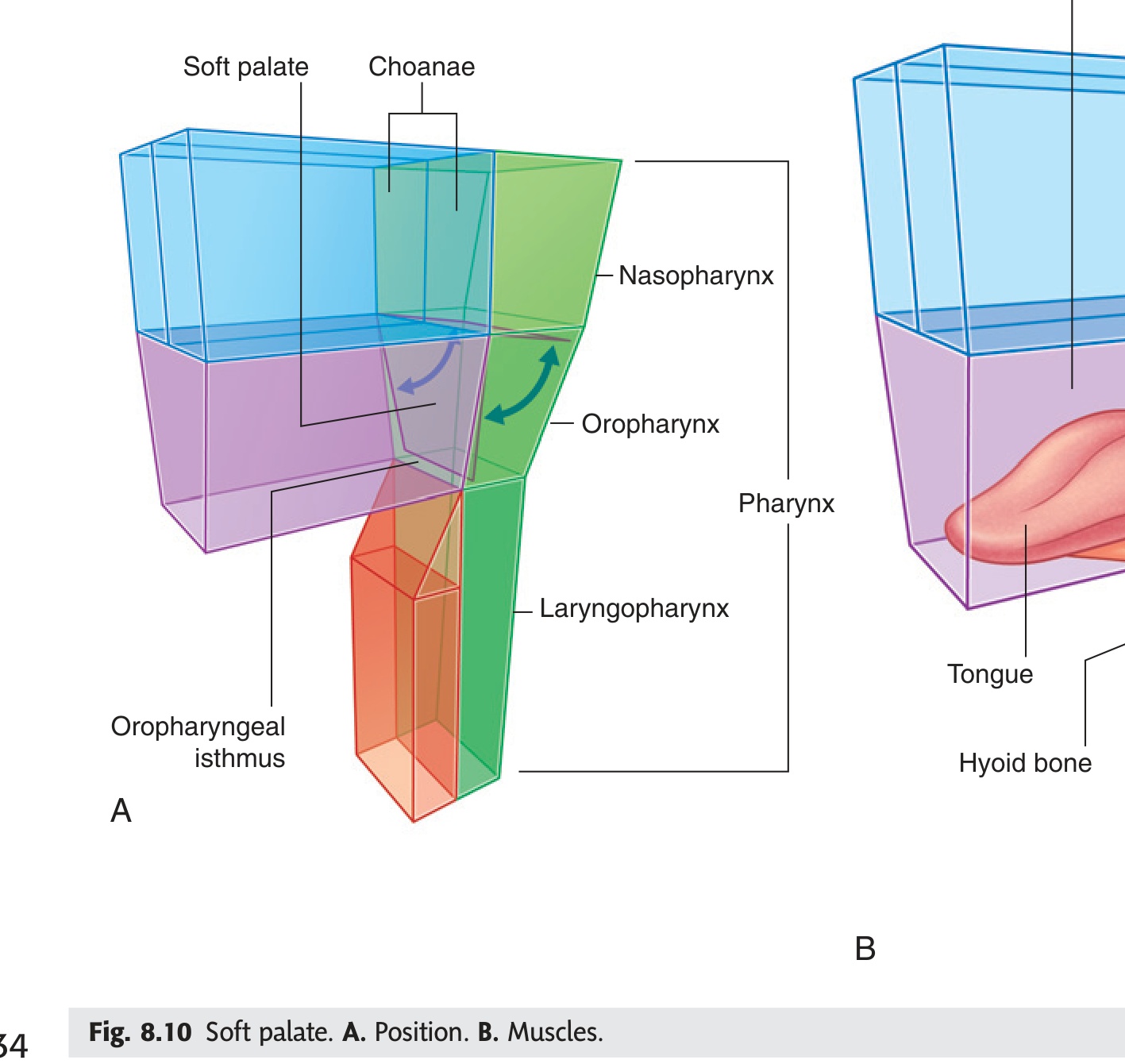
All soft palate muscles are supplied by the vagus nerve (CN X) via the pharyngeal plexus, EXCEPT tensor veli palatini (CN V3).
| Muscle | Action | Nerve |
|---|---|---|
| Tensor veli palatini | Tenses soft palate (stiffens it); opens the auditory (Eustachian) tube during swallowing and yawning | Medial pterygoid nerve (branch of V3) |
| Levator veli palatini | Elevates soft palate; closes nasopharyngeal isthmus during swallowing (prevents nasal regurgitation) | CN X (vagus) via pharyngeal plexus |
| Palatoglossus | Depresses soft palate OR elevates tongue; forms anterior pillar of fauces | CN X via pharyngeal plexus (NOTE: despite being named "glossus," it is NOT CN XII) |
| Palatopharyngeus | Elevates pharynx + larynx; forms posterior pillar of fauces; closes nasopharynx | CN X via pharyngeal plexus |
| Musculus uvulae | Elevates + shortens uvula; bulges mucosa of soft palate | CN X via pharyngeal plexus |
In unilateral CN X palsy (e.g., vagal nerve injury, posterior fossa tumour): uvula deviates AWAY from the side of the lesion (intact levator veli palatini on the healthy side pulls it across). Also: loss of the gag reflex (CN IX afferent, CN X efferent) on the affected side. Bilateral CN X palsy → nasal regurgitation, dysarthria, dysphagia.
- Which soft palate muscle is NOT innervated by CN X? → Tensor veli palatini (CN V3 — medial pterygoid branch)
- What is tensor veli palatini's function besides stiffening the palate? → Opens the Eustachian tube during swallowing/yawning
- Why do cleft palate children get glue ear? → Abnormal tensor veli palatini cannot open the Eustachian tube → middle ear effusion
- Palatoglossus is named "glossus" — which nerve supplies it? → CN X (pharyngeal plexus), NOT CN XII — classic exam trap
- Uvula deviates to which side in unilateral CN X palsy? → Away from the lesion (intact side pulls it across)
Ready to test yourself on Unit 04?
Practice MCQs, T/F and essay questions — timed and scored.
Pelvic Floor & Perineal Muscles ★
If you removed the pelvis and looked up from below, you would see the pelvic floor as a hammock of muscle spanning the bony ring, pierced by three openings: the urethra, vagina (in females), and anal canal. This hammock — the pelvic diaphragm — consists primarily of levator ani, a broad sheet with three named parts (pubococcygeus, puborectalis, iliococcygeus), plus coccygeus posteriorly. The critical part is puborectalis: its U-shaped sling wraps behind the anorectal junction, creating the anorectal angle of roughly 90°. Faecal continence depends on this angle — the kink means that any increase in intra-abdominal pressure (coughing, straining) compresses the anorectal junction closed rather than forcing it open. During defecation, puborectalis relaxes and the angle straightens to 135°, allowing passage. Damage to puborectalis from obstetric injury or pudendal neuropathy abolishes this angle and causes faecal incontinence even with intact sphincters.
The pelvic diaphragm is the muscular floor of the pelvic cavity, forming a funnel-shaped sheet. It consists of levator ani (three parts) and coccygeus posteriorly.
| Muscle | Origin → Insertion | Action | Nerve |
|---|---|---|---|
| Pubococcygeus (part of levator ani) | Pubis (body) → coccyx + perineal body | Supports pelvic viscera; elevates pelvic floor; constricts anorectal junction | S3–S4 (perineal branch) + inferior rectal nerve |
| Puborectalis (part of levator ani) | Pubis → loops behind anorectal junction (U-shaped sling) | Creates anorectal angle (~90°) — essential for faecal continence. Relaxes during defecation to straighten anorectal canal. | S3–S4 |
| Iliococcygeus (part of levator ani) | Tendinous arch (obturator fascia) → coccyx + anococcygeal raphe | Supports pelvic floor; elevates anorectal canal | S3–S4 |
| Coccygeus (ischiococcygeus) | Ischial spine → coccyx + lower sacrum | Supports pelvic floor; flexes coccyx; same plane as sacrospinous ligament | S4–S5 |
Pelvic organ prolapse: weakening of levator ani (multiparity, menopause, chronic straining) → uterine prolapse, cystocoele (anterior vaginal wall/bladder), rectocoele (posterior/rectum). Faecal incontinence: puborectalis damage (obstetric tear, pudendal neuropathy) → loss of anorectal angle. Hirschsprung disease: absence of ganglion cells in internal anal sphincter → failure of relaxation. Episiotomy: surgical cut through skin + perineal membrane + bulbospongiosus to widen vaginal outlet during delivery — heals in layers.
- What three structures pierce the pelvic diaphragm? → Urethra, vagina (females), anal canal
- Puborectalis creates what angle — and why does it maintain continence? → ~90° anorectal angle; increased abdominal pressure compresses the junction closed rather than open
- Which nerve supplies most of levator ani? → S3–S4 (perineal branch of pudendal + direct branches)
- Coccygeus lies in the same plane as which ligament? → Sacrospinous ligament
- What causes pelvic organ prolapse structurally? → Weakening of levator ani (multiparity, menopause, chronic straining) → uterine, bladder (cystocoele), or rectal (rectocoele) descent
The perineum is the diamond-shaped region below the pelvic diaphragm. A transverse line between the two ischial tuberosities divides it into the urogenital triangle anteriorly (urethra and vagina/penis) and the anal triangle posteriorly. The perineal body (central tendon of the perineum) is the fibromuscular knot at the centre of this region where eight muscles converge; it is the anchor that holds the perineum together. Obstetric tears are graded by how far back through this region they extend: a 4th-degree tear reaches the internal anal sphincter and requires careful surgical layered repair. The external urethral sphincter is skeletal muscle under voluntary pudendal nerve control — this is why a spinal cord injury at or above S2 causes spastic retention (sphincter cannot relax), while a lower motor neuron lesion causes stress incontinence (sphincter cannot contract). Ischiocavernosus and bulbospongiosus drive erection and ejaculation by compressing the venous outflow of the corpora — pudendal nerve anaesthetic block for perineal surgery will temporarily abolish these reflexes.
| Muscle | Location | Action | Nerve |
|---|---|---|---|
| External urethral sphincter | Surrounds membranous urethra (deep perineal pouch) | Voluntary urinary continence | Pudendal nerve (S2–S4) — perineal branch |
| External anal sphincter | Surrounds anal canal (subcutaneous + superficial + deep parts) | Voluntary faecal continence | Inferior rectal nerve (branch of pudendal S2–S4) |
| Bulbospongiosus | Perineal body → male: corpus spongiosum; female: clitoris | Male: empties urethra, erection; Female: compresses vaginal orifice, clitoral erection | Pudendal nerve (perineal branch) |
| Ischiocavernosus | Ischial tuberosity/ramus → corpus cavernosum | Maintains erection by compressing crus (prevents venous drainage) | Pudendal nerve |
| Superficial + Deep transverse perinei | Ischial ramus → perineal body | Stabilises perineal body (central tendon of perineum — insertion point for 8 muscles) | Pudendal nerve |
External anal sphincter (EAS): skeletal muscle; voluntary; pudendal nerve (S2–S4). Damaged in obstetric third/fourth degree tears → faecal incontinence. Repaired surgically (sphincteroplasty).
- What is the perineal body and why does it matter? → Central fibromuscular tendon where 8 perineal muscles meet; anchor of perineal integrity — obstetric tears disrupt it
- External urethral sphincter: nerve supply and muscle type? → Pudendal nerve (S2–S4); skeletal (voluntary) muscle
- Upper vs lower motor neuron bladder: which causes retention vs incontinence? → UMN (spinal cord injury ≥S2) → spastic retention; LMN (cauda equina) → flaccid stress incontinence
- Internal anal sphincter: smooth or skeletal, voluntary or involuntary? → Smooth, involuntary; autonomic supply (sympathetic = contraction; parasympathetic = relaxation)
- Hirschsprung disease: what is absent and what is the result? → Absent ganglion cells in IAS (and above) → permanent contraction → functional obstruction