The Skull & Joints
Overview & Skull Bones
The skull is not a solid dome — it is a precisely engineered composite of 22 interlocking bones, each contributing specific structural or functional properties. The brain gets protection from the neurocranium, while the face and airways are shaped by the viscerocranium — these two divisions develop separately, with the neurocranium from mesenchyme and cartilage and the viscerocranium largely from neural crest cells. The classic examination trap here is the inferior nasal concha: unlike the superior and middle conchae which are genuine extensions of the ethmoid, the inferior concha is an entirely independent bone of the viscerocranium.
The skull is the bony framework of the head composed of 22 bones divided into the neurocranium (8 bones — protects the brain) and the viscerocranium (14 bones — facial skeleton forming orbits, nasal cavity, and jaws).
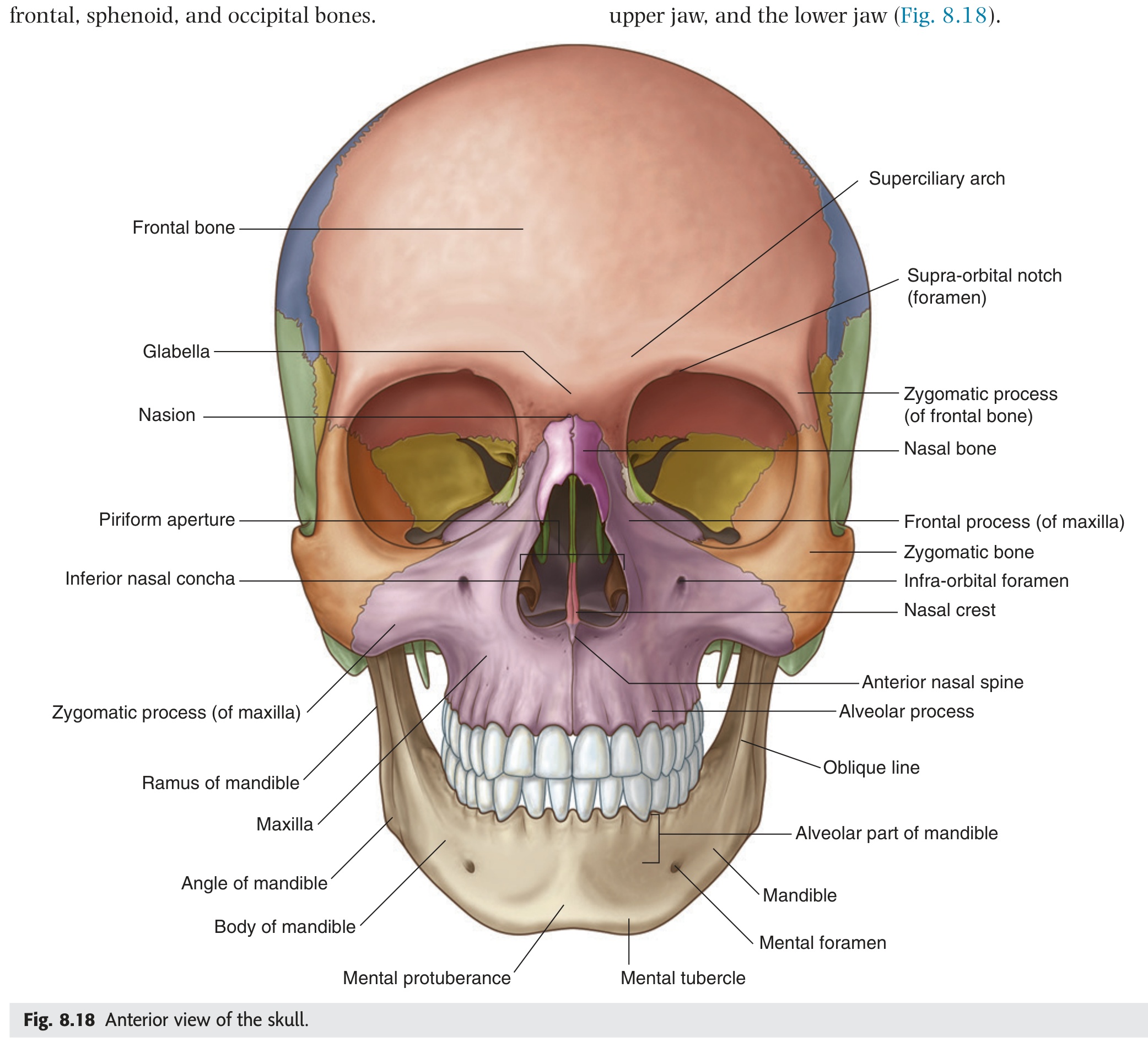
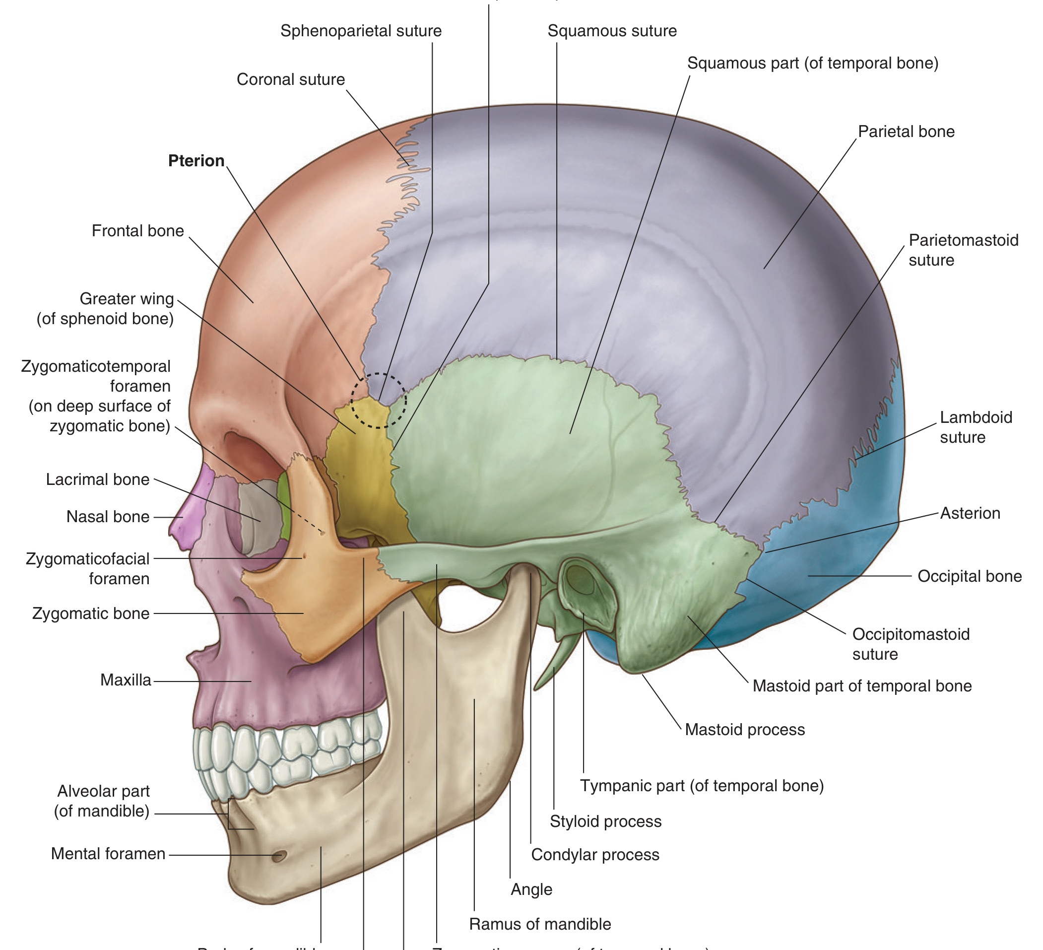
| Bone | Count | Position / Key Feature |
|---|---|---|
| Frontal | 1 | Forehead, orbital roof, anterior cranial fossa floor |
| Parietal | 2 | Upper lateral walls and vault; sagittal suture between them |
| Occipital | 1 | Posterior/inferior skull; foramen magnum |
| Temporal | 2 | Lower lateral skull; houses inner ear |
| Sphenoid | 1 | Central skull base; bat-shaped; sella turcica |
| Ethmoid | 1 | Anterior cranial fossa floor; cribriform plate; nasal septum |
"Flat Plate Of True Skull Encasing Brains"
Frontal · Parietal ×2 · Occipital · Temporal ×2 · Sphenoid · Ethmoid (count: 1+2+1+2+1+1 = 8)
- Q: Name the 8 neurocranial bones. — Frontal, 2× parietal, occipital, 2× temporal, sphenoid, ethmoid (mnemonic: "Flat Plate Of True Skull Encasing Brains").
- Q: Is the inferior nasal concha part of the ethmoid? — No. It is an independent viscerocranial bone; only the superior and middle conchae belong to the ethmoid.
- Q: Which bone articulates with all other cranial bones? — Sphenoid — the "keystone" of the skull base.
- Q: What is the difference between neurocranium and viscerocranium? — Neurocranium (8 bones) protects the brain; viscerocranium (14 bones) forms the facial skeleton — orbits, nasal cavity, jaws.
- Q: How many bones in the viscerocranium and name three. — 14 bones; includes mandible, 2× maxilla, 2× zygomatic, 2× nasal, 2× lacrimal, vomer, etc.
Sutures & Fontanelles
Sutures begin as fibrous joints — narrow seams of connective tissue that allow the skull to flex during birth and to expand rapidly in the first two years as the brain triples in volume. By adulthood most fuse into rigid synarthroses, but their landmarks persist as critical surgical reference points. The coronal suture runs across the top of the head; bregma (where coronal meets sagittal) is the anterior fontanelle site in the neonate — the soft pulsating spot a parent feels on an infant's head — and remains a neurosurgical landmark. Premature fusion of any suture (craniosynostosis) forces the growing brain to expand in the only remaining direction, producing characteristic skull deformities that neurosurgeons must correct in infancy.
Sutures are fibrous joints between skull bones allowing moulding in infancy and gradually ossifying in adulthood. They are immovable (synarthroses) in adults.
| Suture | Bones Joined | Junction Landmark |
|---|---|---|
| Coronal | Frontal + both Parietals | Bregma — junction with sagittal suture (anterior fontanelle site) |
| Sagittal | Right Parietal + Left Parietal | Midline; runs from bregma → lambda |
| Lambdoid | Both Parietals + Occipital | Lambda — junction with sagittal suture (posterior fontanelle site) |
| Squamous | Parietal + Temporal | Weakest skull wall — overlies middle meningeal artery |
| Sphenoparietal | Sphenoid + Parietal | Part of Pterion region (anterolateral skull) |
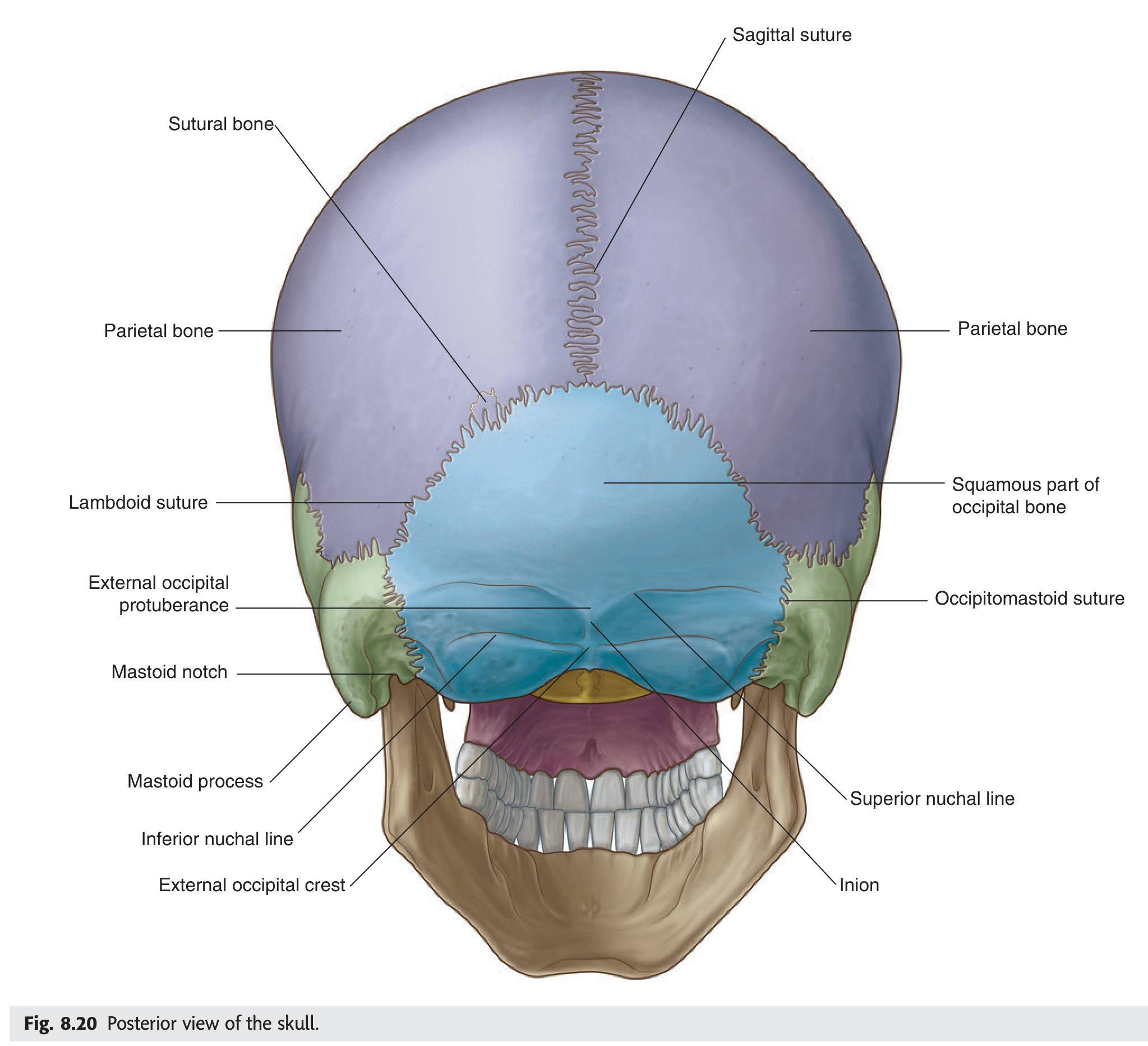
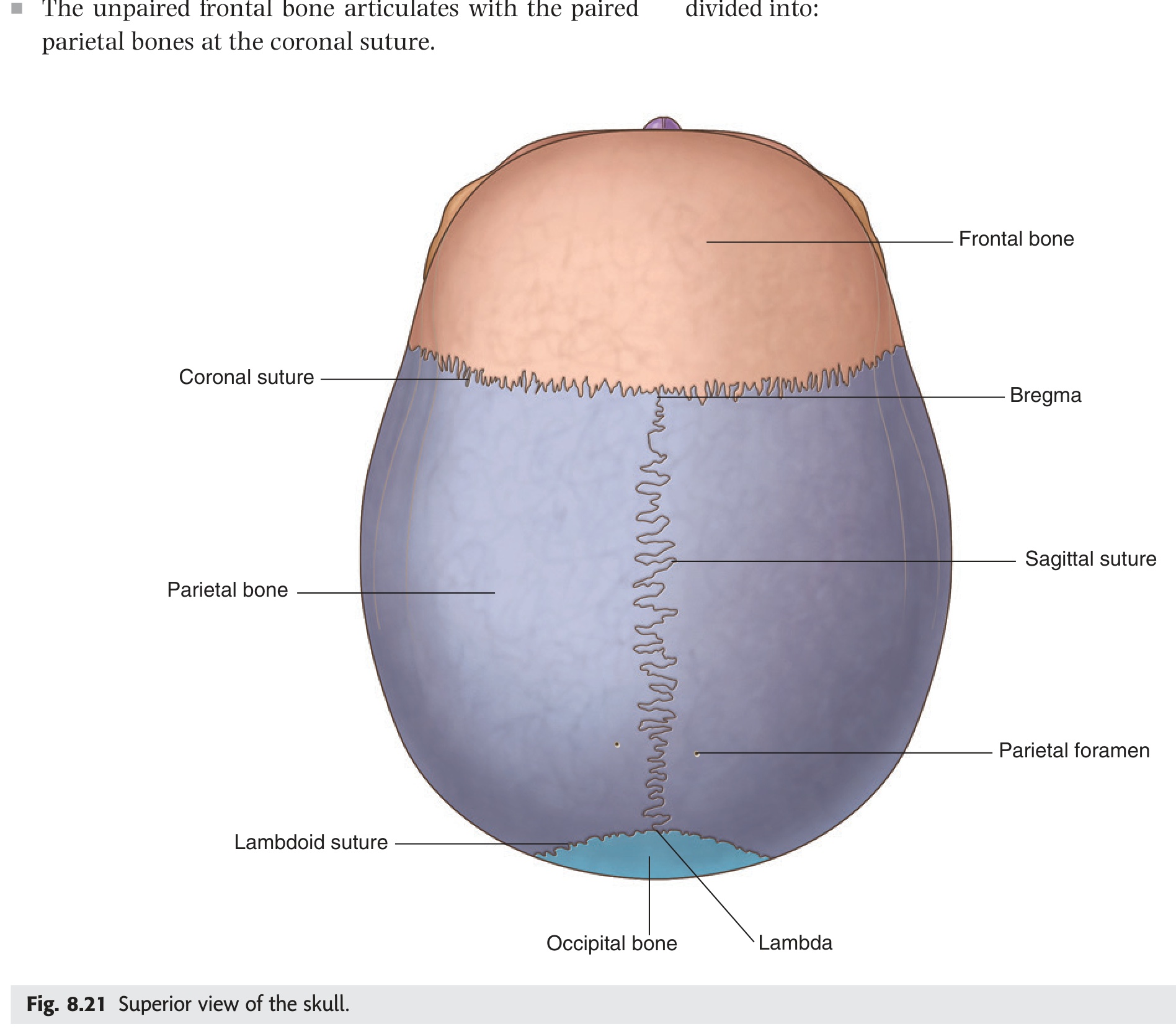
- What landmark lies at the junction of the coronal and sagittal sutures? Bregma — the site of the anterior fontanelle in the neonate.
- What landmark lies at the junction of the sagittal and lambdoid sutures? Lambda — the site of the posterior fontanelle.
- Which suture overlies the middle meningeal artery and is the weakest skull wall? Squamous suture (parietal meets temporal) — also the region of pterion.
- What is craniosynostosis and what is its consequence? Premature fusion of one or more sutures — forces the growing brain to expand in the only remaining direction, causing characteristic skull deformity.
- Which sutures meet at pterion? Frontal, parietal, temporal, and sphenoid — forming an H-shaped junction over the thinnest cranial vault point.
In a newborn, the skull bones haven't fully fused — six membranous gaps called fontanelles allow the rigid skull to compress and overlap during passage through the birth canal, then spring apart afterward. Feel the anterior fontanelle on a crying infant: it bulges with each heartbeat and rises when the infant is supine — a physiological finding that becomes pathological if persistently tense, signalling raised intracranial pressure from meningitis, hydrocephalus, or intracranial haemorrhage. The posterior fontanelle closes within three months, but the large diamond-shaped anterior fontanelle remains open until about 18 months, providing a crucial acoustic window for neonatal brain ultrasound scanning when it is still open.
Fontanelles are membranous gaps between incompletely ossified skull bones in the neonate, allowing skull moulding during birth and accommodating rapid brain growth in infancy.
| Fontanelle | Location | Closes | Clinical Note |
|---|---|---|---|
| Anterior (largest; diamond) | Bregma — coronal + sagittal | 18 months | Bulging = raised ICP; sunken = dehydration |
| Posterior (triangular) | Lambda — lambdoid + sagittal | 2–3 months | Neonatal ultrasound window |
| Sphenoidal (anterolateral) | Pterion region | 2–3 months | — |
| Mastoid (posterolateral) | Asterion region | 12–18 months | — |
- Q: When does the anterior fontanelle close? — ~18 months; the posterior fontanelle closes at 2–3 months.
- Q: Where is bregma? — Junction of coronal and sagittal sutures; site of anterior fontanelle in the neonate.
- Q: Bulging fontanelle = ? — Raised intracranial pressure (meningitis, hydrocephalus). Sunken = dehydration.
- Q: Which fontanelle provides the neonatal brain ultrasound window? — Anterior fontanelle (largest; still open at birth scan).
Pterion is the danger zone of the lateral skull — the thinnest point of the entire cranial vault, where four bones converge in an H-shaped sutural junction with the middle meningeal artery running in a groove immediately beneath. A cricket ball, steering wheel, or assault blow to the temple can fracture this paper-thin point without requiring enormous force; what follows is arterial bleeding — brisk, filling the extradural space with a rapidly expanding haematoma. The characteristic "lucid interval" — a brief period of consciousness between the initial LOC and secondary deterioration — occurs because arterial blood accumulates steadily and must compress enough cortical tissue before herniation begins, giving emergency teams a critical window for intervention if the diagnosis is made promptly.
Pterion is the H-shaped sutural junction on the anterolateral skull where four bones meet: the frontal, parietal, squamous temporal, and greater wing of sphenoid. It is the thinnest part of the skull.
Deep to Pterion: The anterior branch of the middle meningeal artery (branch of the maxillary artery, entering via foramen spinosum) runs in a groove on the inner skull surface at this point.
A blow to the temple fractures the thin pterion → lacerates the middle meningeal artery → arterial blood accumulates in the extradural space (between skull and dura).
Classic presentation: Initial LOC → lucid interval (brief recovery) → rapid deterioration as haematoma expands → transtentorial herniation → death if untreated.
CT: Biconvex (lens-shaped) hyperdense collection. Does NOT cross suture lines.
Treatment: Emergency burr hole or craniotomy + haematoma evacuation.
- Q: Four bones at pterion? — Frontal, parietal, squamous temporal, greater wing of sphenoid.
- Q: What artery is torn, and where does it enter the skull? — Anterior branch of middle meningeal artery; enters via foramen spinosum.
- Q: CT appearance of extradural haematoma? — Biconvex (lens-shaped) hyperdense collection; does NOT cross suture lines.
- Q: What is the lucid interval? — Brief recovery period between initial LOC and secondary deterioration as arterial haematoma steadily expands.
- Q: Treatment of extradural haematoma? — Emergency craniotomy or burr hole + haematoma evacuation.
Temporal Bone ★★
The temporal bone is perhaps the most anatomically complex bone in the body — a paired structure that in its petrous part alone houses the cochlea, three semicircular canals, the facial nerve canal, and the carotid canal, all within a few cubic centimetres of dense "rock-like" bone. Understanding its four parts matters clinically because each generates distinct pathology: a petrous fracture can shear CN VII (facial palsy) or CN VIII (sensorineural deafness), cause CSF otorrhoea, or damage the ICA; mastoid infection tracks from the middle ear through mastoid air cells to the sigmoid sinus (risk of sinus thrombosis and meningitis); and the stylomastoid foramen — where the facial nerve exits between styloid and mastoid — is the surgical target for CN VII decompression in severe Bell's palsy.
The temporal bone is a complex paired bone forming the lower lateral cranial wall. It houses the organ of hearing (cochlea) and organ of balance (semicircular canals) within its dense petrous part, and transmits the facial nerve (CN VII).
- Squamous part — flat, lateral skull wall; zygomatic process → zygomatic arch; mandibular fossa (articular surface for TMJ) on its inferior surface.
- Petrous part — pyramidal, dense "rock-like" bone projecting anteromedially; contains cochlea + semicircular canals; internal acoustic meatus (IAM) transmits CN VII + CN VIII + labyrinthine artery.
- Mastoid part — posterior; mastoid process (palpable behind ear; SCM attachment); mastoid air cells (connected to middle ear); stylomastoid foramen between styloid and mastoid processes — CN VII exits here.
- Tympanic part — C-shaped plate forming the anterior, floor, and posterior walls of the external acoustic meatus; gives the styloid process (stylohyoid, styloglossus, stylopharyngeus muscles).
Bell's palsy: Inflammation of CN VII within the narrow facial canal (petrous bone) → LMN facial nerve palsy (ipsilateral complete facial weakness including forehead). Nerve exits at stylomastoid foramen.
Mastoiditis: Spread of otitis media to mastoid air cells → mastoid tenderness + displaced pinna; risk of sigmoid sinus thrombosis, meningitis.
Acoustic neuroma (vestibular schwannoma): At IAM → CN VIII symptoms (tinnitus, sensorineural hearing loss) → later CN VII (facial weakness), CN V (facial numbness) as tumour enlarges into CPA.
- Q: What three structures pass through the internal acoustic meatus? — CN VII, CN VIII, and the labyrinthine (internal auditory) artery.
- Q: Which temporal bone part is "rock-like" and houses the cochlea? — Petrous part.
- Q: Where does CN VII exit the skull? — Stylomastoid foramen (between styloid and mastoid processes).
- Q: What are mastoid air cells connected to, and why does this matter? — Connected to the middle ear; otitis media can spread to mastoid air cells → mastoiditis → risk of sigmoid sinus thrombosis and meningitis.
- Q: Earliest symptom of a vestibular schwannoma at the IAM? — Sensorineural hearing loss / tinnitus (CN VIII affected first); CN VII palsy is a later sign as tumour enlarges.
Sphenoid Bone ★★
If you could make the skull transparent and look at only one bone that touches every other cranial bone, you would see the sphenoid — a solitary bat-shaped structure spanning the entire skull base and forming the keystone of the cranial floor. Its body cradles the pituitary gland in the sella turcica above the sphenoidal sinus below — a geography exploited every time a neurosurgeon approaches the pituitary through a trans-sphenoidal route, entering the nose, crossing the sphenoidal sinus, and opening directly into the sella without touching the brain. The greater wings carry three foramina in sequence — rotundum (V2), ovale (V3), spinosum (middle meningeal artery) — so a midface fracture running through the greater wing can simultaneously knock out jaw sensation and trigger an intracranial bleed.
The sphenoid is an unpaired, bat-shaped bone occupying the central skull base. It is unique in articulating with all other cranial bones. Its body contains the sphenoidal sinuses and the sella turcica housing the pituitary gland.
- Body — central cube; contains sphenoidal sinuses; superior surface bears the sella turcica (hypophyseal fossa housing pituitary, bounded by tuberculum sellae anteriorly and dorsum sellae posteriorly).
- Greater wings (×2) — lateral extensions forming middle cranial fossa floor; three key foramina: foramen rotundum (V2), foramen ovale (V3), foramen spinosum (middle meningeal a.).
- Lesser wings (×2) — horizontal plates; posterior floor of anterior cranial fossa; the optic canal lies between lesser wing root and body (CN II + ophthalmic artery); superior orbital fissure between lesser and greater wings.
- Pterygoid processes (×2) — project inferiorly; medial and lateral pterygoid plates; medial pterygoid, lateral pterygoid, tensor veli palatini muscles attach.
The pituitary gland sits in the sella turcica, directly above the sphenoidal sinus (used for transsphenoidal surgical approach). A pituitary macroadenoma expanding superiorly compresses the optic chiasm → bitemporal hemianopia (loss of both temporal visual fields). Lateral expansion → cavernous sinus invasion → CN III, IV, VI palsies (ophthalmoplegia) ± Horner's syndrome.
- Q: What is the sella turcica and what does it contain? — A concave depression on the superior sphenoid body; contains the pituitary gland. Bounded by tuberculum sellae (anterior) and dorsum sellae (posterior).
- Q: Name the three foramina in the greater wing and their contents. — Rotundum → V2; Ovale → V3 + lesser petrosal nerve; Spinosum → middle meningeal artery.
- Q: What passes through the optic canal? — CN II (optic nerve) + ophthalmic artery (branch of ICA). Located between lesser wing root and body.
- Q: Pituitary macroadenoma expanding superiorly compresses what structure, causing what visual defect? — Optic chiasm → bitemporal hemianopia (loss of both temporal fields).
- Q: Which bone articulates with all other cranial bones? — Sphenoid — hence "keystone" of the skull base.
Frontal · Parietal · Occipital · Ethmoid
The frontal, parietal, occipital, and ethmoid bones are easily dismissed as "the rest of the skull," but each has high-yield clinical anatomy attached to it. The ethmoid is the most fragile and most surgical — its cribriform plate is the slimmest partition between brain and nose, and tearing it during an anterior fossa fracture or endoscopic sinus surgery produces CSF rhinorrhoea (glucose-positive nasal drip confirmed by β2-transferrin). The occipital bone encloses the foramen magnum, through which the medulla continues as the spinal cord — tonsillar herniation compressing here is an immediate threat to respiratory drive. The external occipital protuberance (inion) is not just a palpable landmark; internally it overlies the confluence of dural venous sinuses, making it the neurosurgeon's navigation point for posterior fossa approach.
• Supraorbital notch/foramen — supraorbital nerve (V1) + artery
• Glabella — smooth midline elevation between superciliary arches
• Frontal sinuses — within superciliary arch; drain to middle nasal meatus
• Nasion — junction with nasal bones; surface landmark
• Superior temporal line — upper limit of temporalis fascia
• Inferior temporal line — upper attachment of temporalis muscle
• Parietal foramen — emissary vein (connects scalp to superior sagittal sinus)
• Inner surface grooved by middle meningeal artery branches
• Foramen magnum — spinal cord + meninges, vertebral arteries, anterior + posterior spinal arteries, CN XI spinal roots
• Occipital condyles — articulate with atlas (atlanto-occipital joint; nodding movement)
• External occipital protuberance (inion) — palpable midline landmark; confluence of sinuses internally
• Hypoglossal canal — above occipital condyle; CN XII
• Superior nuchal line — trapezius, SCM, splenius capitis attachment
• Cribriform plate — horizontal; roof of nasal cavity; multiple foramina for CN I (olfactory) filaments
• Crista galli — midline projection superior to cribriform; attachment of falx cerebri
• Perpendicular plate — upper nasal septum
• Superior + middle nasal conchae — lateral nasal wall (inferior concha = separate bone)
Fracture of cribriform plate/orbital plates → raccoon eyes (periorbital ecchymosis), CSF rhinorrhoea (CSF drains into nasal cavity — glucose-positive; β2-transferrin confirms), anosmia (CN I shearing at cribriform foramina).
- Q: Clinical triad of anterior cranial fossa fracture? — Raccoon eyes (periorbital ecchymosis), CSF rhinorrhoea, anosmia (CN I shearing at cribriform foramina).
- Q: What passes through the cribriform foramina? — CN I olfactory filaments (from nasal mucosa to olfactory bulb in anterior cranial fossa).
- Q: What is the crista galli? — Midline ethmoid projection superior to the cribriform plate; attachment point of the falx cerebri.
- Q: What is at the external occipital protuberance (inion) on the internal surface? — Confluence of dural venous sinuses (superior sagittal, straight, transverse sinuses meet here).
- Q: What passes through the hypoglossal canal? — CN XII (hypoglossal nerve); supplies all intrinsic tongue muscles except palatoglossus.
Internal Cranial Fossae ★★★
Think of the intracranial floor as three stepped tiers — the frontal lobes sit highest in the anterior fossa, the temporal lobes and pituitary occupy the middle fossa, and the cerebellum-brainstem unit fills the lowest posterior fossa. Each fossa has its own floor bones and therefore its own characteristic fracture patterns and nerve injuries: a middle fossa fracture can simultaneously injure V2 (foramen rotundum), V3 (foramen ovale), the ICA (carotid canal), and trigger a middle meningeal artery bleed (foramen spinosum) depending on the fracture line. The posterior fossa is the most surgically treacherous — masses expanding here compress the brainstem and cerebellum directly, and herniation through the foramen magnum is rapidly fatal.
The internal skull base is divided into three stepped fossae — anterior (highest), middle, posterior (lowest) — each housing different parts of the brain and transmitting specific cranial nerves and vessels through their foramina.
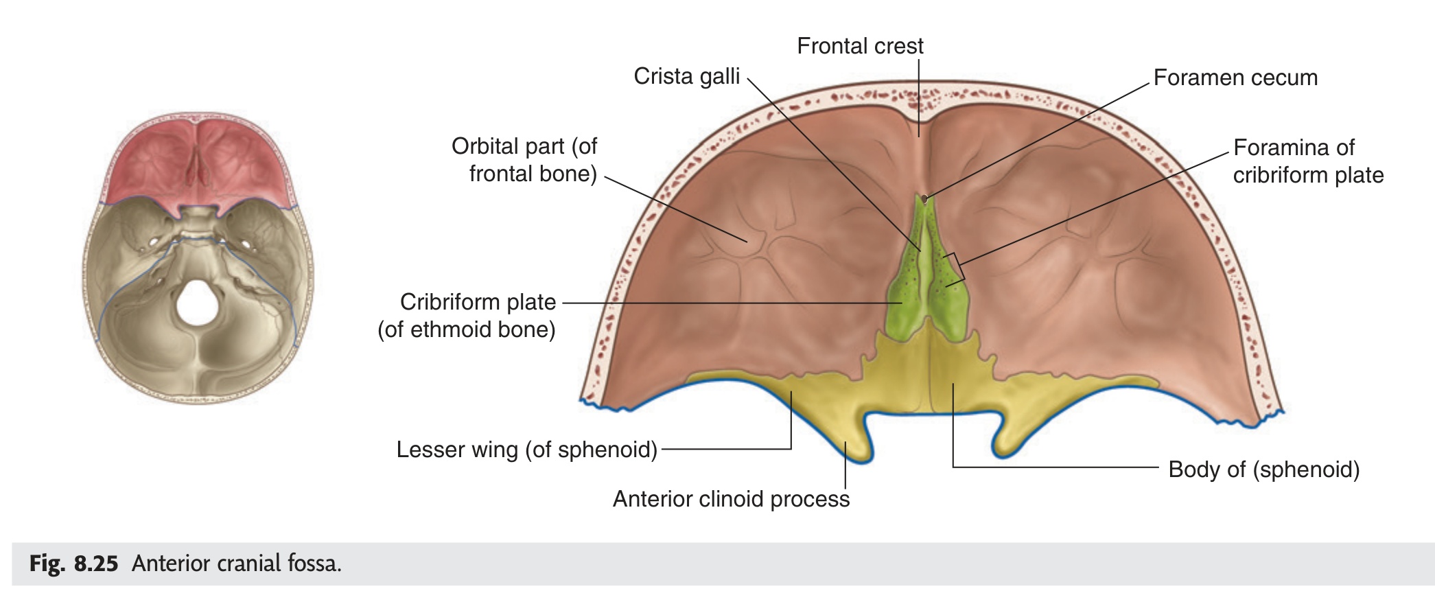
| Fossa | Floor Bones | Brain Contents | Key Foramina & Contents |
|---|---|---|---|
| Anterior cranial fossa | Orbital plates of frontal · Cribriform plate of ethmoid · Lesser wings + anterior body of sphenoid | Frontal lobes · Olfactory bulbs & tracts | Cribriform foramina (CN I) · Foramen cecum (emissary vein) · Optic canal (CN II + ophthalmic a.) |
| Middle cranial fossa | Greater wings of sphenoid · Squamous temporal · Anterior face of petrous temporal · Sphenoid body (sella) | Temporal lobes · Pituitary · Cavernous sinus | Optic canal (CN II) · SOF (III, IV, V1, VI) · Foramen rotundum (V2) · Foramen ovale (V3) · Foramen spinosum (middle meningeal a.) · Foramen lacerum (fibrocartilage plug; ICA passes OVER) |
| Posterior cranial fossa | Petrous + mastoid temporal · Occipital bone · Basilar sphenoid | Cerebellum · Pons · Medulla oblongata | IAM (CN VII + VIII) · Jugular foramen (IX, X, XI + IJV) · Hypoglossal canal (CN XII) · Foramen magnum (spinal cord, vertebral aa., CN XI spinal) |
SOF → III, IV, V1, VI
Foramen Rotundum → V2 (Round, two letters in "Ro")
Foramen Ovale → V3 + lesser petrosal
Foramen Spinosum → Middle meningeal Artery
Foramen Lacerum → Filled with cartilage; ICA passes over, not through
- Q: Name the three fossae and what each contains. — Anterior (frontal lobes, olfactory bulbs); Middle (temporal lobes, pituitary, cavernous sinus); Posterior (cerebellum, pons, medulla).
- Q: What passes through the jugular foramen? — CN IX, X, XI (cranial root) + sigmoid sinus → internal jugular vein.
- Q: What is special about foramen lacerum? — Filled with fibrocartilage in life; the ICA passes OVER it (not through it). Classic MCQ trap.
- Q: Name the four structures that pass through the SOF. — CN III, IV, V1, VI + superior/inferior ophthalmic veins.
- Q: What passes through the IAM? — CN VII + CN VIII + labyrinthine artery. Located in the posterior cranial fossa (petrous temporal).
External Cranial Base & Foramina
The external base of the skull is the undersurface you see when you remove the brain and look down — a mosaic of foramina through which every cranial nerve and the great vessels pass. Knowing the exact foramen for each nerve is not academic trivia; it is how you localise a basal skull lesion before imaging. A patient with right-sided tongue weakness (CN XII lesion) has pathology at the right hypoglossal canal; combined CN IX, X, XI palsy on one side points to the jugular foramen — a "jugular foramen syndrome" caused by a glomus jugulare tumour or metastasis. The petrous bone here is also traversed by the carotid canal — a fracture tearing the ICA can produce a traumatic carotico-cavernous fistula presenting as pulsatile exophthalmos.
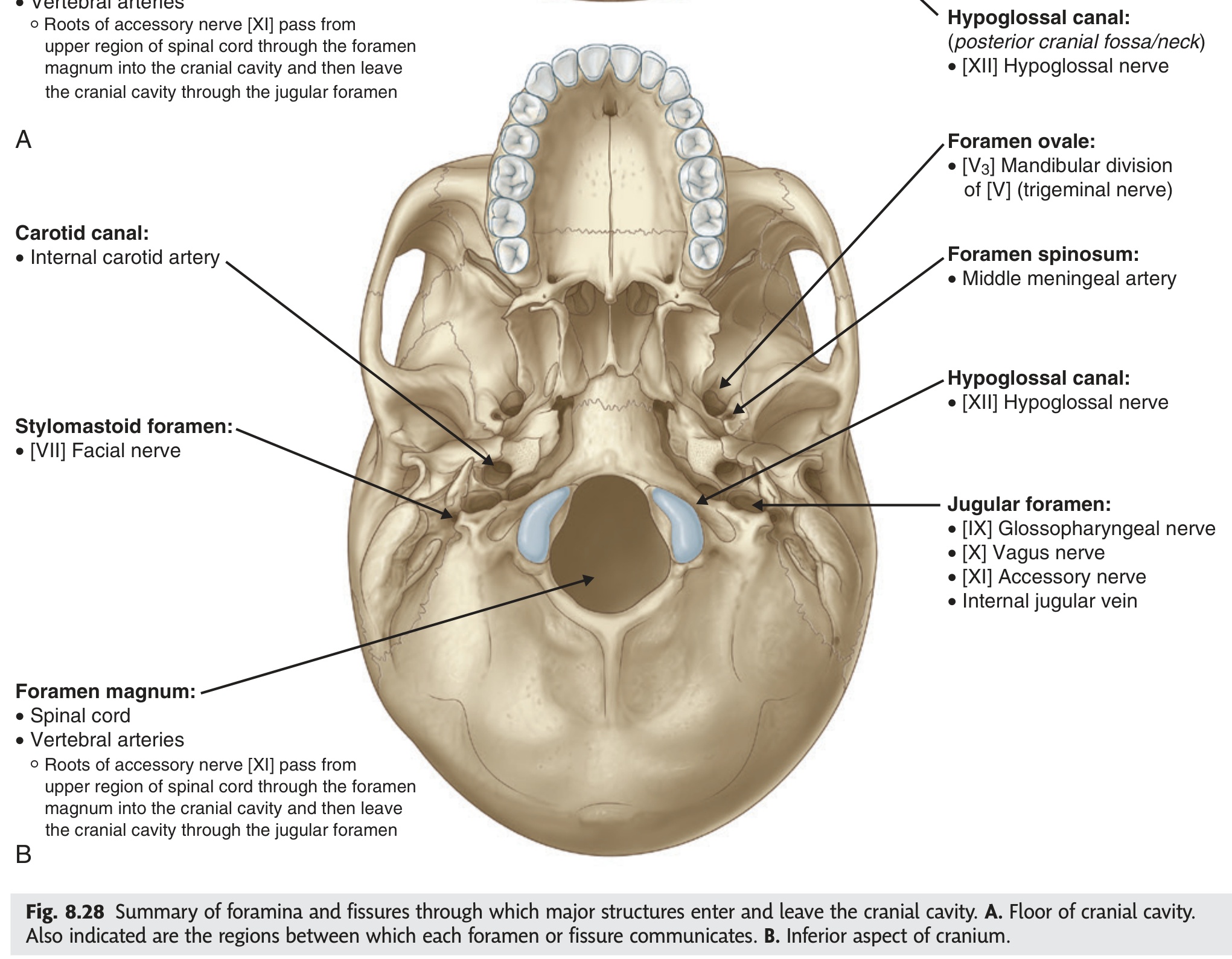
| Foramen | Location | Contents |
|---|---|---|
| Foramen magnum | Occipital bone | Spinal cord + meninges · Vertebral arteries · Anterior + posterior spinal aa. · CN XI spinal roots |
| Jugular foramen | Petro-occipital junction | CN IX, X, XI (cranial root) · Sigmoid sinus → internal jugular vein |
| Carotid canal | Petrous temporal | Internal carotid artery · Sympathetic carotid plexus |
| Stylomastoid foramen | Between styloid + mastoid | CN VII (facial nerve) exits skull |
| Hypoglossal canal | Above occipital condyle | CN XII (hypoglossal nerve) |
| Foramen ovale | Greater wing of sphenoid | CN V3 · Lesser petrosal nerve · Accessory meningeal artery |
| Foramen spinosum | Greater wing of sphenoid | Middle meningeal artery · Meningeal branch of V3 |
| Greater palatine foramen | Lateral hard palate | Greater palatine nerve + artery (V2) |
| Incisive fossa | Anterior hard palate (maxilla) | Nasopalatine nerve · Greater palatine artery |
- Q: What exits the stylomastoid foramen? — CN VII (facial nerve); then enters the parotid gland and divides into five terminal branches.
- Q: What exits the hypoglossal canal? — CN XII (hypoglossal nerve); supplies all intrinsic and extrinsic tongue muscles except palatoglossus.
- Q: What passes through the carotid canal? — Internal carotid artery + sympathetic carotid plexus; located in the petrous temporal bone.
- Q: Name the four structures that exit the jugular foramen. — CN IX, CN X, CN XI (cranial root) + internal jugular vein (continuation of sigmoid sinus).
- Q: What is the content of foramen magnum? — Spinal cord + meninges, vertebral arteries, anterior and posterior spinal arteries, CN XI spinal roots.
Mandible & Temporomandibular Joint ★★
The mandible is the only movable bone of the skull — a horseshoe-shaped jaw that joins itself at the midline symphysis menti and articulates laterally at the TMJ. Its clinical richness lies in its neurovascular geography: the inferior alveolar nerve and artery enter the medial ramus through the mandibular foramen, guarded anteriorly by the lingula — which is precisely where a dental anaesthetist aims the needle for an inferior alveolar nerve block to anaesthetise lower molars. The mental foramen on the external body surface (level with the second premolar) is the exit point of the mental nerve (V3) and the landmark for anterior dental blocks and chin surgery.
The mandible is the only movable bone of the skull. It consists of a horizontal body (bearing lower teeth) and two vertical rami, each with a coronoid process (temporalis insertion) and a condylar process (head for TMJ).
| Part | Key Features |
|---|---|
| Body | Mental protuberance (chin) · Mental foramen (mental nerve V3 exits — dental block site) · Alveolar part (lower teeth) |
| Ramus | Angle of mandible · Coronoid process (temporalis) · Condylar process (head for TMJ) |
| Mandibular foramen | Medial ramus; inferior alveolar nerve + artery enter; lingula guards its anterior lip (sphenomandibular ligament attachment) |
| Mandibular notch | Between coronoid + condylar processes; masseteric vessels + nerve pass through |
- Which is the only movable bone of the skull? The mandible — articulates at the TMJ bilaterally.
- What enters the mandibular foramen, and what guards its anterior lip? Inferior alveolar nerve and artery; the lingula guards the anterior margin (sphenomandibular ligament attachment).
- Where is the mental foramen, and what exits through it? External surface of the mandibular body, level with the second premolar; the mental nerve (V3) exits here — landmark for inferior dental blocks and chin surgery.
- What inserts onto the coronoid process of the mandible? Temporalis muscle.
- Which nerve is blocked to anaesthetise all lower molar teeth? Inferior alveolar nerve — block achieved by injecting near the mandibular foramen on the medial ramus, just anterior to the lingula.
The temporomandibular joint is unlike any other synovial joint in the body: it is bicondylar (both TMJs must work as one functional unit), its articular surfaces are fibrocartilage rather than hyaline cartilage, and an articular disc physically divides it into two separate synovial compartments with different movements — gliding above (protrusion) and hinging below (opening). Clinically, if a patient's jaw locks open after a wide yawn or vomit, the condyle has slid anterior to the articular tubercle and cannot return — the reduction technique is to wrap your thumbs, press down on the lower molars to depress the condyle below the tubercle level, then guide posteriorly, directly reversing the joint mechanics. The articular surface is fibrocartilage, not hyaline — examiners test this fact specifically because every other synovial joint uses hyaline.
The TMJ is a synovial, bicondylar joint between the mandibular condyle and the mandibular fossa + articular tubercle of temporal bone. An articular disc divides it into two separate compartments allowing gliding and hinge movements.
- Articular surfaces — mandibular condyle below; mandibular fossa + articular tubercle above. Both surfaces covered by fibrocartilage (not hyaline cartilage — this is a TMJ-specific fact examiners test).
- Articular disc — biconcave fibrocartilaginous disc; divides joint into two compartments; moves anteriorly with the condyle during wide mouth opening.
- Upper (superior) compartment — between disc and temporal bone; gliding movement (protrusion and retrusion of mandible); driven by bilateral lateral pterygoid contraction.
- Lower (inferior) compartment — between disc and condyle; hinge movement (first 20 mm of mouth opening, rotation of condyle under disc).
- Lateral (temporomandibular) ligament — strongest ligament; reinforces the lateral capsule; prevents posterior dislocation.
- Sphenomandibular ligament — medial accessory; from spine of sphenoid → lingula of mandible.
- Stylomandibular ligament — posterior accessory; from styloid process → angle of mandible.
Depression (wide opening): Lateral pterygoid (both heads pull condyle + disc forward)
Protrusion: Bilateral lateral pterygoid
Retrusion: Posterior fibres of temporalis
Lateral excursion: Unilateral lateral pterygoid (ipsilateral)
Anterior TMJ dislocation: Condyle passes anterior to articular tubercle during wide opening (yawning, vomiting) and locks there → patient cannot close mouth. Reduction: press down on lower molars (thumbs wrapped) to depress condyle below tubercle, guide posteriorly.
Inferior alveolar nerve block: For lower molar anaesthesia, the inferior alveolar nerve (V3) is blocked at the mandibular foramen on the medial ramus. The lingula is the landmark. Needle enters medial to ramus, just above the foramen level.
- Q: Type of cartilage covering TMJ articular surfaces? — Fibrocartilage (NOT hyaline) — distinguishes TMJ from every other synovial joint.
- Q: Which compartment glides and which hinges? — Upper = gliding (protrusion/retrusion); Lower = hinge (rotation, first 20 mm of opening).
- Q: Nerve supply of the TMJ? — Auriculotemporal nerve (V3) + masseteric nerve (V3).
- Q: What is the lingula and why does it matter? — Bony lip guarding the anterior mandibular foramen; landmark for inferior alveolar nerve block.
- Q: Anterior TMJ dislocation — reduction technique? — Thumbs on lower molars, press down to depress condyle below tubercle, then guide posteriorly.
Orbital Cavity & Walls
The orbit is a four-walled pyramid whose anatomy is best learnt through its fracture patterns: the floor is the thinnest wall — a punch to the eye transmits hydraulic pressure inward and the orbital floor cracks downward (blowout fracture), trapping orbital fat and the inferior rectus in the maxillary sinus below. The medial wall is paper-thin (lamina papyracea of the ethmoid), which is why a child with ethmoid sinusitis can develop an orbital cellulitis within hours as pus tracks straight through. The apex funnels every important orbital nerve and vessel into two tight openings — the optic canal for CN II and the ophthalmic artery, and the superior orbital fissure for CN III, IV, V1, and VI — so a retrobulbar mass at the apex can paralyse all ocular movements simultaneously.
The orbit is a four-walled pyramidal bony cavity housing the eyeball, extraocular muscles, lacrimal gland, fat, and neurovascular structures. It is formed by 7 bones. The apex points posteromedially; the base (orbital rim) faces anterolaterally.
| Wall | Bones (main contributor first) | Key structures / Clinical |
|---|---|---|
| Roof (superior) | Orbital plate of frontal (mainly) + lesser wing of sphenoid (posterior) | Lacrimal fossa (anterior lateral roof) = lacrimal gland. Separates orbit from anterior cranial fossa. Thin — meningioma, frontal sinus mucocele can erode through. |
| Floor (inferior) | Orbital plate of maxilla (mainly) + zygomatic + palatine (small) | Thinnest wall. Separates orbit from maxillary sinus. Blow-out fracture: punch/ball to eye → increased intraorbital pressure → floor fractures inferiorly → orbital fat + inferior rectus herniate into maxillary sinus → enophthalmos + vertical diplopia. |
| Medial wall | Ethmoid (orbital plate / lamina papyracea — paper-thin) + lacrimal + frontal process of maxilla + sphenoid | Lamina papyracea: extremely thin → ethmoidal sinusitis spreads medially into orbit (orbital cellulitis) — most common route of infection spread to orbit. Contains lacrimal groove (nasolacrimal duct). |
| Lateral wall | Zygomatic (anterior) + greater wing of sphenoid (posterior) | Strongest wall. SOF between lateral wall and roof (posterior apex). Lateral orbital rim palpable through skin. |
| Opening | Location | Contents |
|---|---|---|
| Optic canal | Apex; lesser wing of sphenoid | CN II (optic nerve) + ophthalmic artery (branch of ICA) |
| Superior orbital fissure (SOF) | Between lesser + greater wings of sphenoid | CN III, IV, V1 (lacrimal + frontal + nasociliary branches), CN VI; superior + inferior ophthalmic veins |
| Inferior orbital fissure (IOF) | Between maxilla + greater wing of sphenoid; floor–lateral wall junction | CN V2 (infraorbital nerve) + infraorbital artery; inferior ophthalmic vein → pterygoid plexus |
| Lacrimal fossa | Medial wall (lacrimal bone + maxilla) | Lacrimal sac → continues as nasolacrimal duct → drains into inferior nasal meatus |
| Supraorbital notch/foramen | Supraorbital rim (frontal) | Supraorbital nerve (V1) + artery |
| Infraorbital foramen | Anterior face of maxilla, 1 cm below orbital rim | Infraorbital nerve (V2) — anaesthetic landmark for maxillary nerve block |
Preseptal (periorbital) cellulitis: infection anterior to orbital septum — no proptosis, no restricted EOM; treat with oral antibiotics. Orbital (postseptal) cellulitis: infection posterior to orbital septum — proptosis, chemosis, painful restricted EOM, visual threat; most common source = ethmoid sinusitis spreading through paper-thin lamina papyracea. Emergency: IV antibiotics + CT orbit/sinuses; urgent surgical drainage if subperiosteal abscess. Complication: cavernous sinus thrombosis (retrograde spread via ophthalmic veins).
- Q: Which orbital wall is thinnest and fractures in a blowout? — Floor (orbital plate of maxilla); orbital fat + inferior rectus herniate into maxillary sinus → enophthalmos + vertical diplopia.
- Q: What is the lamina papyracea? — Paper-thin orbital plate of the ethmoid (medial wall); allows ethmoid sinusitis to spread into the orbit as orbital cellulitis.
- Q: What passes through the optic canal? — CN II (optic nerve) + ophthalmic artery (branch of ICA).
- Q: What passes through the superior orbital fissure? — CN III, IV, V1 (lacrimal + frontal + nasociliary), CN VI + superior and inferior ophthalmic veins.
- Q: Distinguish preseptal from orbital cellulitis. — Preseptal: anterior to septum, no proptosis, no restricted EOM, treat oral antibiotics. Orbital: posterior to septum, proptosis, restricted painful EOM, visual threat, IV antibiotics ± surgery.
Paranasal Sinuses
The paranasal sinuses are air-filled cavities that lighten the skull, warm and humidify inhaled air, and — from a clinical standpoint — generate a wide variety of headache and infection pathology almost entirely dictated by their drainage. The maxillary sinus, despite being the largest, drains through an ostium high on its medial wall rather than at the floor, meaning gravity works against secretion clearance when you sit upright — hence its disproportionate infection rate. The sphenoidal sinus, tucked in the sphenoid body, sits in immediate proximity to the pituitary above, cavernous sinus laterally, and optic chiasm anterosuperiorly — sphenoid sinusitis can therefore present with sudden visual loss or ophthalmoplegia without any nasal symptoms at all.
| Sinus | Bone | Drainage (meatus) | Clinical |
|---|---|---|---|
| Maxillary | Maxilla (largest sinus) | Middle meatus (hiatus semilunaris); ostium is in the UPPER medial wall — poor gravity drainage = sinusitis prone | Most commonly infected sinus; floor = root of upper molar teeth (dental abscess → maxillary sinusitis); "water's view" X-ray (occipitomental); antral lavage via inferior meatus |
| Ethmoid | Ethmoid (multiple air cells) | Anterior + middle cells → middle meatus; posterior cells → superior meatus | Spreads to orbit via lamina papyracea; optic nerve at risk in posterior ethmoiditis |
| Frontal | Frontal bone | Middle meatus (via frontonasal duct / infundibulum) | Absent at birth, develops by age 7; frontal sinusitis → pott's puffy tumour (osteomyelitis of frontal bone); complications: meningitis, cerebral abscess (posterior wall = anterior cranial fossa) |
| Sphenoidal | Sphenoid body | Sphenoethmoidal recess (above superior meatus) | Relations: pituitary above, optic chiasm, cavernous sinus laterally, ICA; sphenoid sinusitis → cavernous sinus thrombosis; transsphenoidal hypophysectomy enters via nasal cavity → sphenoidal sinus |
Middle meatus: Maxillary + Frontal + anterior/middle Ethmoid (= MFE). Superior meatus: posterior Ethmoid. Sphenoethmoidal recess: Sphenoid. Inferior meatus: nasolacrimal duct (not a sinus, but opens here).
- Q: Why is maxillary sinusitis so common? — Ostium is high on the medial wall (not at the floor) → gravity opposes drainage when upright → secretions pool and stagnate.
- Q: Where does the sphenoidal sinus drain? — Sphenoethmoidal recess (above the superior meatus).
- Q: Which sinuses drain into the middle meatus? — Maxillary, frontal, anterior + middle ethmoid cells (mnemonic: MFE → Middle meatus).
- Q: Trans-sphenoidal hypophysectomy passes through which sinus? — Sphenoidal sinus (nasal cavity → sphenoidal sinus → sella turcica).
- Q: Complication of frontal sinusitis eroding the posterior wall? — Intracranial spread → meningitis or cerebral abscess (posterior wall = anterior cranial fossa floor).
Cavernous Sinus ★★★
The cavernous sinus is a trabeculated venous pool in the dura beside the sella — what makes it the highest-stakes sinus in anatomy is the number of crucial structures either running inside it or in its lateral wall. The ICA and CN VI run inside the blood-filled sinus; CN III, IV, V1, and V2 travel in the lateral dural wall — meaning a cavernous sinus thrombosis (CST) can affect all of them simultaneously. The earliest clinical signal of CST is CN VI palsy (failure to abduct the eye, diplopia on lateral gaze), because CN VI lies within the sinus rather than protected in the wall and is compressed first by rising venous pressure. Because the ophthalmic veins are valveless, even a squeezed pimple in the danger triangle of the face (upper lip to nasal bridge) can spread retrograde into this sinus — which is why "never squeeze pimples in the danger area" is a genuine clinical rule.
The cavernous sinus is a paired venous sinus within the dura mater of the middle cranial fossa, on either side of the sella turcica. It is uniquely traversed by cranial nerves and the internal carotid artery — making it clinically critical.
| Structure | Position within/on sinus | Note |
|---|---|---|
| Internal carotid artery (ICA) | Runs through the sinus (surrounded by venous blood) | S-shaped siphon within sinus; carotico-cavernous fistula: pulsatile exophthalmos |
| CN VI (Abducens) | Within sinus (medial to ICA) — most medially placed nerve | Most vulnerable to raised sinus pressure → diplopia (lateral rectus palsy) is earliest CN sign in cavernous sinus pathology |
| CN III (Oculomotor) | In lateral dural wall — superior | Palsy → ptosis + down-and-out eye + mydriasis |
| CN IV (Trochlear) | In lateral dural wall — below CN III | Palsy → vertical diplopia; superior oblique affected |
| CN V1 (Ophthalmic) | In lateral dural wall — below CN IV | Sensory loss over forehead + corneal reflex lost |
| CN V2 (Maxillary) | In lateral dural wall — most inferior | Sensory loss over cheek + upper teeth |
O TOM CAT (lateral wall top to bottom, then in sinus):
Lateral wall: Oculomotor (III) · Trochlear (IV) · Ophthalmic V1 · Maxillary V2
Within sinus: Carotid (ICA) · Abducens (VI)
Or simply: 3, 4, V1, V2 in wall; ICA + 6 within sinus.
| Connection | Clinical significance |
|---|---|
| Superior + inferior ophthalmic veins (from orbit) — valveless | Facial/orbital infection spreads to cavernous sinus (danger triangle of face) |
| Superficial middle cerebral vein | Cortical venous thrombosis can extend |
| Pterygoid venous plexus (via emissary veins) | Dental infection pathway |
| Drains via superior + inferior petrosal sinuses → IJV / sigmoid sinus | Posterior drainage route |
| Intercavernous sinuses connect left and right | Bilateral involvement in cavernous sinus thrombosis (CST) |
The danger triangle is the roughly triangular area of the face bounded by the corners of the mouth and the bridge of the nose (nasolabial fold region + nasal dorsum). It is dangerous because facial veins here are valveless and communicate directly with the cavernous sinus via the angular vein → ophthalmic veins → cavernous sinus. Infection in this zone (furuncle, squeezed pimple, infected nasal vestibule) can spread retrogradely into the cavernous sinus, causing cavernous sinus thrombosis. Key rule: Never squeeze pimples in the danger triangle.
Source: Facial infections in the "danger area" (upper lip, nose) → angular/ophthalmic vein → cavernous sinus. Also: sphenoid sinusitis, orbital cellulitis, dental abscess.
Features: Fever, headache, proptosis (exophthalmos), chemosis, periorbital oedema, ophthalmoplegia (CN III, IV, VI palsies), sensory loss over forehead/cheek (V1/V2). Initially unilateral → bilateral (via intercavernous sinus).
Earliest CN sign: CN VI palsy (lateral rectus weakness → failure of abduction → diplopia on lateral gaze) — because CN VI lies within the sinus, most exposed to venous pressure.
Treatment: IV antibiotics + anticoagulation (heparin) + treat source. High mortality without treatment.
- Q: Which CN lies within the sinus (not in the wall)? — CN VI (abducens) + ICA run inside; CN III, IV, V1, V2 are in the lateral dural wall.
- Q: Order of CNs in lateral wall top to bottom? — CN III → IV → V1 → V2 (mnemonic: O TOM — Oculomotor, Trochlear, Ophthalmic, Maxillary).
- Q: Why is CN VI the first nerve affected in CST? — It lies within the sinus, directly bathed in venous blood — most exposed to rising venous pressure.
- Q: What is the danger triangle of the face? — Area from lip corners to nasal bridge; valveless facial veins communicate retrograde with the cavernous sinus via angular → ophthalmic veins.
- Q: Name three sources of cavernous sinus thrombosis. — Facial infection (danger triangle), sphenoid sinusitis, orbital cellulitis, dental abscess via pterygoid plexus.
Cranial Nerves → Skull Foramina (I → XII)
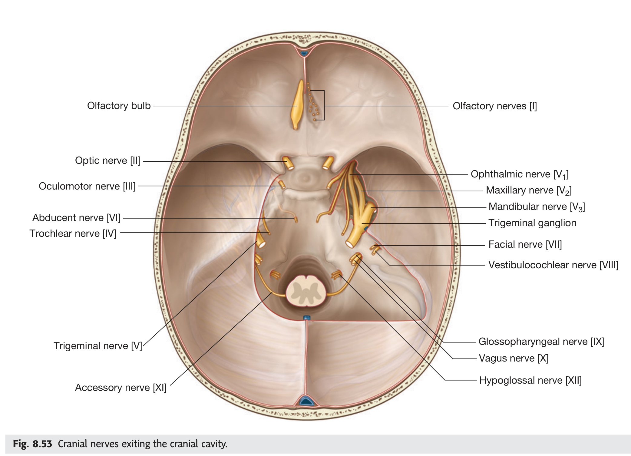
Every cranial nerve exits (or enters) the skull through a named foramen, and knowing which foramen belongs to which nerve lets you read a CN deficit as a precise anatomical address. The three foramina you must own for examinations are: (1) the superior orbital fissure — through which pass CN III, IV, V1, and VI; (2) the internal acoustic meatus — CN VII enters here and runs through the petrous bone to exit at the stylomastoid foramen, while CN VIII enters but never exits; and (3) the jugular foramen — CN IX, X, and XI exit together alongside the sigmoid sinus forming the internal jugular vein. A total unilateral SOF syndrome (CN III + IV + V1 + VI palsied together) means pathology at the orbital apex or posterior SOF, while a selective CN VI palsy alone points to the cavernous sinus or Dorello's canal where CN VI makes a sharp bend.
Each cranial nerve exits the skull through a specific foramen. This is one of the most tested topics in anatomy finals — learn them in Roman numeral order.
- CN I — Olfactory: Multiple cribriform foramina of ethmoid bone (filaments from nasal mucosa → olfactory bulb in anterior cranial fossa)
- CN II — Optic: Optic canal (lesser wing of sphenoid) — also transmits ophthalmic artery; opens into orbit
- CN III — Oculomotor: Superior orbital fissure (SOF) → orbit; controls all extraocular muscles except SO and LR
- CN IV — Trochlear: SOF → orbit; smallest CN; only CN that exits dorsal brainstem; controls superior oblique (SO4)
- CN V — Trigeminal (3 divisions): V1 → SOF; V2 (maxillary) → foramen rotundum; V3 (mandibular) → foramen ovale
- CN VI — Abducens: SOF → orbit; controls lateral rectus only (LR6)
- CN VII — Facial: Enters via internal acoustic meatus (IAM) → runs through facial canal within petrous temporal → exits via stylomastoid foramen
- CN VIII — Vestibulocochlear: IAM — does NOT exit skull; terminates in cochlea and vestibular apparatus within petrous temporal
- CN IX — Glossopharyngeal: Jugular foramen (with CN X + XI cranial root)
- CN X — Vagus: Jugular foramen — exits with CN IX + XI; supplies pharynx, larynx, thoracic + abdominal viscera
- CN XI — Accessory: Spinal roots enter skull via foramen magnum (from C1–C5/6), then exit with CN IX + X via jugular foramen
- CN XII — Hypoglossal: Hypoglossal canal (above occipital condyle); supplies all tongue muscles except palatoglossus
SOF group: CN III, IV, V1, VI + ophthalmic veins (= "3, 4, V1, 6 all through SOF")
IAM group: CN VII (enters + runs through petrous) + CN VIII (enters, stays in temporal bone)
Jugular group: CN IX + X + XI cranial root + internal jugular vein
Unique canals: CN I = cribriform · CN II = optic canal · CN XII = hypoglossal canal
- Q: Which four cranial nerves pass through the SOF? — CN III, IV, V1 (lacrimal + frontal + nasociliary), VI + ophthalmic veins.
- Q: CN VII — entry foramen vs exit foramen? — Enters via IAM → runs through facial canal in petrous temporal → exits via stylomastoid foramen.
- Q: Which CN enters the IAM but does NOT exit the skull? — CN VIII (vestibulocochlear); it terminates in the cochlea and vestibular apparatus within the petrous temporal bone.
- Q: Which three CNs exit via the jugular foramen? — CN IX (glossopharyngeal), X (vagus), XI cranial root — plus the internal jugular vein.
- Q: CN XI spinal roots — where do they enter the skull and where do they exit? — Enter via foramen magnum (from C1–C5/6 spinal cord), then exit via jugular foramen with CN IX + X.
Le Fort Fractures of the Midface ★★
All three Le Fort fractures share one pathognomonic finding — fracture of the pterygoid plates — because the pterygoid plates form the posterior structural pillar of the midface; no matter how high or low the fracture line runs anteriorly, it always snaps through these plates posteriorly. The level of midface detachment determines the type: Le Fort I detaches only the alveolus (floating palate), Le Fort II detaches the central face pyramid (floating maxilla), and Le Fort III separates the entire midface from the cranial base (floating face — craniofacial dysjunction). CSF rhinorrhoea in Le Fort II and III signals that the fracture has reached the anterior cranial fossa floor through the cribriform plate or orbital roof, confirming intracranial communication — a significant escalation in management requiring neurosurgical involvement.
Le Fort fractures are horizontal fractures of the midface caused by high-velocity blunt trauma. All three types share one feature: fracture of the pterygoid plates of the sphenoid. Clinically tested by grasping the upper teeth and pulling anteriorly — positive mobility indicates a Le Fort fracture.
| Type | Fracture Line | What Floats? | Mechanism |
|---|---|---|---|
| Le Fort I (Horizontal) |
Horizontal line through the maxilla just above the teeth — separates the maxillary alveolus + hard palate from the rest of the midface. Passes through: lower maxilla + lower nasal septum + pterygoid plates (base) | "Floating palate" — only the lower maxilla (alveolar process + teeth + hard palate) moves | Horizontal blow to the upper lip / maxilla |
| Le Fort II (Pyramidal) |
Pyramidal line through: medial orbital walls + infraorbital rims + across the nasal bones + inferior orbital floor + pterygoid plates. Naso-orbital-ethmoid region involved | "Floating maxilla" — central midface pyramid (maxilla + nasal bones + medial orbits) moves | Blow to the central midface / nasal region |
| Le Fort III (Craniofacial dysjunction) |
Transverse line through: zygomatic arches + lateral orbital walls + frontozygomatic sutures + nasal bones + pterygoid plates. Complete separation of the entire midface from the cranial base | "Floating face" — the entire midface is detached from the skull | High-velocity (road traffic accident, high-energy impact to upper face) |
Le Fort I = lowest (above the teeth) • Le Fort II = middle (through orbital rims = pyramidal) • Le Fort III = highest (through zygomatic arches = entire face off).
All share: fracture of pterygoid plates of sphenoid (pathognomonic) + bilateral + symmetric.
Clinical test: Grasp the upper incisor teeth and apply an anterior force — movement of the midface relative to the rest of the skull confirms a Le Fort fracture. Level of movement identifies the type.
Common features of all Le Fort fractures:
• Bilateral periorbital ecchymosis ("panda eyes" / "raccoon eyes")
• Midface oedema + lengthening ("dish face")
• Malocclusion (teeth don't align)
• Mobile midface on examination
Le Fort II + III specific: CSF rhinorrhoea (fracture communicates with anterior cranial fossa via cribriform plate or orbital roof involvement — β2-transferrin positive).
Le Fort III specific: Risk of meningitis, pneumocephalus, carotid-cavernous fistula.
Treatment: Airway first (bleeding + oedema may cause obstruction → intubate early). Open reduction + internal fixation (ORIF) with titanium mini-plates once swelling subsides.
- Q: What do ALL three Le Fort fracture types share? — Fracture of the pterygoid plates of the sphenoid (pathognomonic posterior component).
- Q: Le Fort I — what "floats"? — Floating palate: only the alveolar process + lower maxilla + hard palate detaches (fracture just above the teeth).
- Q: Which Le Fort types cause CSF rhinorrhoea and why? — Le Fort II and III — their fracture lines reach the cribriform plate or orbital roof (anterior cranial fossa floor), allowing CSF into the nasal cavity.
- Q: Clinical test for Le Fort fracture? — Grasp upper incisor teeth and apply anterior force — mobile midface confirms a Le Fort fracture.
- Q: Le Fort III specific risk not present in I or II? — Meningitis, pneumocephalus, carotid-cavernous fistula (complete craniofacial dysjunction with intracranial communication).
Past Paper Q&A — Skull
Anterior: Floor = orbital plates of frontal + cribriform plate of ethmoid + lesser wings/anterior sphenoid body. Contains frontal lobes + olfactory bulbs. Foramina: cribriform (CN I), foramen cecum.
Middle: Floor = greater wings of sphenoid + squamous temporal + anterior petrous face + sphenoid body. Contains temporal lobes + pituitary (sella) + cavernous sinus. Foramina: optic canal (CN II), SOF (III, IV, V1, VI), foramen rotundum (V2), foramen ovale (V3), foramen spinosum (middle meningeal a.), foramen lacerum (fibrocartilage; ICA passes over).
Posterior: Floor = petrous/mastoid temporal + occipital. Contains cerebellum, pons, medulla. Foramina: IAM (CN VII + VIII), jugular foramen (IX, X, XI + IJV), hypoglossal canal (XII), foramen magnum (spinal cord, vertebral aa., CN XI spinal).
Clinical significance: Thinnest part of the skull. The anterior branch of the middle meningeal artery grooves its inner surface. A blow to the temple fractures pterion → ruptures middle meningeal artery → extradural haematoma. Classic presentation: lucid interval → rapid deterioration. CT: biconvex hyperdense collection. Treatment: craniotomy/burr hole + evacuation.
Articular surfaces: Mandibular condyle (head) + mandibular fossa/articular tubercle of temporal bone — covered by fibrocartilage (not hyaline).
Disc: Biconcave fibrocartilaginous; divides joint into two separate synovial compartments.
Upper compartment (disc–temporal) = gliding (protrusion/retrusion).
Lower compartment (disc–condyle) = hinge (opening/closing).
Ligaments: (1) Lateral — strongest, prevents posterior dislocation; (2) Sphenomandibular; (3) Stylomandibular.
Nerve supply: Auriculotemporal nerve (V3) + masseteric nerve (V3).