Bones of Trunk & Joints
Vertebral Column — Overview
A 25-year-old rugby player is tackled and hits the ground head-first. His neck snaps into hyperextension and he immediately reports both arms going numb. The paramedics immobilize his cervical spine before moving him. Everything about that response — why the cervical region is most vulnerable, why immobilization is the right first step, why arm numbness is the warning sign — makes sense only when you understand the vertebral column as an engineering structure. It is not simply a stack of bones; it is a segmented column that must simultaneously be rigid enough to protect the spinal cord and flexible enough to allow a human range of motion. That contradiction is resolved by dividing the column into five regions, each with a different bone morphology, a different range of movement, and a different clinical risk profile.
The column has 33 vertebrae in the fetus, but many fuse by adulthood. Think of it as a 26-unit structure in adults (the sacral and coccygeal segments fuse into solid blocks). Each of the five regions solves the same basic engineering problem — stacking a ring-shaped bone on top of another, separated by a hydrostatic disc — but each makes different design trade-offs between mobility and stability.
| Region | Count | Distinguishing features | Key clinical |
|---|---|---|---|
| Cervical | 7 (C1–C7) | Smallest bodies; foramen transversarium in each transverse process (VA passes through C1–C6); triangular vertebral foramen; bifid spinous processes (C3–C6) | Whiplash, dens fracture, cervical stenosis |
| Thoracic | 12 (T1–T12) | Heart-shaped body; costal facets (demifacets) on body + transverse process facets for rib articulation; long downward-sloping spinous processes; circular vertebral foramen | Osteoporotic compression fractures, Scheuermann's kyphosis |
| Lumbar | 5 (L1–L5) | Largest kidney-shaped bodies (weight-bearing); horizontal spinous processes; no rib facets; no foramen transversarium; triangular vertebral foramen; mammillary processes | Disc herniation, spondylolisthesis, lumbar stenosis, LP |
| Sacral | 5 → 1 fused sacrum | Triangular; base up, apex down; 4 pairs of sacral foramina; auricular surface for SIJ; sacral hiatus | Sacral stress fracture, caudal block, SIJ dysfunction |
| Coccygeal | 3–5 → 1 coccyx | Vestigial; 4 segments; attachment for pelvic floor | Coccydynia (fall onto tailbone) |
- How many vertebrae are in each region? Cervical 7, Thoracic 12, Lumbar 5, Sacrum 5 (fused → 1), Coccyx 3–5 (fused → 1).
- Which region has the largest vertebral foramina relative to the cord, and which has the smallest? Cervical (largest — triangular); Thoracic (smallest — circular); the mismatch in thoracic explains why cord compression there is most dangerous.
- Which vertebral region has foramen transversarium and what passes through it? Cervical (C1–C6) — vertebral arteries ascend through the foramen transversarium bilaterally.
- What is spondylolisthesis and at which lumbar level is it most common? Anterior slip of one vertebra over the one below (bilateral pars fractures); most common at L4–L5 and L5–S1.
- What clinical procedure uses the sacral hiatus and which region is it in? Caudal epidural anaesthesia — the sacral hiatus is the inferior opening of the sacral canal used for obstetric and paediatric epidural blocks.
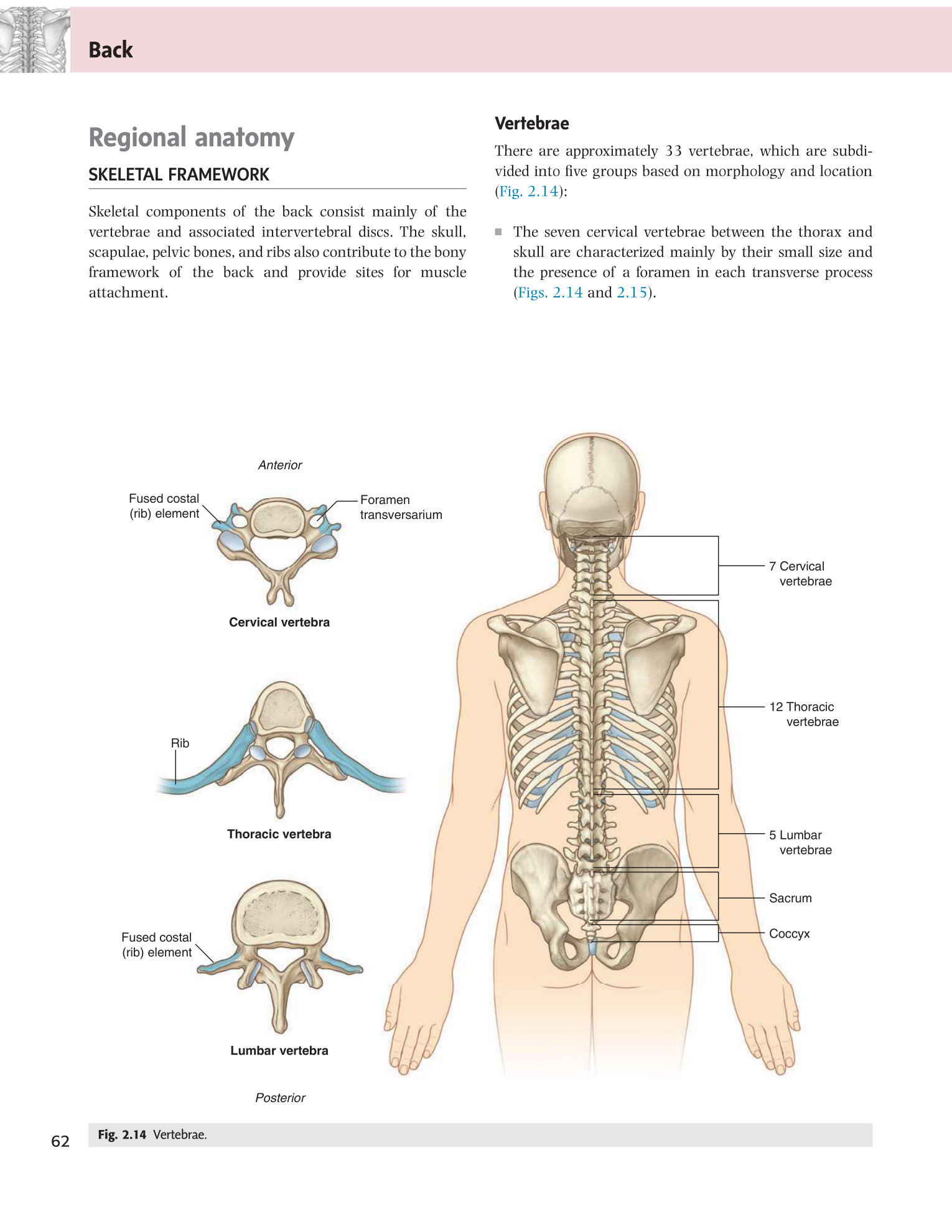
- How many vertebrae in the adult spine (functional units)? 26 — 7 cervical, 12 thoracic, 5 lumbar, 1 fused sacrum, 1 coccyx (33 vertebrae in the fetus fuse to 26 in the adult).
- Which region has foramen transversarium in each transverse process, and what passes through them? Cervical vertebrae (C1–C6) — vertebral arteries.
- Which vertebral region has the largest bodies and why? Lumbar — bears the greatest compressive load; each lumbar vertebra supports the cumulative weight of the trunk above it.
- Which spinal region is most vulnerable to cord injury and why? Cervical — smallest canal relative to cord diameter, greatest mobility (highest torque on trauma), and the vertebral arteries supplying the posterior brain pass through it.
- What are the distinguishing features of thoracic vertebrae that no other region shares? Costal facets (demifacets) on the vertebral body AND transverse process facets for rib articulation.
Every fetus has a single C-shaped curve — the whole column bows posteriorly (kyphosis) because the infant is curled in the fetal position. After birth, two more curves develop in response to new mechanical demands: when the infant begins lifting its head (3–4 months), the cervical lordosis develops; when it stands and walks (12–18 months), the lumbar lordosis develops. This produces the adult S-shape. The key distinction is primary (present at birth, fetal curves: thoracic + sacral = kyphotic) vs secondary (postnatal, compensatory: cervical + lumbar = lordotic). When posture deteriorates or when disease strikes, these curves exaggerate — producing scoliosis, kyphosis, or lordosis.
| Curve | Direction (convexity) | Type | When it forms |
|---|---|---|---|
| Cervical | Anterior (lordosis) | Secondary | When infant begins to lift head (3–4 months) |
| Thoracic | Posterior (kyphosis) | Primary | Present in fetal life; the original C-shaped fetal curve |
| Lumbar | Anterior (lordosis) | Secondary | When infant begins to walk (12–18 months) |
| Sacral | Posterior (kyphosis) | Primary | Present in fetal life |
C + L = Lordosis (concave posteriorly). T + S = kyphoSiS. Primary = Thoracic + Sacral (fetal). Secondary = Cervical + Lumbar (postnatal).
Scoliosis: lateral (side-to-side) curvature of the spine in the coronal plane, often with rotation. Idiopathic scoliosis accounts for 80% (adolescent females, right thoracic convexity most common). Adam's forward bend test reveals a rib hump (rotation). Cobb angle >40° = surgery indicated. Kyphosis: exaggerated thoracic posterior convexity. Causes: Scheuermann's disease (osteochondrosis of vertebral endplates in adolescents), osteoporotic compression fractures (dowager's hump), ankylosing spondylitis. Lordosis: exaggerated lumbar anterior convexity; associated with obesity, pregnancy, hip flexion contracture.
• When does the lumbar lordosis develop and why? → When the infant walks (12–18 months) — mechanical response to upright posture
• Scoliosis involves which plane, and what test reveals the rotational component? → Coronal plane; Adam's forward bend test reveals the rib hump (rotational deformity)
• Cobb angle threshold for surgical intervention? → >40°
A Typical Vertebra
Think of a typical vertebra as a castle design: the solid cylindrical body is the wall facing the enemy (compressive load), and the arch behind it (formed by two pedicles + two laminae) is the defensive ring that protects the garrison (spinal cord) inside. The entire vertebral canal is formed by these rings lined up in a column. Every named part of the vertebra makes logical sense once you understand this: the pedicles are the bridges connecting the body to the arch; the laminae close the ring posteriorly; the spinous and transverse processes are the attachment points for the muscles and ligaments that hold the castle together.
The most clinically important structure to understand is the intervertebral foramen — the opening between adjacent pedicles through which each spinal nerve exits. This is where disc herniation or bone spur (osteophyte) can compress the nerve root. The level of the foramen tells you exactly which nerve root is at risk, which dermatomal territory goes numb, and which reflex disappears. Most vertebrae (C3–C7, T1–T12, L1–L5) share this common structural plan; C1, C2, and the sacrum are atypical.
| Part | Description & Function | Clinical significance |
|---|---|---|
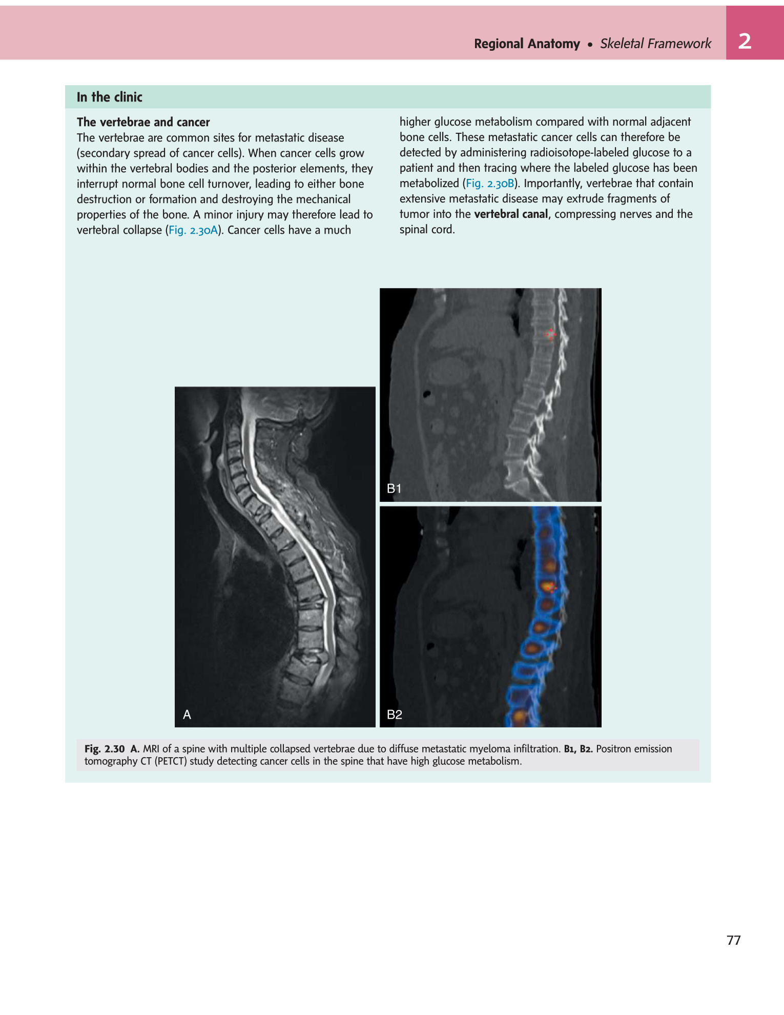
| Vertebral body | Anterior cylindrical mass; cancellous bone with thin cortex; weight-bearing element; connected to adjacent bodies by intervertebral discs | Site of osteoporotic compression fractures; primary bone tumour metastases; vertebroplasty/kyphoplasty |
| Pedicles | Short thick pillars projecting posteriorly from body; vertebral notches superior + inferior form intervertebral foramina between adjacent pedicles | Pedicle screws used in spinal fusion; pedicle erosion on X-ray = "winking owl" sign of metastasis |
| Laminae | Flat plates forming posterior wall of vertebral arch; meet at midline to form spinous process | Laminectomy (removal of lamina) = decompress spinal canal in stenosis |
| Vertebral foramen | Space enclosed by body anteriorly + arch posteriorly; all foramina aligned = vertebral (spinal) canal | Spinal cord + meninges + vessels run within canal; stenosis compresses cord/roots |
| Intervertebral foramen | Formed between pedicle notches of adjacent vertebrae; each transmits one spinal nerve + vessels | Disc herniation or osteophyte narrows IVF → nerve root compression → radiculopathy |
| Spinous process | Posteriorly projecting bony spine; palpable surface landmark; attachment for ligamentum nuchae, supraspinous lig., back muscles | Used to count vertebral levels for LP, epidural, nerve blocks |
| Transverse processes | Project laterally; muscle + ligament attachment; in thoracic: facets for rib tubercle | Transverse process fractures = high-energy trauma, associated with renal injury (L1–L2) |
| Articular processes | Superior (face posterosuperiorly) + inferior (face anteroinferiorly); form facet (zygapophyseal) joints | Facet joint OA = lower back pain; may narrow IVF laterally |
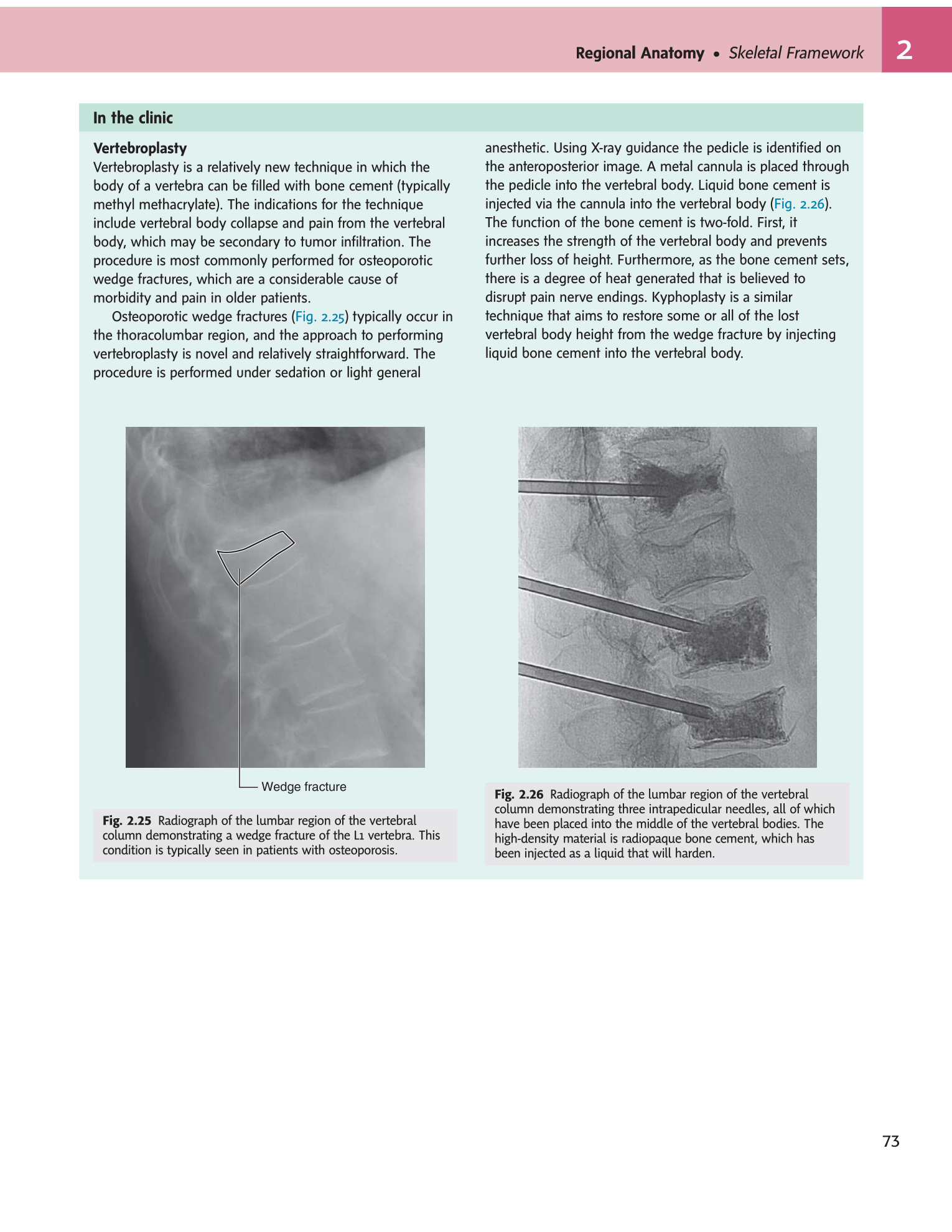
• "Winking owl" sign on X-ray means what? → Erosion of a pedicle by metastatic tumour
• What is a laminectomy and why does it decompress the cord? → Surgical removal of laminae; widens the vertebral canal, relieving pressure on the spinal cord
• C1 and C2 are atypical — what do they lack that typical vertebrae have? → C1 (atlas) has no body and no spinous process; C2 (axis) has the dens (odontoid process) projecting upward from its body
- Name the two components that form the vertebral arch. Two pedicles (bridge body to arch) + two laminae (close the ring posteriorly) — together enclosing the vertebral foramen.
- What is the functional difference between the spinous and transverse processes? Both are lever arms for muscle and ligament attachment; spinous = posterior midline (palpable landmarks for LP); transverse = lateral (rib facets in thoracic, foramen transversarium in cervical).
- Why are pedicle screws used as the main fixation in spinal fusion surgery? Pedicles are the strongest cortical bone in the vertebra — screws placed through them anchor the construct into the vertebral body, resisting flexion-extension forces.
- "Winking owl" sign on plain X-ray indicates what? Pedicle erosion from metastatic tumour — one pedicle appears absent (the "closed eye") compared to the normal contralateral side.
- Which part of the vertebra is most vulnerable to osteoporotic fracture and why? Vertebral body — cancellous bone with thin cortex; under compressive load from body weight, wedge fractures occur anteriorly when the cortex fails.
Individual Vertebral Groups
The cervical spine is the most mobile region of the vertebral column — and therefore the most vulnerable to injury. Its small vertebral bodies, near-horizontal facet joints, and modest ligaments all allow the enormous range of motion the head needs. But that mobility comes at a cost: the spinal canal is narrowest relative to the cord here, and the vertebral arteries (supplying the posterior brain) thread through the foramen transversarium of C1–C6. A posterior cervical disc herniation does not just compress a nerve root; in severe cases it compresses the cord itself (myelopathy), causing weakness and spasticity in all four limbs. C1 and C2 are so radically different from the rest that they deserve separate memorisation.
All cervical vertebrae: foramen transversarium (C1–C6: vertebral artery; C7: vertebral veins only), bifid spinous process (C3–C6), triangular vertebral foramen, small body, uncinate processes (lateral body hooks that form uncovertebral joints of Luschka).
| Vertebra | Special features | Clinical |
|---|---|---|
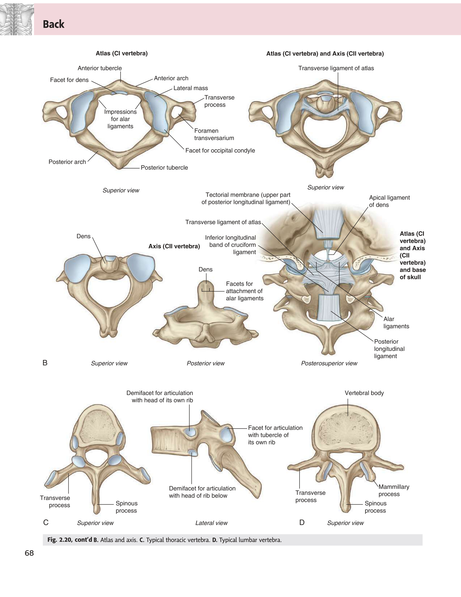
| C1 — Atlas | Ring-shaped; NO body; NO spinous process; 2 lateral masses with superior + inferior articular facets; anterior arch (anterior tubercle) + posterior arch (posterior tubercle); anterior facet articulates with dens of axis | Jefferson fracture: burst fracture of atlas (axial load, e.g. diving injury); "spreading" of lateral masses on odontoid view X-ray |
| C2 — Axis | Dens (odontoid process): finger-like projection from body superiorly; represents embryological body of atlas; held against anterior arch of atlas by transverse ligament; pivot for rotation | Dens fracture (Type II at base most common); hangman's fracture = bilateral pedicle fractures at C2 (hyperextension) |
| C3–C6 | Typical cervical morphology; bifid spinous processes; vertebral artery passes through foramen transversarium | Most common level for cervical disc herniation: C5/C6 (C6 root) and C6/C7 (C7 root) |
| C7 — Vertebra prominens | Longest non-bifid spinous process; easily palpated at base of neck; foramen transversarium contains vertebral veins only (NOT artery) | Surface landmark for counting vertebral levels |
In the cervical spine, a herniated disc at C5/C6 compresses the C6 root (exits below C5 pedicle). C6 root: lateral forearm, thumb, index finger; weakness of biceps + wrist extensors; diminished biceps reflex. C7 root (C6/C7 disc): middle finger; weakness of triceps + wrist flexors; diminished triceps reflex. In cervical spine: the nerve exits ABOVE its numbered pedicle — C5 exits above C5 vertebra (i.e., between C4 and C5).
• Jefferson fracture: mechanism and X-ray appearance? → Axial load (diving injury); burst fracture of atlas; lateral masses spread outward on open-mouth odontoid view
• C5/C6 disc herniation: which root, which reflex, which weakness? → C6 root; biceps reflex; biceps + wrist extensor weakness
• What is the vertebra prominens and how do you find it? → C7 — the most prominent non-bifid spinous process, palpable at the base of the neck
The thoracic vertebrae are uniquely constrained: each one articulates with a pair of ribs, and the rib cage braces the entire thoracic column like a cylinder. This dramatically reduces the range of motion (especially rotation and lateral flexion) compared with the cervical and lumbar regions. The design trade-off is stability over mobility — the thoracic spine rarely herniates discs because it barely moves. The clinical price is that when a tumour metastasises here, there is almost no escape route for the swelling: the circular vertebral foramen is already the smallest, and any expansion compresses the cord very quickly. This is why malignant spinal cord compression most commonly presents at the thoracic level.
| Feature | Detail |
|---|---|
| Body | Heart-shaped; progressively larger T1→T12 to bear increasing weight |
| Costal facets on body | Superior + inferior demifacets (T2–T9); full facets on T1 (for 1st rib head), T10, T11, T12 (for own rib head only) |
| Transverse process facets | For rib tubercle (T1–T10); absent on T11–T12 |
| Spinous processes | Long, slender, angled steeply downward (T5–T8 most angled); overlap vertebra below — limits thoracic extension |
| Vertebral foramen | Circular; smallest canal of all regions — spinal cord is tightest here |
The thoracic spinal canal is the smallest and least vascular region. Metastatic tumours to thoracic vertebrae (breast, lung, prostate) commonly cause malignant spinal cord compression (MSCC) — acute back pain, then progressive paraparesis, sensory level, and sphincter dysfunction. Oncological emergency — requires high-dose dexamethasone + urgent MRI + radiotherapy/surgery.
- What unique feature do thoracic vertebrae have that no other region shares? Costal facets — demifacets on the vertebral body for rib head articulation, plus transverse process facets (T1–T10) for rib tubercle articulation.
- Why is the thoracic spinal canal the most dangerous region for cord compression? It has the smallest vertebral foramen (circular, most narrow) AND the poorest blood supply, so any expanding lesion (tumour, haematoma) compresses the cord rapidly with little reserve.
- Why is disc herniation rare in the thoracic spine? The rib cage braces the thoracic column, dramatically restricting motion — minimal flexion/rotation means minimal disc stress.
- What is malignant spinal cord compression (MSCC) and how does it present? Metastatic tumour (breast, lung, prostate) compresses the thoracic cord → acute back pain + progressive paraparesis + sensory level + sphincter dysfunction — oncological emergency.
- The long angled spinous processes of T5–T8 mean what when doing spinal procedures? Needle must angle steeply cranially to enter the interspinous space — unlike the near-horizontal spinous processes of the lumbar spine.
The lumbar vertebrae carry more body weight than any other vertebral region, so they are the largest. Their thick horizontal spinous processes are a direct invitation for the lumbar puncture needle — you can slip a needle between them relatively easily, which is why LP is performed here rather than in the thoracic or cervical regions. Below the conus medullaris (end of spinal cord at L1–L2 in adults), the subarachnoid space contains only the cauda equina — a bundle of nerve roots floating freely in CSF. Piercing this space with a needle at L3–L4 is safe because the roots float out of the way of the needle tip. The anatomy of this region also explains the two most feared lumbar emergencies: disc herniation compresses individual roots (leg pain, reflex loss); a central herniation compresses the entire cauda equina simultaneously (bilateral weakness + saddle anaesthesia + sphincter loss = surgical emergency).
| Feature | Detail |
|---|---|
| Body | Largest; kidney-shaped; weight-bearing; no rib or transverse process facets; no foramen transversarium |
| Spinous process | Short, thick, horizontal — easily accessible for LP |
| Vertebral foramen | Triangular |
| Mammillary processes | Small bony projections on superior articular processes — unique to lumbar; muscle attachment |
| Pars interarticularis | Narrow isthmus of bone between superior and inferior articular processes — stress fracture site |
The conus medullaris (end of spinal cord) lies at L1–L2 in adults (lower in neonates: L3). Below this, the subarachnoid space contains the cauda equina (nerve roots) floating in CSF. LP is performed at L3–L4 (line joining iliac crests = Tuffier's line = L4 spinous process level) or L4–L5. Needle trajectory (layers penetrated): skin → subcutaneous fat → supraspinous ligament → interspinous ligament → ligamentum flavum (pop/resistance loss) → epidural space → dura mater → subdural space → arachnoid mater → subarachnoid space (CSF obtained). Normal opening pressure: 7–18 cmH₂O.
Spondylolysis: stress fracture of the pars interarticularis (most common at L4–L5 and L5–S1). Common in gymnasts, fast bowlers. X-ray "Scotty dog" sign — fracture of the dog's "neck." Spondylolisthesis: anterior slip of one vertebral body over the one below (bilateral pars fractures). Graded I–IV by % slip. Symptoms: low back pain, tight hamstrings, buttock pain. Severe slip can cause L5 nerve root compression (foot drop). Retrolisthesis: posterior slip (degenerative).
Compression of multiple lumbar and sacral nerve roots below the conus (L2 and below). Causes: large central disc herniation (L4/L5 or L5/S1), tumour, haematoma, abscess. Red flags: bilateral leg weakness/numbness, saddle anaesthesia (inner thighs/perineum), bladder retention or incontinence (overflow), loss of anal tone. Surgical emergency — decompression within 48 hours to prevent permanent sphincter dysfunction.
• LP layers from skin to CSF in order? → Skin → fat → supraspinous lig → interspinous lig → ligamentum flavum (pop) → epidural space → dura → subdural → arachnoid → subarachnoid space
• Tuffier's line landmarks which vertebral level? → L4 spinous process (line connecting the tops of the iliac crests)
• Cauda equina syndrome red flag triad? → Saddle anaesthesia + bilateral leg weakness + bladder/bowel dysfunction → emergency surgery within 48 hrs
Sacrum & Coccyx
The sacrum is where the vertebral column hands off its load to the pelvis. The sacroiliac joints (SIJ) are the connection points, and they are among the strongest joints in the body — braced by massive posterior sacroiliac ligaments that can only be disrupted by very high-energy force. Clinically, the sacrum has two important access points you must know: the anterior sacral foramina (transmit the sacral ventral rami that form the sacral plexus, relevant for pelvic nerve blocks) and the sacral hiatus (the inferior opening used for caudal epidural anaesthesia, particularly in obstetric and paediatric practice).
| Feature | Detail | Clinical relevance |
|---|---|---|
| Structure | 5 fused sacral vertebrae; triangular; base (S1 superior) + apex (S5 inferior) | Fuses completely by ~25 years of age; before that, S1–S2 may show incomplete fusion (spina bifida occulta) |
| Sacral promontory | Anterior projecting margin of S1 body; creates lumbosacral angle (≈130°) | Key obstetric landmark: conjugate diameter of pelvic inlet measured from promontory to pubic symphysis (obstetric conjugate >10 cm needed for vaginal delivery) |
| Anterior sacral foramina | 4 pairs; transmit ventral rami of S1–S4 | Routes for sacral nerve root blocks; visible on pelvic X-ray |
| Posterior sacral foramina | 4 pairs; transmit dorsal rami | Approach for sacral neuromodulation |
| Sacral canal | Continuation of lumbar vertebral canal; ends at sacral hiatus | Contains cauda equina (dural sac ends at S2), filum terminale, epidural fat + venous plexus |
| Sacral hiatus | Inferior opening of sacral canal; covered by sacrococcygeal ligament; created by non-fusion of S4–S5 laminae | Caudal epidural anaesthesia: needle inserted through hiatus into sacral canal; used for perianal surgery, paediatric circumcision, labour analgesia |
| Auricular surface | Ear-shaped rough area on lateral surface; articulates with ilium at sacroiliac joint (SIJ) | SIJ = diarthrodial joint anteriorly + fibrous posteriorly; strong ligaments; SIJ dysfunction causes posterior pelvic pain (mimics sciatica) |
- 3–5 (usually 4) fused coccygeal vertebrae; vestigial tail
- Articulates with sacral apex at sacrococcygeal joint (fibrocartilaginous symphysis)
- Provides attachment for: coccygeus muscle, levator ani (pubococcygeus), anococcygeal ligament, gluteus maximus (partial), sacrotuberous ligament
- Coccydynia: pain from fall onto the coccyx or difficult childbirth; managed conservatively (donut cushion, local injection); coccygectomy in refractory cases
• Caudal epidural: which structure is the access point and what covers it? → Sacral hiatus (non-fusion of S4–S5 laminae); covered by sacrococcygeal ligament
• Obstetric conjugate: measured from where to where? → Sacral promontory to posterior superior pubic symphysis; must be >10 cm for vaginal delivery
• SIJ pain mimics which common condition? → Sciatica (posterior thigh/leg pain) — important to distinguish clinically
Joints & Ligaments of the Vertebral Column
The top two vertebrae are unlike any others because they face a unique mechanical challenge: supporting the weight of the skull while allowing the head to nod and rotate independently. The atlanto-occipital joint (skull-on-C1) solves the first problem: it is a condyloid joint that allows nodding (the “yes” movement), with the skull rocking forward and back on the atlas like a ball in a shallow cup. The atlantoaxial joint (C1 rotating around C2's dens) solves the second: a pivot joint allowing ~90° of total head rotation. The key ligament holding this whole system together is the transverse ligament of the atlas, which wraps around the dens to prevent it flying backward into the spinal cord. When this ligament fails — in trauma, rheumatoid arthritis, or Down syndrome — the result can be sudden death.
| Joint | Type | Surfaces | Movement | Key ligaments |
|---|---|---|---|---|
| Atlanto-occipital | Synovial condyloid (ellipsoidal) | Occipital condyles (skull) + superior articular facets of atlas (C1) | Nodding (flexion/extension = "yes" movement); slight lateral flexion | Anterior + posterior atlanto-occipital membranes; articular capsules |
| Median atlantoaxial | Synovial pivot (trochoid) | Anterior arch of atlas + dens of axis (anterior facet); dens + transverse ligament (posterior facet) | Rotation ("no" movement, ~45° each side); C1 rotates around the stationary dens | Transverse ligament of atlas (most important); alar ligaments (dens to occipital condyles); apical ligament |
| Lateral atlantoaxial | Synovial plane | Inferior articular facets of C1 + superior articular facets of C2 | Gliding during rotation | Articular capsules |
The transverse ligament of the atlas holds the dens of the axis against the anterior arch of C1. If this ligament is ruptured (trauma) or attenuated (rheumatoid arthritis, Down syndrome, os odontoideum), the dens can sublux posteriorly into the spinal canal, compressing the cervicomedullary junction. This is life-threatening — can cause sudden death or quadriplegia. Down syndrome patients need pre-anaesthetic cervical X-ray to assess ADI (atlanto-dens interval; normal <3 mm adult, <5 mm child).
- What movement does the atlanto-occipital joint produce? Nodding ("yes" movement) — flexion/extension of the skull on C1; it is a condyloid (ellipsoidal) synovial joint.
- What movement does the atlantoaxial joint produce and what type of joint is it? Rotation ("no" movement, ~45° each side) — C1 rotates around the dens of C2; it is a pivot (trochoid) synovial joint.
- Which ligament is most critical for preventing dens subluxation into the spinal cord? Transverse ligament of the atlas — holds the dens against the anterior arch of C1.
- Name three conditions that can rupture or attenuate the transverse ligament. Trauma (odontoid fracture/ligament rupture), rheumatoid arthritis (ligament erosion by pannus), Down syndrome (ligamentous laxity — requires pre-anaesthetic cervical X-ray).
- What is the atlanto-dens interval (ADI) and what is the upper normal limit? Distance between the anterior arch of C1 and the dens on lateral flexion X-ray; normal <3 mm in adults, <5 mm in children.
The intervertebral disc is the hydraulic cushion of the spine — picture a water balloon (the nucleus pulposus) sealed inside a tough, multi-layered rubber sleeve (the anulus fibrosus). When the spine is loaded, the nucleus does not compress; instead it distributes force equally in all directions, like water in a balloon, pushing outward against the anulus. The anulus fibres, running at alternating 30° angles in each layer, resist this outward pressure in both rotation and flexion simultaneously — an elegant engineering solution. The system fails when the anulus tears: the nucleus herniates through the tear and compresses whatever is nearby — the exiting nerve root posterolaterally, or the entire cauda equina centrally.
The IVD is a fibrocartilaginous joint (secondary cartilaginous / symphysis) between C2–S1 vertebral bodies. It consists of an outer anulus fibrosus and a central nucleus pulposus. Discs account for ~25% of total vertebral column height (more in lumbar).
| Component | Structure | Function | Pathology |
|---|---|---|---|
| Nucleus pulposus | Central gelatinous core; remnant of notochord; 70–90% water at birth (decreases with age to ~70% in adults); type II collagen + proteoglycans (aggrecan, versican) | Hydraulic shock absorber; distributes compressive load evenly across endplates; swells under compression; dehydrates and rehydrates with activity (diurnal variation — taller in morning) | Herniation through annular tears; degeneration (black disc on MRI T2) |
| Anulus fibrosus | 10–20 concentric lamellae of fibrocartilage; type I + II collagen; fibres of alternate lamellae run at ±30° to horizontal (optimal for torsional resistance); posterior portion thinner and weaker | Resists tension during flexion, rotation; contains nucleus; prevents excessive spinal motion | Tears (annular fissures) allow nucleus herniation; posterolateral due to narrow PLL |
| Cartilaginous endplates | Thin hyaline cartilage covering superior and inferior surfaces of disc; fused to vertebral body | Diffusion of nutrients into avascular disc (IVD is the largest avascular structure in the body) | Endplate fractures in heavy loading; Schmorl's nodes (nucleus herniates through endplate superiorly/inferiorly) |
Nucleus pulposus herniates most commonly posterolaterally (PLL is narrow here; anulus is thinnest). Most common levels and affected roots:
• L4/L5 disc → L5 root: weakness of dorsiflexion of ankle + great toe extension (EHL); numbness over dorsum of foot and lateral leg; no specific reflex loss
• L5/S1 disc → S1 root: weakness of plantarflexion; numbness of lateral foot and sole; diminished or absent ankle jerk (S1–S2)
• C5/C6 disc → C6 root: lateral forearm + thumb/index; weakness of biceps; diminished biceps + brachioradialis reflex
• C6/C7 disc → C7 root: middle finger; triceps weakness; diminished triceps reflex
Central (massive) herniation at lumbar level → cauda equina syndrome (bilateral symptoms + sphincter involvement) — surgical emergency.
• Why does disc herniation occur posterolaterally rather than centrally? → Posterior anulus is thinner; PLL is narrow at disc levels; flexion pushes nucleus posteriorly
• L5/S1 disc herniation: reflex lost + weakness? → Ankle jerk (S1); plantarflexion weakness
• Schmorl's node = ? → Nucleus herniates vertically through the cartilaginous endplate into the vertebral body
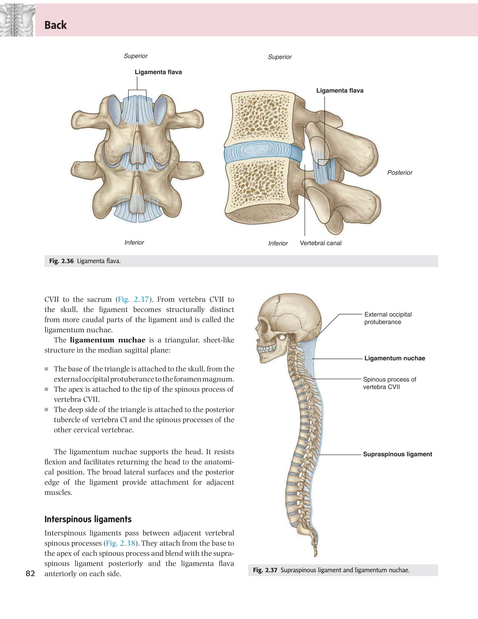
The spinal ligaments form a three-column passive support system. The anterior longitudinal ligament (ALL) prevents hyperextension; the posterior longitudinal ligament (PLL) and posterior elements prevent hyperflexion. Think of them as seatbelts on either side of a car seat — each belt only becomes taut when the column moves beyond its neutral position. This is why the ligamentum flavum — which runs between laminae and must stretch every time the spine flexes — is uniquely composed of 80% elastic fibres (yellow, hence flavum). It is this elastic recoil that snaps the spine back to neutral from flexion. When it thickens with age, it buckles inward and narrows the canal — one cause of lumbar stenosis.
| Ligament | Location / Extent | Movement limited | Clinical note |
|---|---|---|---|
| Anterior longitudinal (ALL) | Anterior surface of vertebral bodies + discs; occiput to sacrum; widest and thickest in thoracic | Hyperextension; prevents anterior vertebral body separation | Ruptures in extension injuries (hyperextension fracture-dislocation); calcification in DISH (diffuse idiopathic skeletal hyperostosis) |
| Posterior longitudinal (PLL) | Posterior surface of bodies within vertebral canal; C2 to sacrum; narrows at disc levels laterally | Flexion; reinforces posterior disc | Narrow lateral extent = posterolateral disc herniation; ossification of PLL (OPLL) in East Asian populations — cervical myelopathy |
| Ligamentum flavum | Connects laminae of adjacent vertebrae; left and right halves meet at midline; 80% elastic fibres (yellowish) | Flexion; elastic recoil assists return to neutral posture | Thickens and buckles in flexion → contributes to lumbar spinal stenosis; felt as resistance then give during LP needle passage |
| Interspinous | Between spinous processes | Flexion | Ruptured in "chance" fracture (lap-belt injury); palpable tenderness in posterior instability |
| Supraspinous | Tips of spinous processes; C7 to sacrum; in neck = ligamentum nuchae (septum between paraspinal muscles) | Flexion | Ligamentum nuchae: large elastic structure in ruminants for head support; in humans relatively small |
| Intertransverse | Between transverse processes | Contralateral lateral flexion | Less well-defined than other ligaments |
- Which ligament prevents hyperextension of the spine? Anterior longitudinal ligament (ALL) — runs along the anterior surface of vertebral bodies from occiput to sacrum.
- Why is the ligamentum flavum uniquely elastic (80% elastic fibres)? It must stretch with every spinal flexion and snap the spine back to neutral — constant stretch/recoil demands high elasticity; its yellow colour (flavum = yellow) reflects the elastic fibres.
- Why does ligamentum flavum thickening cause lumbar stenosis specifically in extension? In extension the ligament buckles inward (doesn't stretch) — in flexion it flattens and the canal opens, which is why neurogenic claudication is relieved by leaning forward.
- Which ligament is narrow at disc levels posterolaterally, contributing to disc herniation direction? Posterior longitudinal ligament (PLL) — narrow at disc levels leaves the posterolateral disc unprotected.
- What is DISH (diffuse idiopathic skeletal hyperostosis) and which ligament is involved? Calcification/ossification of the ALL producing flowing osteophytes bridging ≥4 contiguous vertebrae — causes stiffness but preserves disc height (distinguishes it from spondylosis).
The orientation of the facet joints determines the type of movement available in each spinal region — this is the key principle. Cervical facets are near-horizontal, which is why the cervical spine can rotate almost 90° in each direction. Lumbar facets are oriented near-sagittally (facing medially), which blocks rotation almost completely and explains why the lumbar spine flexes and extends well but rotates poorly. This sagittal locking of the lumbar facets is a protective adaptation — it prevents the large lumbar discs from being torn by twisting forces. When these facet joints become arthritic, the resulting capsular thickening and osteophyte formation narrows the spinal canal from behind, contributing to lumbar stenosis.
| Region | Facet orientation | Primary movements | Clinical |
|---|---|---|---|
| Cervical | Near horizontal (45°); face superoposteriorly | Flexion, extension, rotation, lateral flexion — most mobile region | Whiplash = sudden extension then flexion; facet injury + disc herniation |
| Thoracic | Near coronal (60°); face posterolaterally | Rotation; flexion/extension limited by rib cage | Compression fractures; Scheuermann's |
| Lumbar | Near sagittal (facing posteromedially) | Flexion/extension; rotation greatly restricted (important for stability) | Facet joint OA (can be treated with intraarticular steroid injection or medial branch block/radiofrequency ablation) |
Narrowing of the lumbar spinal canal by a combination of: disc bulge, ligamentum flavum hypertrophy, osteophytes from facet joints. Produces neurogenic claudication: bilateral leg pain/numbness/weakness that worsens with walking or standing (lumbar extension narrows canal), relieved by sitting or leaning forward (flexion widens canal). Distinguish from vascular claudication (worsens with walking, relieved by standing). Treatment: physiotherapy → epidural steroids → decompressive laminectomy ± fusion.
• Why does the lumbar spine resist rotation? → Sagittally oriented facets block rotational movement — protective against disc tearing
• Neurogenic vs vascular claudication: one key differentiator? → Neurogenic relieved by flexion/sitting; vascular relieved by simply stopping (standing is enough)
• What does the ligamentum flavum contribute to lumbar stenosis? → It thickens with age and buckles inward during extension, narrowing the posterior canal
Thoracic Cage — Ribs, Sternum & Apertures
The thoracic cage does two contradictory things simultaneously: it is rigid enough to protect the heart and lungs from blunt trauma, and flexible enough to move with every breath. That flexibility is built into the costal cartilages (which deform elastically) and the two different rib movement mechanisms (pump handle and bucket handle). When that flexibility is destroyed — as in a flail chest where multiple ribs fracture in two places each — the chest wall becomes a paradoxical piston: it sucks inward during inspiration (instead of expanding) because the segment is no longer coupled to the rest of the thorax. The resulting inadequate ventilation, combined with the underlying pulmonary contusion, is why flail chest carries significant mortality.
The thoracic cage is formed by 12 thoracic vertebrae (posterior), 12 pairs of ribs + costal cartilages (lateral), and the sternum (anterior). It protects the heart and lungs, and its movements drive ventilation.
- What gives the thoracic cage its respiratory flexibility despite being a bony structure? Costal cartilages (elastic deformation) + two rib movement mechanisms (pump handle and bucket handle).
- What is flail chest and why is it dangerous? Multiple rib fractures in two places create a floating segment that moves paradoxically (sucked in on inspiration) — inadequate ventilation + underlying pulmonary contusion = significant mortality.
- Name the three components of the thoracic cage. 12 thoracic vertebrae (posterior) + 12 pairs of ribs and costal cartilages (lateral) + sternum (anterior).
- What is the sternal angle (angle of Louis) and why is it clinically important? Junction of manubrium and sternal body — marks T4/T5, the 2nd rib cartilage, the aortic arch, tracheal bifurcation, and the upper mediastinal landmarks.
- Which ribs are "true," "false," and "floating," and what defines each? True (1–7): attach directly to sternum via own cartilage; False (8–10): attach via 7th costal cartilage; Floating (11–12): no anterior attachment at all.
| Part | Features & Landmarks | Clinical |
|---|---|---|
| Manubrium | Superior; widest part; jugular (suprasternal) notch at superior margin (T2 level); clavicular notches bilaterally; articulates with 1st costal cartilage (synchondrosis) + 2nd at sternal angle | Central venous catheter: subclavian/internal jugular access; manubrium is site of sternal bone marrow biopsy |
| Body (gladiolus) | Longest part; four sternebrae fused; articulates with 2nd–7th costal cartilages; xiphomanubrial angle opens posteriorly | Sternotomy for cardiac surgery; sternal fracture (airbag, steering wheel) |
| Xiphoid process | Inferior; smallest; initially cartilaginous, ossifies by 40–50 years; often bifid; may project anteriorly (xiphodynia) | CPR hand position: lower sternum (NOT xiphoid); xiphoid tip = T9; marks inferior mediastinum; may be palpated during abdominal surgery |
Palpable transverse ridge at the manubriosternal joint. Clinically, it marks: (1) level of T4/T5 intervertebral disc; (2) attachment of 2nd costal cartilage → used to count rib spaces; (3) bifurcation of trachea (carina); (4) arch of aorta (begins and ends); (5) division of superior and inferior mediastinum.
| Type | Ribs | Anterior attachment | Note |
|---|---|---|---|
| True (vertebrosternal) | 1–7 | Own costal cartilage to sternum directly | 1st rib: atypical (short, flat, no costal groove, scalene tubercle) |
| False (vertebrochondral) | 8–10 | Costal cartilage joins cartilage of rib above → forms costal margin | Costal margin = clinical landmark for liver, spleen percussion |
| Floating (vertebral) | 11–12 | No anterior attachment; free cartilaginous tips in abdominal wall musculature | Short; no neck; no tubercle; no costal groove; may be mistaken for fracture on X-ray |
| Part | Description | Articulation |
|---|---|---|
| Head | Two articular facets: superior for vertebra above, inferior for own vertebra; separated by crest of head | Costovertebral joint (synovial plane): head articulates with two adjacent vertebral demifacets + intervening disc |
| Neck | Short flat bar connecting head to tubercle; costotransverse ligament runs from here to transverse process | — |
| Tubercle | Articular facet (medial) for costotransverse joint; non-articular part for ligament attachment | Costotransverse joint (synovial plane): tubercle facet + transverse process facet (T1–T10 only) |
| Angle | Bend in shaft; where rib turns anterolaterally; iliocostalis attachment | — |
| Shaft (body) | Curved; costal groove on inferior inner border; contains VAN (from superior to inferior: Vein, Artery, Nerve) | — |
The intercostal neurovascular bundle in the costal groove runs: VAN (Vein above, Artery middle, Nerve below). Insert intercostal drain / needle above the upper border of the LOWER rib to avoid the bundle. "Avoid the groove."
| Rib | Atypical features | Clinical |
|---|---|---|
| 1st | Shortest; flattest; broadest; single articular facet (articulates only with T1); scalene tubercle (anterior scalene attachment); grooves for subclavian artery (posterior to scalene tubercle) and vein (anterior) | Thoracic outlet syndrome: neurovascular compression between 1st rib + clavicle; cervical rib (accessory rib arising from C7) is a cause |
| 2nd | Longer than 1st; has tuberosity for serratus anterior attachment; two articular facets (T1 and T2) | Landmark for counting ribs (identified at sternal angle) |
| 10th | Single articular facet (only with T10); no costotransverse joint in some individuals | — |
| 11th & 12th | Single articular facet; no neck/tubercle/costotransverse joint; no costal groove; short | Floating ribs; sometimes fractured by renal surgery incisions |
Most fractured: ribs 4–9 (long, poorly protected). 1st rib fracture: high-energy only; associated with brachial plexus injury, subclavian vessel injury. Flail chest: ≥3 consecutive ribs fractured in two places — segment moves paradoxically (inward in inspiration, outward in expiration due to intrathoracic pressure changes) → inadequate ventilation → respiratory failure. Underlying pulmonary contusion worsens gas exchange. Management: positive pressure ventilation, analgesia (epidural).
| Aperture | Boundaries | Contents passing through |
|---|---|---|
| Superior thoracic aperture (inlet) | T1 (posterior), 1st ribs (lateral), manubrium/1st costal cartilage (anterior); tilted anteriorly ≈30° | Trachea, oesophagus, subclavian vessels, common carotid arteries, jugular veins, brachial plexus, thoracic duct, vagus nerves, phrenic nerves, sympathetic trunks |
| Inferior thoracic aperture (outlet) | T12 (posterior), 11th + 12th floating ribs, costal margin (7th–10th cartilages), xiphoid; closed by diaphragm | Aorta (T12), inferior vena cava (T8), oesophagus (T10) pass through the diaphragm |
| Mechanism | Ribs affected | Effect |
|---|---|---|
| Pump handle movement | Upper ribs (1–5/6) | Anterior end of rib elevates (like a pump handle); increases anteroposterior (AP) diameter of thorax |
| Bucket handle movement | Lower ribs (6–10) | Middle of rib shaft moves laterally and upward; increases transverse diameter of thorax |
| Caliper movement | Floating ribs (11–12) | Move outward; support diaphragm as it contracts downward |
C3 — Hyoid bone level; bifurcation of common carotid artery; beginning of internal/external carotid
C4 — Thyroid cartilage (upper border); junction of pharynx/oesophagus; referred shoulder pain from diaphragm (phrenic C3–C5)
C6 — Cricoid cartilage; larynx ends / trachea begins; oesophagus begins; vertebral artery enters C6 foramen transversarium
T2 — Jugular notch of manubrium sterni
T4/T5 — Sternal angle (angle of Louis); tracheal bifurcation (carina); aortic arch begins + ends; superior/inferior mediastinum junction; azygos drains into SVC
T8 — IVC + right phrenic nerve through diaphragm
T9 — Xiphoid process
T10 — Oesophagus + left + right vagus through diaphragm; hepatic portal vein
T12 — Aorta + thoracic duct + azygos through diaphragm; coeliac trunk origin
L1 — Renal hila; conus medullaris ends (adult); iliohypogastric + ilioinguinal nerves emerge from lateral border of psoas; SMA origin; deep inguinal ring
L2 — Testicular/ovarian arteries from aorta; IMA origin (~L3 in some texts)
L3 — IMA origin; umbilicus surface mark; 3rd part of duodenum crosses aorta
L4 — Iliac crest (Tuffier's line = L4 spinous process); bifurcation of aorta into common iliac arteries; preferred LP level
L5 — Common iliac bifurcation into external/internal iliac (at L5/S1 junction)
S2 — Posterior superior iliac spine (PSIS) dimples; dural sac ends; sacral hiatus begins
Mnemonic for diaphragm: "I 8 (ate) 10 Eggs At 12" → T8 IVC, T10 oesophagus, T12 aorta
• Where does the VAN bundle run and how do you avoid it with a chest drain? → Inferior border of rib; insert ABOVE the upper border of the lower rib
• Pump handle vs bucket handle: which increases AP diameter? → Pump handle (upper ribs, anterior end elevates)
• Flail chest: what defines it and what is paradoxical about its movement? → ≥3 consecutive ribs fractured in 2 places each; segment moves inward during inspiration (paradoxical) instead of expanding
• Diaphragm levels: T8/T10/T12? → T8 = IVC; T10 = oesophagus + vagus; T12 = aorta + thoracic duct
Test your knowledge
12 MCQs + 20 definitions + 2 essays on Bones of Trunk