Normal Constitution of the Human Body
What is Anatomy?
Pick up any clinical textbook and you will find anatomy on every page — not as abstract diagrams, but as the reason behind every examination finding, every surgical incision, and every radiological report. When a surgeon says “the common bile duct runs in the hepatoduodenal ligament,” that is anatomy. When a physician taps the angle between the 12th rib and erector spinae to assess renal tenderness, that is anatomy. Anatomy is not a subject you study and then set aside; it is the language in which all clinical medicine is written.
The word comes from the Greek anatome, meaning dissection — and historically, dissection was the only way to learn it. Today, imaging has become a second dissection, letting you slice through a living patient in any plane. The branches of anatomy below are less separate subjects than different lenses on the same body: gross anatomy gives you the large-scale map, histology zooms in to tissue level, and radiological anatomy translates both into what you will actually see on a scan.
- What does the Greek word "anatome" mean? Dissection — anatomy was originally learned by cutting open cadavers, and the name has stuck even as methods have evolved to include imaging.
- Which branch of anatomy explains why a horseshoe kidney forms? Embryology — it studies development from fertilisation onward, explaining all congenital anomalies.
- What is the difference between systemic and regional anatomy? Systemic = organ-system based (all structures of one system across the body); regional = area-based (all structures — bone, muscle, nerve, vessel — in one body region).
- Why is radiological anatomy inseparable from gross anatomy? Every imaging report describes gross anatomical relationships — interpreting it requires knowing what is where, which is exactly what gross anatomy teaches.
- Name the clinical consequence of not knowing surface anatomy. Misplacing an IV, LP needle, biopsy, or auscultation point — surface anatomy maps external landmarks onto deep structures, enabling safe bedside procedures.
Anatomy (Greek: anatome = dissection) is the science of body structures and the relationships between them. It forms the foundation of clinical medicine — every examination finding, every surgical approach, every imaging report depends on anatomical knowledge.
| Branch | What it studies | Clinical relevance |
|---|---|---|
| Gross / Macroscopic | Structures visible to the naked eye; divided into systemic (organ-based) and regional (area-based) approaches | Surgical anatomy, surface landmarks, examination |
| Histology (Microscopic) | Tissues and cells examined with microscopy; stains (H&E, PAS, Masson's trichrome) | Pathology, cancer diagnosis, tissue typing |
| Embryology | Development from fertilisation through birth; germ layers: ectoderm / mesoderm / endoderm and their derivatives | Explains congenital anomalies (patent ductus, cleft palate, horseshoe kidney) |
| Neuroanatomy | Brain, spinal cord, peripheral nerves, and special sense organs | Localises lesions from clinical signs; e.g. which spinal level from dermatomal loss |
| Radiological anatomy | Structures as they appear in X-ray, CT, MRI, ultrasound | Interpreting imaging without misidentification of normal structures as pathology |
| Surface anatomy | External landmarks that indicate underlying structures | IV access, LP positioning, auscultation points, biopsy guidance |
| Surgical anatomy | Tissue planes, neurovascular bundles, fascial envelopes relevant to operations | Safe dissection, avoiding iatrogenic nerve/vessel injury |
• Branch that links anatomy to pathology reports? → Histology (microscopic anatomy)
• Which branch explains why a horseshoe kidney forms? → Embryology
• Why must anatomy and physiology be learned together? → Structure determines function; clinical findings only make sense when you understand both
Anatomical Position, Planes & Directional Terms
Imagine receiving a radiology report that reads: “A 2 cm hypodense lesion is seen in the posterior segment of the right lobe of the liver, just superior to the right adrenal gland.” Every word of that sentence — posterior, right, superior — is meaningless without a shared reference point. That reference is the anatomical position, and the three cardinal planes that slice through it. Anatomists agreed on this standard position so that “anterior” always means the same thing regardless of whether the patient is standing, lying, or upside-down on an operating table.
Think of the body in anatomical position as a standing map. The planes are like the planes of a GPS coordinate system: one divides you left-right, one front-back, and one top-bottom. Every directional term is simply a compass reading on that map. Get these right once, and reading any clinical document or imaging report becomes automatic.
- Why is a shared reference position essential in anatomy? Without anatomical position, terms like "superior," "anterior," and "lateral" have no fixed meaning — every description would be ambiguous between examiners, surgeons, and radiologists.
- Describe anatomical position precisely. Standing erect, eyes directed horizontally forward, feet together, arms at sides with palms facing anteriorly (forearms fully supinated).
- Which plane is most commonly used in clinical CT scanning? Transverse (axial) plane — divides the body into superior and inferior halves; most abdominal/thoracic CT is acquired and read in this plane.
- A scan is labelled "coronal MRI spine." What does this mean? The image slice is in the coronal (frontal) plane — parallel to the coronal suture, dividing the body into anterior and posterior portions.
- What is the midsagittal (median) plane? The single sagittal plane passing exactly through the midline, dividing the body into two equal right and left halves.
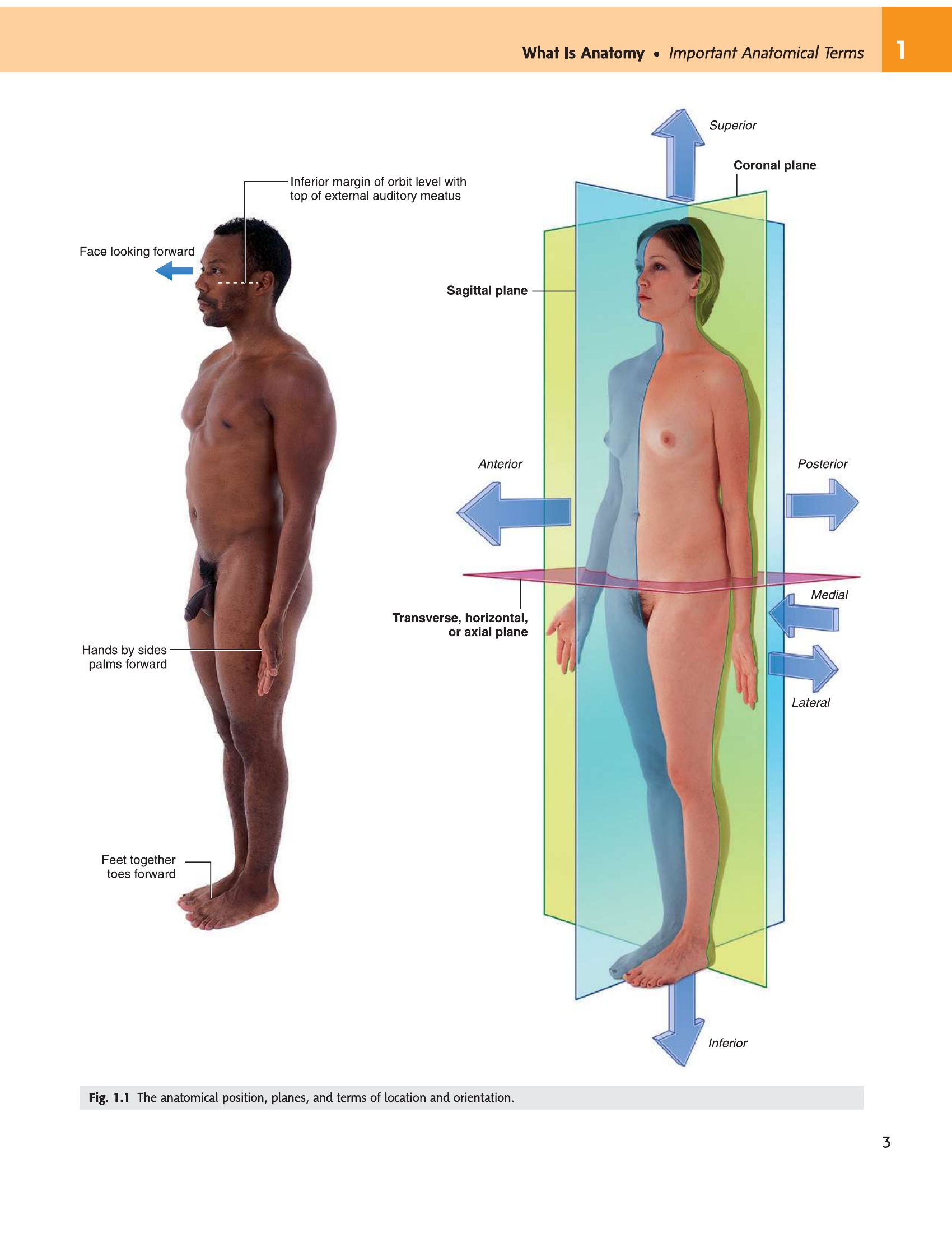
Standing erect, eyes directed forward (horizontal), feet together, arms hanging at sides with palms facing anteriorly (supinated). All anatomical descriptions refer to this standard position — it must be memorised and applied consistently.
- Which plane divides the body into left and right? Sagittal plane (a midsagittal/median plane divides equally; a parasagittal plane is off-centre).
- Which plane divides anterior from posterior? Coronal (frontal) plane — parallel to the coronal suture of the skull.
- Which plane divides superior from inferior? Transverse (horizontal/axial) plane — perpendicular to the long axis of the body.
- On a standard axial CT image, which side of the image is the patient's right? The patient's right is on the viewer's LEFT — convention is "as if you are looking up from the patient's feet."
- What is a parasagittal plane? Any sagittal plane that does NOT pass through the midline — it runs parallel to the median plane but offset to one side.
| Plane | Divides body into | Key imaging plane |
|---|---|---|
| Sagittal | Right and left. Midsagittal (median plane) = two equal halves. Parasagittal = off-centre. | Sagittal MRI: corpus callosum, spinal cord, IV discs |
| Coronal (frontal) | Anterior and posterior; parallel to the coronal suture of the skull | Coronal CT chest: lung parenchyma, mediastinum width |
| Transverse (horizontal/axial) | Superior and inferior; perpendicular to long axis of body | Axial CT abdomen: liver, kidneys, aorta — most common clinical scan plane |
These terms define where one structure sits relative to another — always relative, never absolute. “The sternum is anterior to the heart” remains true whether the patient is upright or lying flat, because anterior means “toward the front of the body in anatomical position.” The pairs you must know cold are the four main axes: superior/inferior (head–foot), anterior/posterior (front–back), medial/lateral (midline–side), and proximal/distal (root–tip, used for limbs). The rest follow logically.
| Term | Meaning | Example |
|---|---|---|
| Superior (cranial) | Toward the head | Heart is superior to the diaphragm |
| Inferior (caudal) | Away from head, toward the feet | Bladder is inferior to the uterus |
| Anterior (ventral) | Toward the front of the body | Sternum is anterior to the heart |
| Posterior (dorsal) | Toward the back | Vertebral column is posterior to the oesophagus |
| Medial | Toward the midline | Ulna is medial to the radius |
| Lateral | Away from the midline | Fibula is lateral to the tibia |
| Proximal | Closer to the trunk / point of attachment | Elbow is proximal to the wrist |
| Distal | Farther from the trunk | Fingers are distal to the elbow |
| Superficial (external) | Toward the body surface | Skin is superficial to muscle |
| Deep (internal) | Away from body surface | Femoral artery is deep to the femoral vein laterally |
| Ipsilateral | Same side | Right hand and right foot are ipsilateral |
| Contralateral | Opposite side | Left cerebral hemisphere controls contralateral (right) limb |
| Prone | Lying face down (ventral surface down) | Posterior spinal surgery, LP position |
| Supine | Lying face up (dorsal surface down) | Standard surgical and examination position |
| Bilateral | Both sides | Bilateral leg oedema |
- The radius is lateral or medial to the ulna? Lateral — the radius sits on the thumb side, the ulna on the little-finger side; lateral = away from midline.
- Is the knee proximal or distal to the ankle? Proximal — closer to the trunk (root of the limb); proximal/distal is used only for limbs, not the body axis.
- Supine vs prone: which position is face-up? Supine = face up (dorsal surface down); prone = face down (ventral surface down).
- What does ipsilateral mean? Same side — e.g., the right hand and right foot are ipsilateral; the left cerebral hemisphere controls the contralateral (right) side.
- Why does "anterior" always mean "toward the front of the body" regardless of patient position? Because all directional terms are defined relative to anatomical position, not to the patient's current posture or the examiner's perspective.
Movement terminology gets confusing quickly unless you anchor each term to a concrete action you already know. Flexion and extension are the simplest: when you bend your elbow, you bring the forearm toward the arm — that is flexion (angle decreases). Straightening it is extension. Supination is the one that catches students: the easiest memory hook is “supination = holding a bowl of soup.” When the palm faces up (anatomical position), the forearm is supinated; when it faces down (as on a keyboard), it is pronated.
| Movement | Definition | Example |
|---|---|---|
| Flexion | Decreases angle between bones | Bending elbow, bringing knee to chest |
| Extension | Increases angle between bones; returns to anatomical position or beyond (hyperextension) | Straightening knee, extending neck |
| Abduction | Movement away from the midline | Raising arm laterally; spreading fingers |
| Adduction | Movement toward the midline | Bringing arm back to side |
| Medial (internal) rotation | Anterior surface rotates toward midline | Turning toes inward |
| Lateral (external) rotation | Anterior surface rotates away from midline | Turning foot outward |
| Circumduction | Combination of flexion, extension, abduction, adduction — circular movement; ball-and-socket joints only | Shoulder, hip |
| Pronation | Medial rotation of forearm; palm faces posteriorly/downward | Typing on a keyboard |
| Supination | Lateral rotation of forearm; palm faces anteriorly/upward (anatomical position) | “Hold a bowl of soup” |
| Dorsiflexion | Ankle: toes move toward shin; decreases angle anteriorly | Walking on heels |
| Plantarflexion | Ankle: toes point downward; increases angle anteriorly | Standing on tip-toes |
| Inversion | Sole of foot faces medially | Sprain mechanism |
| Eversion | Sole of foot faces laterally | Flat-foot posture |
| Elevation / Depression | Upward / downward movement of scapula or mandible | Shrugging (elevation), chewing (depression of mandible) |
| Protraction / Retraction | Forward / backward movement in horizontal plane | Scapular protraction (punching), retraction (pulling back) |
- Mnemonic for supination: "hold a bowl of ___"? Soup — the palm faces up (anteriorly, as in anatomical position) when the forearm is supinated.
- Is dorsiflexion a flexion or extension movement? Flexion — the angle between the dorsum of the foot and the shin decreases; walking on heels requires dorsiflexion.
- Circumduction is only possible at which joint types? Ball-and-socket joints (shoulder and hip) — requires flexion, extension, abduction, adduction all in sequence.
- Inversion vs eversion: which mechanism causes a lateral ankle sprain? Inversion (sole faces medially) — the anterior talofibular ligament is maximally stressed in plantar-flexion + inversion.
- Retraction of the scapula moves it toward or away from the vertebral column? Toward — retraction (pulling the shoulder blades back) approximates the scapulae to the spine.
When a patient points to where their pain is, you need a precise vocabulary to document it. That vocabulary is the 9-region system. The most examined landmark here is McBurney’s point — the tenderness site in acute appendicitis — which lies at the junction of the lateral and middle thirds of a line from the right ASIS to the umbilicus. It falls in the right iliac region. Every region name connects to specific organs you need to know cold, because “epigastric pain radiating to the back” immediately tells you pancreas, while “right hypochondriac pain worse after fatty meals” tells you gallbladder.
| 9-Region System | Key structures |
|---|---|
| Epigastric | Stomach, liver (left lobe), pancreas (head), aorta |
| Right hypochondriac | Liver (right lobe), gallbladder, right colic flexure |
| Left hypochondriac | Spleen, stomach (fundus), left colic flexure, pancreas (tail) |
| Umbilical | Small intestine, transverse colon, aorta bifurcation (L4) |
| Right lumbar (flank) | Ascending colon, right kidney (lower pole) |
| Left lumbar (flank) | Descending colon, left kidney (lower pole) |
| Hypogastric (pubic) | Urinary bladder, uterus, sigmoid colon |
| Right iliac (inguinal) | Caecum, appendix, right ovary/tube |
| Left iliac (inguinal) | Sigmoid colon, left ovary/tube |
Clinicians often use 4 quadrants (RUQ, LUQ, RLQ, LLQ) for quick description. McBurney’s point (appendix) = junction of lateral and middle thirds of a line from ASIS to umbilicus — lies in RLQ / right iliac region. Murphy’s sign (gallbladder) = RUQ / right hypochondriac. Renal angle tenderness = costovertebral angle, posterior to lumbar regions.
• Which plane is most common in clinical CT scanning? → Transverse (axial) — divides superior from inferior
• Supine vs prone? → Supine = face up; prone = face down
• McBurney’s point lies in which region? → Right iliac region (RLQ)
• What is the difference between proximal and superior? → Proximal = nearer the limb root (used for limbs only); superior = toward the head (used for the body axis)
Diagnostic Imaging Modalities
A 60-year-old man arrives in the emergency department with right upper quadrant pain, fever, and tenderness. You need to know what is in that region (gallbladder, liver, right colic flexure) and which imaging tool will show it most clearly. Choosing between ultrasound, CT, and MRI is not a random decision — each modality has a fundamentally different physical basis, and that basis determines what it sees well and what it misses. Ultrasound cannot see through bone; MRI cannot be used if the patient has a metal pacemaker; plain X-ray is useless for soft-tissue detail. Knowing why each modality works the way it does means you will always choose correctly.
The unifying principle is this: every imaging method creates contrast between tissues by exploiting a physical property — X-ray uses differential X-ray absorption (bone absorbs a lot, air almost none), MRI uses the behaviour of protons in a magnetic field (fat and water respond differently), and ultrasound uses echoes from acoustic interfaces. The tool that causes no harm at all from the radiation standpoint is MRI and ultrasound — critical knowledge for choosing imaging in children and pregnant patients.
- Which two imaging modalities use NO ionising radiation? MRI (magnetic fields + radiofrequency) and Ultrasound (sound waves) — both are safe in children and pregnancy.
- Why can ultrasound not image through bone or gas? Bone reflects the sound beam; gas scatters it — both block the signal from reaching deeper structures.
- What physical property does CT exploit to create contrast? Differential X-ray absorption (attenuation) — bone absorbs much more than soft tissue, which absorbs more than air.
- Which modality measures metabolic activity rather than anatomy? Nuclear medicine / PET — radioactive tracers (e.g. ¹⁸F-FDG) highlight metabolically active tissue like tumours.
- Name a contraindication to MRI. Ferromagnetic implants, most pacemakers, cochlear implants — the strong magnetic field can move or heat metallic objects and disrupt electronic devices.
- Ionising radiation; cheap and fast; first-line
- Best for: bone, lung pathology, foreign body, bowel gas
- Density spectrum: air (black) → fat → soft tissue (grey) → bone/contrast (white)
- Two views minimum for fractures (AP + lateral)
- Multiple X-ray beams, computer reconstruction; higher radiation than plain X-ray
- Best for: trauma, chest/abdomen/pelvis, stroke (haemorrhage), bony detail
- Displayed in axial slices (multiplanar reformats available)
- Hounsfield units (HU): air −1000, fat −100, water 0, blood 50–80, bone +1000
- Magnetic fields + radiofrequency pulses; no ionising radiation
- Best for: CNS, spinal cord, cartilage, ligaments, soft tissue tumours
- T1: fat bright, CSF dark (anatomy); T2: fluid/CSF/oedema bright (pathology)
- Contraindicated: ferromagnetic implants, pacemakers (most), cochlear implants
- High-frequency sound waves; real-time; no radiation; bedside use
- Best for: abdominal organs, obstetrics, vascular (Doppler), MSK, pericardial effusion
- Echogenic (bright) = dense; anechoic (black) = fluid; shadowing = calcification/gas
- Operator-dependent; limited by gas and bone
- Radioactive tracer (e.g. 18F-FDG); measures metabolic activity, not just anatomy
- PET-CT: cancer staging, recurrence detection; brain metabolism
- SPECT: myocardial perfusion, bone scan (metastases)
- Higher radiation than CT; results delayed
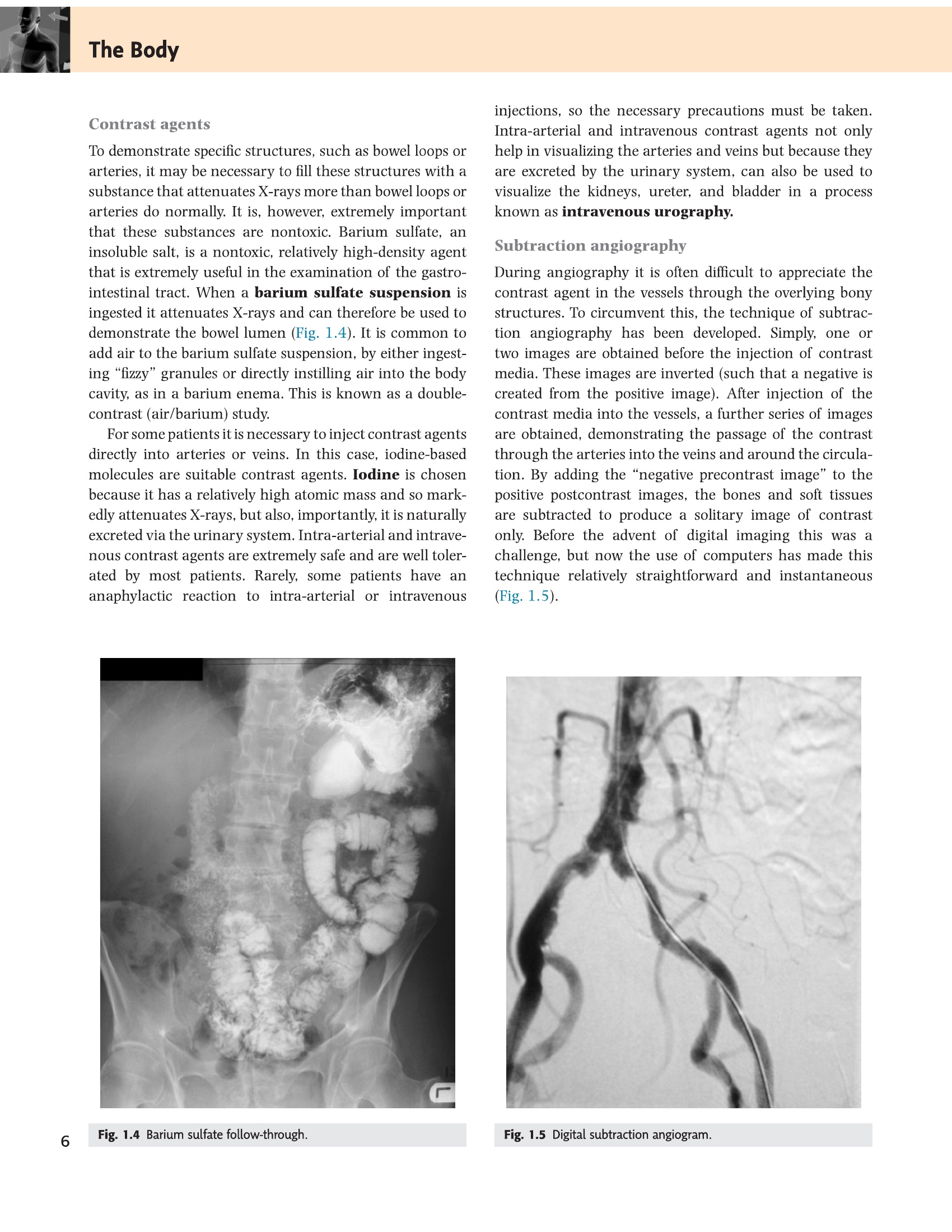
- Iodine (CT/X-ray) or gadolinium (MRI) contrast agents enhance vascular structures
- Barium: GI tract (swallow, follow-through, enema)
- DSA (digital subtraction angiography): gold standard for vascular anatomy and intervention
- Risk: iodinated contrast → nephrotoxicity, anaphylaxis; gadolinium → nephrogenic systemic fibrosis in renal failure
Axial CT/MRI: viewed as if patient is supine, feet toward you — patient’s right is on viewer’s left. Chest X-ray PA view: patient faces the film; again patient’s right = viewer’s left. Lateral CXR: patient’s left side against detector (left lateral) by convention. Never assume left = left without confirming orientation markers.
• Which modality for a suspected ACL tear? → MRI — best for soft tissue (ligaments, cartilage, no radiation)
• T1 vs T2 in MRI: which shows pathology (oedema, fluid)? → T2 (fluid bright)
• Hounsfield units for acute intracranial haemorrhage on CT? → ~60 HU (hyperdense = bright on CT)
• Why is ultrasound preferred in pregnancy? → No ionising radiation; real-time; safe for fetus
Skeletal System
The skeleton is far more than a rigid scaffold — it is a dynamic, living tissue that remodels in response to mechanical load (Wolff’s law), stores 99% of the body’s calcium, and produces every blood cell you have in its marrow. When it fails — through fracture, osteoporosis, or tumour — the consequences cascade through every other system. Understanding bone structure is therefore not academic; it directly explains why certain fractures heal well, why others don’t, and why bone is one of the hardest tissues to permanently damage under normal conditions.
| Division | Components | Count |
|---|---|---|
| Axial skeleton | Skull (22) + auditory ossicles (6) + hyoid (1) + vertebral column (26) + thoracic cage (25 = 12 pairs ribs + sternum) | 80 |
| Appendicular skeleton | Pectoral girdles (4: 2 clavicles + 2 scapulae) + upper limbs (60) + pelvic girdle (2 hip bones) + lower limbs (60) | 126 |
| Total adult skeleton | 206 | |
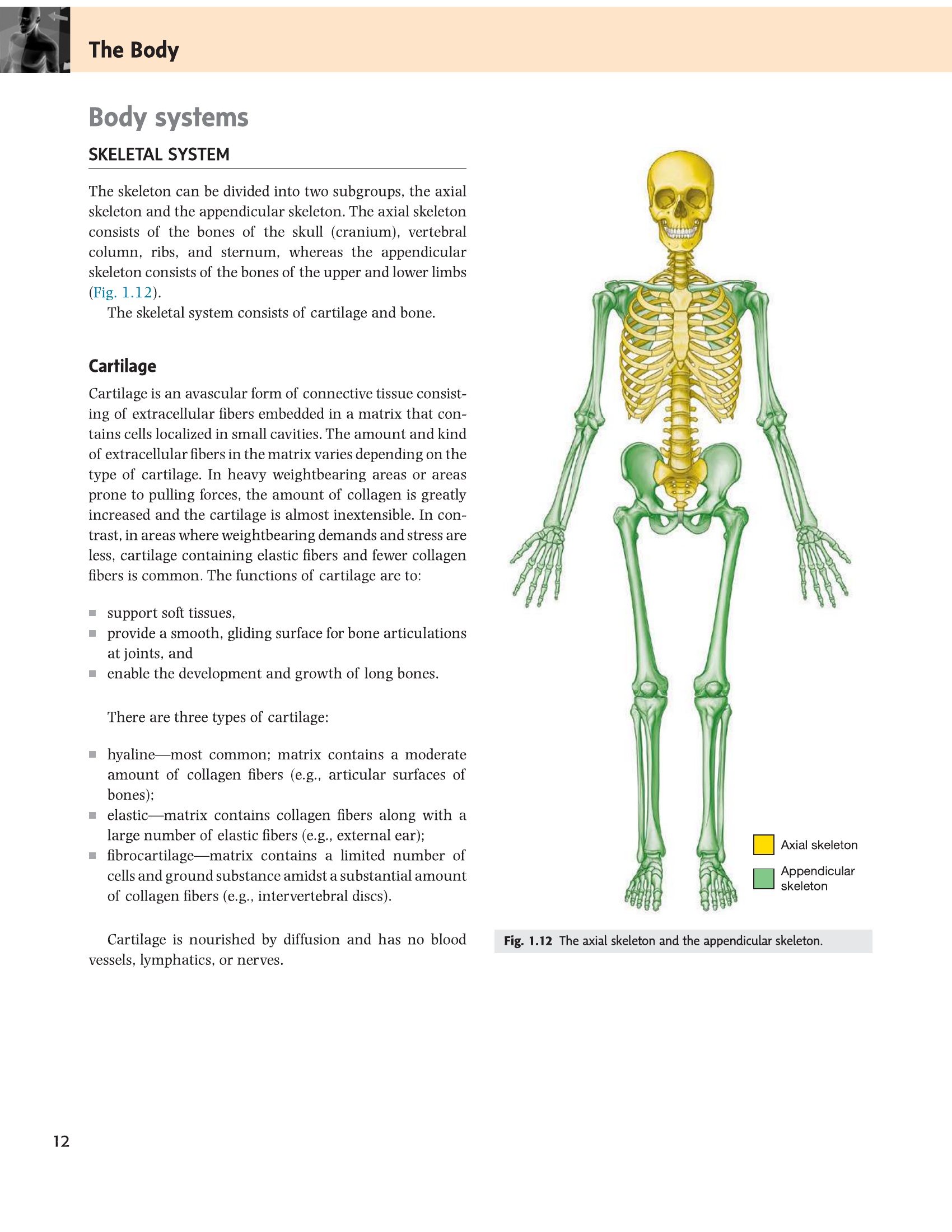
- How many bones does the adult skeleton have, and how are they split? 206 total — axial skeleton 80 (skull, vertebral column, thoracic cage) + appendicular 126 (girdles + limbs).
- What does Wolff's law state? Bone remodels along lines of mechanical stress — loading leads to hypertrophy; disuse leads to atrophy (osteoporosis).
- Where does haematopoiesis occur in the adult? Red marrow in flat bones (sternum, iliac crest, skull) and epiphyses of long bones — NOT the medullary cavity of long bone shafts (yellow/fat marrow).
- What percentage of the body's calcium is stored in bone? ~99% — bone is the primary calcium reservoir and releases it under PTH control when serum levels fall.
- Which is larger: axial or appendicular skeleton? Appendicular (126 bones) > axial (80 bones) — the limbs plus pectoral and pelvic girdles make up the majority.
Every bone shape has a design logic. Long bones are hollow tubes — the tubular shape gives maximum strength with minimum weight, exactly like the aluminium tubes used in aircraft frames. Flat bones (skull, sternum, ilium) are like sandwiches: two hard cortical layers encasing soft cancellous bone full of marrow, the haematopoietic factory. Sesamoid bones develop inside tendons to change their angle of pull — the patella redirects the quadriceps force around the knee, increasing its mechanical advantage. Knowing the bone type predicts both which fractures occur there and how they heal.
| Type | Description | Examples | Clinical note |
|---|---|---|---|
| Long | Length > width; diaphysis (shaft) + epiphyses (ends) + metaphyses; hollow medullary cavity | Femur, humerus, tibia, fibula, radius, ulna | Fractures here heal via endochondral callus; growth plate (physis) at metaphysis |
| Short | Nearly equal dimensions; mostly cancellous bone with thin cortex | Carpals, tarsals | Scaphoid fracture: avascular necrosis risk (proximal pole has poor supply) |
| Flat | Two plates of cortical bone enclosing diploe (cancellous); large surface area | Skull vault (parietal, frontal), sternum, scapula, ribs, ilium | Major site of red marrow (haematopoiesis) in adults |
| Irregular | Complex shape; does not fit other categories | Vertebrae, facial bones, os coxa, calcaneus | Vertebral compression fractures in osteoporosis |
| Sesamoid | Develop within tendons; alter direction of pull + reduce friction | Patella (largest), sesamoids of 1st MTP joint | Bipartite patella (unfused sesamoid) — normal variant, do not mistake for fracture |
- Periosteum — outer fibrous layer (Type I collagen) + inner cellular layer (osteoblasts + osteoprogenitor cells); continuous with tendons via Sharpey’s fibres; sensory nerve supply = periosteum is pain-sensitive (bone pain)
- Compact (cortical) bone — dense; Haversian systems (osteons): concentric lamellae around a central (Haversian) canal carrying blood vessels and nerves; connected by Volkmann’s canals (transverse)
- Cancellous (trabecular/spongy) bone — lattice of trabeculae oriented along stress lines (Wolff’s law); spaces contain red marrow in children and flat bones, yellow marrow (fat) in adult long bones
- Medullary cavity — central canal of long bone diaphysis; yellow marrow (fat) in adults; reverts to red (active) marrow in severe anaemia, leukaemia
- Endosteum — thin cellular lining of medullary cavity and trabeculae; contains osteoblasts and osteoclasts; site of remodelling
Closed vs Open: open = skin broken overlying fracture = contamination risk → emergency surgery. Transverse: direct force. Oblique/Spiral: torsional force. Comminuted: >2 fragments. Greenstick: one cortex breaks, other bends — children only (immature bone is more flexible). Stress fracture: repetitive loading (> bone remodelling capacity) — metatarsals, tibia, femoral neck. Pathological fracture: through diseased bone (tumour, osteoporosis, Paget’s disease).
2. Reparative phase (2 weeks–3 months):
• Soft callus: periosteal and endosteal cells differentiate into fibroblasts and chondroblasts → fibrocartilaginous bridging callus forms around fracture.
• Hard callus: endochondral ossification converts cartilaginous callus → woven bone (visible on X-ray at ~3 weeks). Union = fracture site bridged on all sides.
3. Remodelling phase (months to years): Osteoclasts resorb excess callus; osteoblasts lay lamellar bone along lines of stress (Wolff’s law); medullary canal is re-established; bone returns to original shape.
Primary (direct) bone healing: Only possible with ORIF + anatomical reduction + rigid fixation (zero movement). No callus forms — Haversian cutting cones drill directly across the fracture line.
Local: Infection (osteomyelitis) · poor blood supply (AVN) · interposed soft tissue · excessive movement at fracture site (inadequate immobilisation) · bone loss/comminution · pathological fracture (tumour/Paget’s).
Non-union: fracture fails to heal by 6 months. Delayed union: healing slower than expected. Malunion: heals in incorrect position. Wolff’s law: bone remodels along lines of mechanical stress — disuse → atrophy; loading → hypertrophy.
• Which layer of bone contains osteoblasts and is pain-sensitive? → Periosteum (inner cellular layer)
• Haversian canal runs which way? → Longitudinal (parallel to long axis); Volkmann’s = transverse
• Three stages of fracture healing in order? → Inflammatory → Reparative (soft callus → hard callus) → Remodelling
• Bone type with highest risk of avascular necrosis after fracture? → Short bones: scaphoid (proximal pole end-artery supply)
There are two fundamentally different ways the body builds bone, and knowing which method applies to which bone explains a key clinical fact: skull vault fractures in a newborn behave very differently from limb fractures, because they formed by different processes. Intramembranous ossification — which builds the skull vault, mandible, and clavicle — goes straight from mesenchyme to bone, with no cartilage intermediate. Endochondral ossification (all long bones, vertebrae, ribs) first makes a cartilage blueprint, then replaces it with bone — which is why the growth plate (a cartilage remnant) exists in long bones and not in skull vault bones.
| Type | Template | Process | Bones formed |
|---|---|---|---|
| Intramembranous | Mesenchymal connective tissue membrane (no cartilage template) | Mesenchymal cells → osteoblasts → ossification centres → woven bone → lamellar bone; no epiphyseal plate | Most skull vault bones (frontal, parietal, occipital squama), mandible, clavicle (partly), facial bones |
| Endochondral | Hyaline cartilage model | Cartilage calcifies → vascular invasion → primary ossification centre (diaphysis, 6th–12th wk fetal) → secondary centres (epiphyses, postnatal/adolescent) → growth plate (physis) between them until closure | All long bones, short bones, most of base of skull, ribs, vertebrae |
The growth plate (epiphyseal plate) is hyaline cartilage between epiphysis and metaphysis. Zones (lateral to centre): Reserve → Proliferating → Hypertrophic → Calcified. Closes at end of puberty under sex hormone influence (oestrogen > testosterone for closure). Growth plate fractures (Salter-Harris classification): Type I = through physis only (most common in young children); Type II = through physis + metaphysis (most common overall — better prognosis); Types III–V involve epiphysis → risk of growth disturbance. Because the physis is weaker than ligaments in children, ligamentous-type injuries cause physeal fractures rather than ligament tears.
- Which ossification type forms bone directly from mesenchyme (no cartilage template)? Intramembranous ossification — skull vault, mandible, clavicle (medial part).
- Why does the femur have a growth plate but the parietal bone does not? Femur = endochondral (cartilage blueprint → bone, growth plate between epiphysis and metaphysis); parietal bone = intramembranous (no cartilage step, no physis).
- When does the growth plate close? End of puberty, under sex hormone influence (oestrogen more potent than testosterone for plate closure).
- Salter-Harris Type II fracture: through which structures? Through the growth plate AND the metaphysis — most common overall; better prognosis because the epiphysis (blood supply) is spared.
- Why do ligamentous-type injuries in children cause physeal fractures rather than true ligament tears? The growth plate (hyaline cartilage) is mechanically weaker than ligaments at this age, so the cartilage fails first under the same force.
Cartilage is the body’s most neglected tissue until it is damaged — at which point it becomes one of the hardest problems in orthopaedics. The reason is its fundamental biology: cartilage is avascular, nourished only by diffusion from the perichondrium or synovial fluid. When it is torn or degenerated, no blood supply can bring the repair cells that other tissues rely on. This is why osteoarthritis is irreversible once established and why articular cartilage damage in a young athlete can end a career. The three types differ most importantly in their collagen composition and their biomechanical role.
| Type | Key features | Locations | Clinical |
|---|---|---|---|
| Hyaline | Most common; smooth; type II collagen; glassy appearance; can calcify (enchondral ossification) | Articular surfaces of synovial joints, costal cartilages, larynx (thyroid/cricoid/arytenoids), tracheal rings, fetal skeleton template | Osteoarthritis = hyaline cartilage degeneration. Calcified costal cartilage is normal on CXR. |
| Fibrocartilage | Toughest; type I + II collagen; withstands heavy compression + tension; NO perichondrium; can ossify | Intervertebral discs (annulus fibrosus), pubic symphysis, knee menisci, glenoid and acetabular labra, TMJ disc, sternoclavicular disc | Disc herniation (nucleus pulposus through annulus → nerve root compression). Meniscal tears in athletes. |
| Elastic | Type II collagen + elastic fibres; flexible; returns to shape; yellow on gross specimen | Pinna (auricle), epiglottis, auditory (Eustachian) tube, cuneiform + corniculate laryngeal cartilages | Relapsing polychondritis = autoimmune destruction of elastic cartilage → floppy ears, saddle nose, airway collapse |
The tissue that lines your knee joint (hyaline cartilage) is the same tissue that built your entire skeleton before bone replaced it. Its vulnerability is a direct consequence of its past utility: a tissue designed to be a temporary scaffolding was repurposed for permanent joint cushioning, and it never acquired the blood supply that would allow repair. Every patient with knee pain is living out this evolutionary compromise.
- Why does articular cartilage heal so poorly once damaged? It is avascular — no blood supply delivers repair cells; nutrition depends solely on diffusion from synovial fluid, which cannot support regeneration of a full-thickness defect.
- Which cartilage type lines the articular surfaces of synovial joints? Hyaline cartilage — smooth, glassy, type II collagen; its degeneration is osteoarthritis.
- Where is fibrocartilage found in the spine? Intervertebral discs (annulus fibrosus) — it withstands both compression and tension; also at pubic symphysis and knee menisci.
- Which cartilage type is the most flexible and returns to shape? Elastic cartilage — contains elastic fibres alongside type II collagen; found in the ear pinna and epiglottis.
- Relapsing polychondritis destroys which cartilage type, and what is the clinical consequence? Elastic cartilage — resulting in floppy ears, saddle nose deformity, and life-threatening tracheal/laryngeal collapse.
Joints / Articulations
Every joint in the body is a mechanical compromise between two competing demands: stability and mobility. A fibrous joint (skull suture) sacrifices all movement for rigid protection. A ball-and-socket joint (hip, shoulder) gains enormous range of motion at the cost of vulnerability to dislocation. The classification below is not arbitrary — each category represents a different solution to this stability-mobility trade-off, and understanding it immediately predicts which joints are commonly dislocated, which are most prone to arthritis, and which provide the most functional movement.
| Classification | Joining medium | Movement | Examples |
|---|---|---|---|
| Fibrous | Dense fibrous connective tissue; no joint cavity | None (synarthrosis) or minimal | Cranial sutures (serrated), inferior tibiofibular (syndesmosis), gomphoses (teeth in alveoli) |
| Cartilaginous — Primary (synchondrosis) | Hyaline cartilage | Very slight; temporary (convert to bone) | Epiphyseal plates, 1st sternocostal joint, spheno-occipital synchondrosis (base of skull, fuses ~25 yr) |
| Cartilaginous — Secondary (symphysis) | Fibrocartilage pad between hyaline cartilage surfaces | Slight (amphiarthrosis) | Pubic symphysis (widens in labour), intervertebral discs, manubriosternal joint |
| Synovial | Joint cavity with synovial fluid; articular cartilage; capsule + synovial membrane | Free (diarthrosis); type depends on shape | All major limb joints: hip, knee, shoulder, elbow, wrist, ankle |
- Which joint type is a synarthrosis (immovable)? Fibrous joint — cranial sutures are the classic example; no joint cavity, joined by dense fibrous tissue.
- What type of joint is the pubic symphysis? Secondary cartilaginous joint (symphysis) — a fibrocartilage pad between hyaline cartilage surfaces; allows slight movement; widens in labour.
- What type of joint is the epiphyseal plate? Primary cartilaginous joint (synchondrosis) — hyaline cartilage only; allows minimal movement and converts to bone at closure (end of puberty).
- Which joint type allows the greatest freedom of movement? Synovial joint (diarthrosis) — has a joint cavity with synovial fluid; all major limb joints are synovial.
- What fills the joint cavity of a synovial joint and what does it provide? Synovial fluid — acts as lubricant AND as the sole nutrient source for avascular articular cartilage.
The synovial joint is the joint of movement, and its genius is the small amount of lubricating fluid that reduces friction to nearly zero. Synovial fluid is not just a lubricant — it is also the sole nutrient source for articular cartilage (which has no blood supply). So any disease that destroys the synovial membrane (rheumatoid arthritis, septic arthritis) is simultaneously starving the cartilage and inflaming it. The joint capsule, meanwhile, is the structural envelope that holds everything together; its thickened parts become the named ligaments that prevent excessive movement in specific directions. Understanding this anatomy predicts why certain ligaments are injured by specific forces — the anterior talofibular ligament tears with plantar-flexion inversion (the typical ankle sprain) because it is the weakest part of the capsule and is maximally stressed in that position.
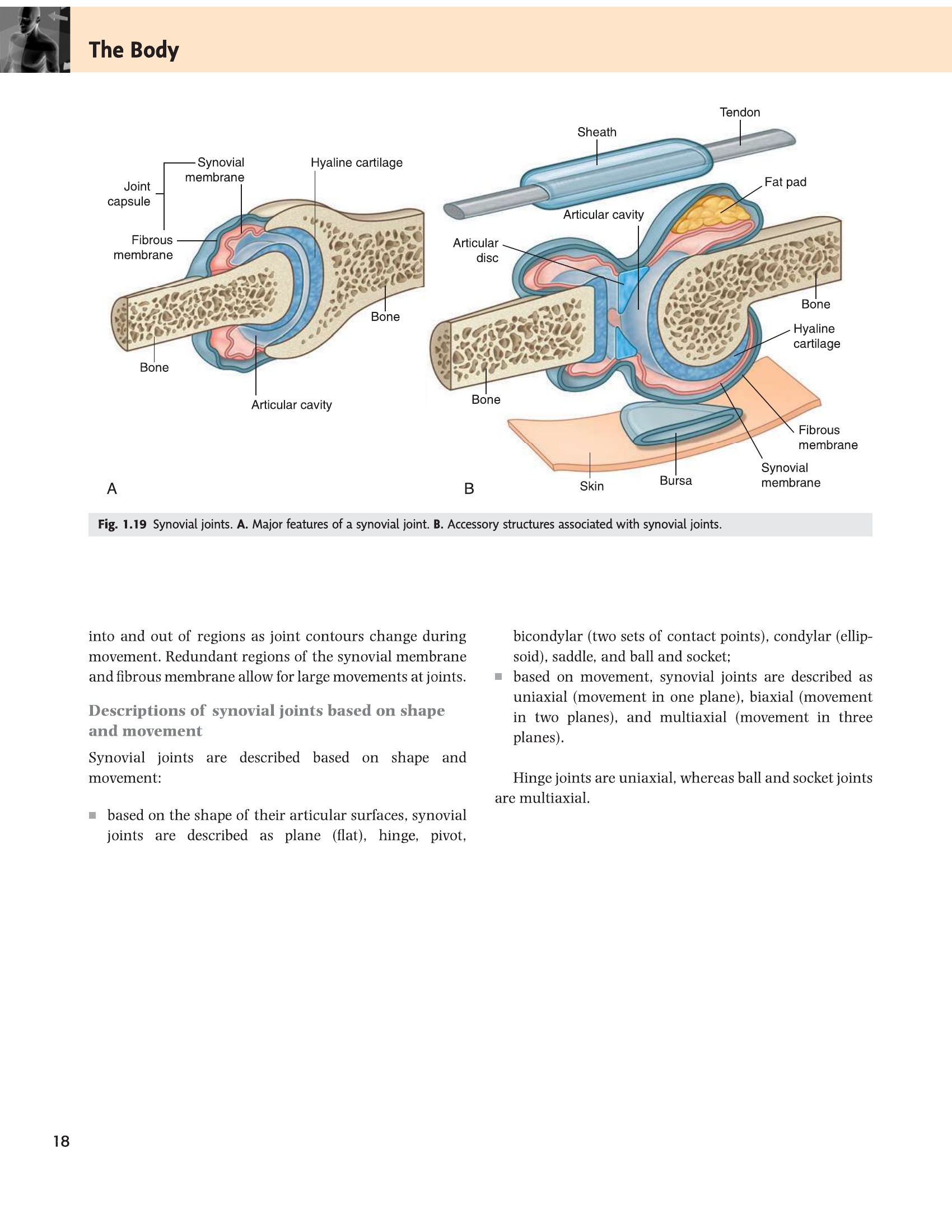
- Articular (hyaline) cartilage — smooth; avascular; aneural; nourished by synovial fluid; damaged cartilage heals very poorly → OA progression
- Fibrous capsule — surrounds joint; continuous with periosteum; thickened locally to form intrinsic ligaments; may fuse with surrounding tendons
- Synovial membrane — inner lining; secretes synovial fluid (hyaluronan + lubricin + water); covers all non-articular surfaces within the capsule; inflamed in RA → pannus formation
- Synovial fluid — viscous, straw-coloured; lubricates (reduces friction 10× more effective than ice on ice); nourishes avascular cartilage; cleared by synovial lining cells
- Ligaments — bone-to-bone; dense regular CT; intracapsular (e.g. cruciate ligaments within knee capsule), capsular (e.g. medial/lateral collateral ligaments as capsule thickenings), extracapsular (e.g. iliotibial band)
- Intra-articular structures — menisci (fibrocartilage, improve congruence), labra (fibrocartilage, deepen socket), articular discs (e.g. TMJ, sternoclavicular)
- Bursae — fluid-filled synovial sacs; reduce friction between tendon/muscle and bone; common bursitis sites: subacromial, prepatellar, olecranon, pes anserinus
| Type | Articular surface | Movements allowed | Examples |
|---|---|---|---|
| Plane (gliding) | Flat or slightly curved | Gliding/sliding only; uniaxial or multiaxial (very limited) | Intercarpal, intertarsal, facet (zygapophyseal) joints, sternoclavicular (with disc) |
| Hinge (ginglymus) | Spool-shaped convexity in concave trough | Flexion/extension only (uniaxial) | Elbow (humeroulnar), interphalangeal joints (PIP, DIP) |
| Pivot (trochoid) | Cylindrical process in ring of bone + ligament | Rotation only (uniaxial) | Atlantoaxial (C1/C2 — dens of axis), proximal radioulnar |
| Condyloid (ellipsoidal) | Oval convex head in elliptical concave socket | Flexion/extension + abduction/adduction (biaxial); no rotation | Wrist (radiocarpal), metacarpophalangeal joints (MCP) |
| Saddle (sellar) | Each surface concave in one plane and convex in perpendicular plane | Flexion/extension + abduction/adduction + circumduction; no axial rotation | 1st carpometacarpal (CMC) joint — thumb; sternoclavicular |
| Ball-and-socket (spheroid) | Spherical head in cup-like socket | All planes + circumduction (multiaxial) | Hip (acetabulum + femoral head), shoulder (glenohumeral) |
Osteoarthritis (OA): degenerative; loss of hyaline articular cartilage; X-ray shows LOSS of joint space, subchondral sclerosis, osteophytes (bony spurs), subchondral cysts. Affects weight-bearing joints (hip, knee) and small hand joints (DIP, CMC of thumb). No systemic features. Rheumatoid Arthritis (RA): autoimmune synovitis (anti-CCP, RF positive); pannus of inflamed synovium erodes cartilage and bone; symmetric small joint involvement (MCP, PIP — spares DIP); erosions on X-ray; systemic features (fatigue, anaemia, extra-articular manifestations).
• Hinge vs pivot: what is the key difference? → Hinge = flexion/extension only (elbow); pivot = rotation only (atlantoaxial, proximal radioulnar)
• What does the synovial fluid provide that articular cartilage cannot get any other way? → Nutrition (cartilage is avascular — no blood supply to articular surface)
• Joint type at the thumb carpometacarpal joint? → Saddle (sellar) — allows circumduction but no axial rotation
• RA attacks which joint layer? → Synovial membrane (pannus formation) → then erodes cartilage and bone
Muscular System
There are three kinds of muscle in your body, and they are built for three completely different lives. Skeletal muscle is built for bursts — fast, strong, under your conscious control, but it tires. Cardiac muscle must contract and relax 100,000 times a day for 80 years without ever stopping, which is why it is striated for power but involuntary and fatigue-resistant. Smooth muscle lines every hollow organ in your body — gut, blood vessels, bladder — moving contents slowly, sustained, without your awareness. The cell biology of each type follows directly from its job description, and the diseases that attack each type follow from its structure.
| Type | Location | Control | Microscopy | Key feature |
|---|---|---|---|---|
| Skeletal (striated voluntary) | Attached to skeleton (and skin/fascia e.g. face, scalp, diaphragm) | Voluntary — somatic motor neurons (alpha motor neurons) | Cross-striations; multinucleated; nuclei at periphery | Rapid, strong, fatigable contraction; over 600 muscles in the body |
| Smooth (non-striated) | Walls of hollow viscera: GI tract, blood vessels, bladder, uterus, iris, arrector pili | Involuntary — autonomic NS + hormones + local paracrine factors | No striations; single central nucleus; fusiform cells; gap junctions | Slow, sustained, non-fatigable; peristalsis, vascular tone |
| Cardiac (striated involuntary) | Heart wall (myocardium) only | Involuntary — intrinsic pacemaker (SA node); modulated by autonomic NS | Striations + intercalated discs (mechanical coupling: fascia adherens + desmosomes; electrical coupling: gap junctions); branched fibres; 1–2 central nuclei | Rhythmic, non-fatigable; action potential without tetanus; highly dependent on aerobic metabolism |
- Which muscle type has intercalated discs and what do they do? Cardiac muscle — intercalated discs contain gap junctions (electrical coupling, allowing synchronised contraction) and fascia adherens + desmosomes (mechanical coupling).
- Where are the nuclei in skeletal muscle fibres? At the periphery (subsarcolemmal) — skeletal muscle fibres are multinucleated with nuclei pushed to the edge by packed myofibrils.
- Which muscle type is BOTH striated AND involuntary? Cardiac muscle — striated (like skeletal) for power, but involuntary (like smooth) controlled by the SA node pacemaker.
- What neurotransmitter acts at the neuromuscular junction and what receptor type does it bind? Acetylcholine (ACh) binds nicotinic ACh receptors on the motor end plate, triggering depolarisation and muscle contraction.
- Myasthenia gravis vs Lambert-Eaton: presynaptic or postsynaptic? MG = postsynaptic (autoantibodies block ACh receptors); Lambert-Eaton = presynaptic (autoantibodies block voltage-gated Ca²⁺ channels, reducing ACh release).
- Origin — proximal or less mobile attachment; typically on the bone that does not move during action
- Insertion — distal or more mobile attachment; the bone that moves
- Agonist (prime mover) — muscle(s) that produce the desired movement
- Antagonist — muscle that opposes the agonist; must relax (reciprocal inhibition) for smooth movement
- Synergist — assists the agonist; may also neutralise unwanted accessory movements
- Fixator (stabiliser) — contracts to hold the origin bone still so the agonist can act effectively (e.g. serratus anterior fixes scapula during shoulder flexion)
- Motor unit — one alpha motor neuron + all the skeletal muscle fibres it innervates. Small motor units (few fibres) = fine control (hand intrinsics, extraocular muscles). Large motor units = power (quadriceps, gastrocnemius).
- Neuromuscular junction (NMJ) — motor end plate; acetylcholine released by nerve terminal binds nicotinic ACh receptors on motor end plate → depolarisation → muscle contraction. Myasthenia gravis: autoantibodies block ACh receptors → fatigable weakness. Lambert-Eaton syndrome: autoantibodies against presynaptic Ca²⁺ channels.
The red meat of a slow-cooking chicken leg and the white breast meat are a direct visual demonstration of fibre types. Red fibres (Type I) are red because they are packed with myoglobin — the oxygen-storing protein they need to sustain aerobic metabolism for postural work all day long. White fibres (Type IIb) have almost no myoglobin, burn glucose anaerobically, and generate enormous power for a short burst. The soleus muscle holding you upright all day is mostly Type I; the gastrocnemius that launches you off the ground on a sprint is predominantly Type II. Training shifts the balance — endurance training increases Type I/IIa capacity, while heavy resistance training hypertrophies Type IIb fibres.
| Property | Type I (Slow-twitch, Red) | Type IIa (Fast-twitch oxidative) | Type IIb/IIx (Fast-twitch glycolytic) |
|---|---|---|---|
| Contraction speed | Slow | Fast | Very fast |
| Fatigue resistance | High (fatigue resistant) | Moderate | Low (fatigues quickly) |
| Metabolism | Aerobic (oxidative phosphorylation) | Aerobic + anaerobic | Anaerobic (glycolytic) |
| Myoglobin content | High (red colour) | High | Low (pale/white) |
| Mitochondria | Many | Many | Few |
| Force generated | Low | Moderate | High |
| Best for | Sustained postural activity, endurance (marathon, standing) | Middle-distance activity | Short bursts of power (sprinting, heavy lifting) |
| Examples | Soleus (90% Type I), back postural muscles | Mixed muscles | Biceps (mainly Type IIa/IIb), extraocular muscles |
When a muscle loses its motor nerve supply (lower motor neuron lesion), it undergoes denervation atrophy within weeks — fibres shrink, EMG shows fibrillations and positive sharp waves. Sustained denervation → replacement by fat and fibrosis. Disuse atrophy (immobilisation, cast) is milder and reversible. Upper motor neuron lesion (e.g. stroke, spinal cord injury above the LMN): increased tone (spasticity), hyperreflexia — muscles do NOT atrophy as quickly because LMN is intact.
• Which fibre type dominates the soleus and why? → Type I (slow, red, fatigue-resistant) — it holds posture all day
• UMN lesion vs LMN lesion: which causes flaccid paralysis? → LMN lesion (nerve or motor unit destroyed); UMN causes spastic paralysis
• What makes cardiac muscle unable to tetanise? → Long absolute refractory period (nearly as long as the contraction itself) — essential so the heart can fill between beats
• Synergist vs fixator? → Synergist assists the prime mover; fixator stabilises the origin bone so the prime mover can act
Fascia & Body Cavities
Fascia is often described as “the connective tissue that wraps everything” — which is accurate but fails to convey why it matters clinically. The critical property of deep fascia is that it is inelastic. It cannot stretch when the contents of a compartment swell. In normal anatomy this is useful: deep fascia creates firm compartments that separate muscle groups and contain neurovascular bundles. But when a tibial fracture causes bleeding into the anterior compartment of the leg, that same inelasticity traps the swelling, pressure builds, and the muscles and nerves inside begin to die from ischaemia. This is compartment syndrome — a surgical emergency that kills tissue within hours and is caused entirely by the anatomy of deep fascia.
| Layer | Composition | Contents / Function |
|---|---|---|
| Superficial fascia (hypodermis) | Loose areolar CT + variable amount of adipose tissue; lies immediately deep to skin | Stores fat (energy, insulation, padding); contains cutaneous nerves, superficial veins, lymphatics; allows skin to move over deep fascia. In face/scalp/perineum: includes muscle fibres (platysma, SMAS, dartos). |
| Deep fascia | Dense fibrous CT; inelastic; few fat cells; firmly attached to underlying structures | Invests muscles and forms intermuscular septa (divides limbs into compartments); surrounds neurovascular bundles; forms retinacula (holds tendons); named examples: fascia lata (thigh), crural fascia (leg), thoracolumbar fascia, pectoralis fascia, axillary sheath |
| Subserous fascia | Loose CT; lines body cavities between deep fascia and serous membranes | Extraperitoneal (retroperitoneal) fat; allows organs to move; contains vessels and nerves supplying organs |
Deep fascia is inelastic. When swelling (haemorrhage, oedema) occurs within a tight fascial compartment, intracompartmental pressure rises. When pressure exceeds capillary perfusion pressure (~30 mmHg), ischaemia occurs to muscles and nerves within the compartment. The 6 Ps: Pain (severe, disproportionate, worse on passive stretch) → Pressure → Paraesthesia → Paralysis → Pallor → Pulselessness (late, indicates arterial compromise). Treatment: emergency fasciotomy — surgical release of deep fascia. Most common: anterior compartment of leg (after tibial fracture), forearm (Volkmann’s ischaemic contracture).
- What property of deep fascia makes compartment syndrome possible? Inelasticity — deep fascia is dense fibrous CT that cannot stretch; when contents swell, pressure builds with nowhere to go.
- At what intracompartmental pressure does ischaemia begin? ~30 mmHg (when compartment pressure approaches or exceeds capillary perfusion pressure); fasciotomy is indicated at >30 mmHg or within 30 mmHg of diastolic BP.
- What is the first sign of compartment syndrome (1st of the 6 Ps)? Pain — disproportionate to the injury, and worsened by passive stretch of the muscles in the compartment.
- Name two common sites for compartment syndrome. Anterior compartment of the leg (after tibial fracture) and the anterior forearm (after supracondylar fracture → Volkmann's contracture).
- What is the difference between superficial and deep fascia? Superficial fascia (hypodermis) = loose CT + fat, contains cutaneous nerves and superficial veins; deep fascia = dense, inelastic, invests muscles and forms compartment walls and retinacula.
Body cavities are not empty spaces — they are precisely controlled microenvironments lined by serous membranes that allow organs to move freely against each other while preventing friction. The two-layer serous membrane design (parietal layer lining the cavity wall + visceral layer directly on the organ, with a thin film of fluid between them) is the same in the pleura, pericardium, and peritoneum. When this system breaks down — infection, cancer, cardiac failure — the small fluid film becomes a large effusion, and the cavity that was designed for freedom of movement becomes a compressive trap.
| Cavity | Lining (serous membrane) | Contents |
|---|---|---|
| Thoracic cavity | Pleura (parietal + visceral) lines each lung; pericardium lines heart | Two pleural cavities (right + left lungs), mediastinum (heart, great vessels, trachea, oesophagus, thoracic duct, lymph nodes, nerves) |
| Abdominopelvic cavity | Peritoneum (parietal + visceral); potential space = peritoneal cavity | Abdominal cavity: stomach, small intestine, large intestine, liver, gallbladder, spleen, pancreas (partly), kidneys (retroperitoneal). Pelvic cavity: bladder, rectum, uterus/ovaries (♀), seminal vesicles/prostate (♂) |
| Pericardial cavity | Fibroserous pericardium (fibrous outer + serous inner); 20–50 mL fluid normally | Heart + proximal great vessels. Accumulation of fluid = pericardial effusion; large amount compresses heart = cardiac tamponade |
• First P in the 6 Ps of compartment syndrome? → Pain (disproportionate, worse on passive stretch of the muscle)
• Normal volume of pericardial fluid? → 20–50 mL; excess = effusion; enough to compress heart = tamponade
• Which organs are retroperitoneal (not covered by peritoneum)? → Kidneys, ureters, pancreas, aorta, IVC, adrenals (SADPUCKER)
• Superficial vs deep fascia: which contains cutaneous nerves and superficial veins? → Superficial fascia (hypodermis)
Organ Systems of the Human Body
No organ system works in isolation. The kidney filters blood that the cardiovascular system delivers; the endocrine system regulates the kidney’s filtration rate; the nervous system controls the cardiovascular system’s output; and so on. The 11 systems below are a convenient organisational fiction that makes it easier to study each subsystem in depth — but in every patient you will ever treat, disease in one system will ripple through several others. Heart failure (cardiovascular) causes breathlessness (respiratory), leg swelling (lymphatic), and reduced kidney perfusion (urinary). Memorise the 11 systems, but always think in terms of how they interact.
| # | System | Major components | Primary function |
|---|---|---|---|
| 1 | Integumentary | Skin (epidermis + dermis + hypodermis), hair follicles, nails, sweat and sebaceous glands, mammary glands | Physical protection; thermoregulation; sensation; vitamin D synthesis; barrier to infection |
| 2 | Skeletal | 206 bones, cartilages, ligaments, joints | Support; locomotion; protection of vital organs; haematopoiesis (red marrow); calcium/phosphate reservoir |
| 3 | Muscular | >600 skeletal muscles; smooth and cardiac muscle | Movement and locomotion; posture; heat production (shivering thermogenesis); visceral peristalsis; cardiac pumping |
| 4 | Nervous | CNS (brain + spinal cord); PNS (cranial and spinal nerves); ANS (sympathetic + parasympathetic) | Rapid communication and integration; sensation; voluntary movement; reflexes; higher cognitive function |
| 5 | Endocrine | Pituitary (master gland), hypothalamus, thyroid, parathyroids, adrenals, pancreatic islets, gonads, pineal, thymus | Hormonal regulation of metabolism, growth, development, reproduction, stress response, homeostasis |
| 6 | Cardiovascular | Heart, systemic arteries, capillaries, systemic veins, pulmonary circuit | Delivery of O₂ + nutrients; removal of CO₂ + waste; transport of hormones; thermoregulation; haemostasis |
| 7 | Lymphatic / Immune | Lymph vessels, lymph nodes, spleen, thymus, tonsils, MALT, bone marrow | Fluid homeostasis (returns ~3 L/day interstitial fluid to blood); immunity (innate + adaptive); fat absorption (lacteals) |
| 8 | Respiratory | Nasal cavity, pharynx, larynx, trachea, bronchi, lungs (alveoli) | Gas exchange (O₂ in, CO₂ out); acid-base regulation (bicarbonate buffer); speech; smell |
| 9 | Digestive | GI tract (mouth→anus); liver; gallbladder; pancreas; salivary glands | Ingestion; mechanical and chemical digestion; absorption; elimination of waste |
| 10 | Urinary | Kidneys (2), ureters (2), urinary bladder, urethra | Filtration; excretion of metabolic waste (urea, creatinine); water, electrolyte and acid-base balance; erythropoietin production |
| 11 | Reproductive | Male: testes, epididymis, vas deferens, prostate, seminal vesicles, penis. Female: ovaries, uterine tubes, uterus, vagina, external genitalia | Gamete production; sex hormone secretion; fertilisation; gestation; lactation |
“I Smell More Neurons Energising Cardiac Lymphocytes Riding Desperately Under Rain”
Integumentary · Skeletal · Muscular · Nervous · Endocrine · Cardiovascular · Lymphatic · Respiratory · Digestive · Urinary · Reproductive
• Which system is responsible for haematopoiesis? → Skeletal (red marrow in flat bones and epiphyses)
• Two systems the pancreas belongs to? → Endocrine (insulin/glucagon from islets) + Digestive (enzymes from acinar cells)
• Which systems are affected by heart failure? → Cardiovascular (pump failure) + Respiratory (pulmonary oedema) + Urinary (reduced renal perfusion) + Lymphatic (peripheral oedema)
• What distinguishes the lymphatic from the cardiovascular system structurally? → Lymphatic is one-way (no circuit back from lymph nodes); no pump equivalent to the heart; starts as blind-ended capillaries
Test your knowledge
12 MCQs + 25 definitions + 2 essays on Normal Constitution