Amino Acids & Peptides
Why the course starts here
Biochemistry begins with amino acids for the same reason a language course begins with the alphabet. Twenty small molecules, joined end to end in different orders, produce every enzyme, every receptor, every antibody and every structural protein in your body. Nothing else in the subject makes sense until you can see why these twenty, and why their differences matter.
And the differences are entirely in one place. Every amino acid in a protein has the same backbone — an α-carbon carrying an amino group, a carboxyl group and a hydrogen. The only thing that varies is the fourth attachment, the R group or side chain. So when you are asked why a particular residue sits in a particular place in a protein, the answer is always about its R group: is it charged, is it greasy, can it hydrogen bond, is it big?
Structure is chemistry, and chemistry is the side chain. A hydrophobic side chain will be buried in the protein's core away from water; a charged one will face outward or form a salt bridge; cysteine's –SH will look for another cysteine to form a disulfide bond. Every level of protein structure you meet in the next three units is the consequence of twenty side chains seeking their most comfortable environment.
- What is the only part of an amino acid that varies between the twenty? → The R group (side chain)
- What four groups attach to the α-carbon? → Amino group, carboxyl group, hydrogen, and the R group
- Why does the side chain determine protein folding? → It decides whether a residue prefers water or the protein's core
The general structure ★★
Every amino acid found in proteins is an L-α-amino acid, and both halves of that phrase are examinable. α means the amino group is attached to the carbon immediately next to the carboxyl group. L refers to the configuration around that carbon — and it is a striking fact of biology that proteins use only the L form, never the D.
An α-carbon bearing four attachments: an amino group (–NH₃⁺), a carboxyl group (–COO⁻), a hydrogen, and a variable R group.
Because the four attachments differ, the α-carbon is chiral — with one exception.
Two more points the slides make explicitly. Mammals do contain some free D-amino acids — D-serine and D-aspartate occur in brain tissue — and certain bacterial peptides and antibiotics contain them too. But proteins are built exclusively from L-α-amino acids.
Answer "20" for the count; mention selenocysteine to show you have read around it.
- What does 'L-α-amino acid' mean? → Amino group on the carbon next to the carboxyl, in the L configuration
- Which amino acid is achiral, and why? → Glycine — its R group is a hydrogen, so the α-carbon has two identical substituents
- Which is a secondary (imino) acid? → Proline — its side chain bonds back to the α-amino nitrogen
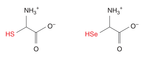
- Name the 21st protein amino acid? → Selenocysteine — selenium replaces the sulfur of cysteine
Classifying the twenty
There are several ways to sort the twenty, and the useful one is by what the side chain does in water, because that is what predicts where the residue sits in a folded protein.
| Class | Amino acids | Behaviour |
|---|---|---|
| Non-polar, aliphatic | Glycine · Alanine · Valine · Leucine · Isoleucine · Proline · Methionine | Hydrophobic — buried in the protein core |
| Aromatic | Phenylalanine · Tyrosine · Tryptophan | Bulky, largely hydrophobic; absorb UV at 280 nm |
| Polar, uncharged | Serine · Threonine · Cysteine · Asparagine · Glutamine | Hydrogen bond; Ser/Thr/Tyr are phosphorylation sites; Cys forms disulfides |
| Acidic (negative) | Aspartate · Glutamate | Negatively charged at pH 7 — low pI |
| Basic (positive) | Lysine · Arginine · Histidine | Positively charged at pH 7 — high pI; histidine buffers near physiological pH |
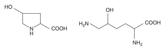
Two further groupings are worth having. The nutritionally essential amino acids — Harper's counts ten that humans cannot make in quantities adequate for infant growth or adult health — must come from the diet. And a number of amino acids are modified after the protein is made: hydroxylation gives 4-hydroxyproline and 5-hydroxylysine in collagen, and methylation, acetylation, prenylation and phosphorylation all extend what a protein can do.
Proline and lysine are hydroxylated after incorporation into procollagen, by enzymes that need vitamin C as a cofactor. Without it, collagen cannot form the hydroxyproline cross-links that give it tensile strength.
The result is scurvy — bleeding gums, loose teeth, poor wound healing, perifollicular haemorrhages. Every one of those signs is connective tissue failing, and the lesion is a post-translational modification of two amino acids.
- Which three amino acids are aromatic? → Phenylalanine, tyrosine, tryptophan
- Which are acidic and which basic at pH 7? → Acidic: aspartate, glutamate · Basic: lysine, arginine, histidine
- How many amino acids are nutritionally essential? → Ten
- Which two modified residues are found in collagen? → 4-hydroxyproline and 5-hydroxylysine
- Why does vitamin C deficiency cause scurvy? → It is the cofactor for prolyl and lysyl hydroxylase in collagen synthesis
Zwitterions ★★★
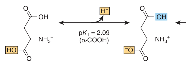
An amino acid carries two ionisable groups that behave in opposite directions: a carboxyl group that wants to lose a proton, and an amino group that wants to gain one. At the pH of blood, both have done so — the carboxyl is –COO⁻ and the amino is –NH₃⁺. The molecule therefore carries a positive and a negative charge at once, and no net charge overall.
A molecule that contains an equal number of positively and negatively charged groups and therefore bears no net charge.
Amino acids exist as zwitterions in blood and most tissues.
Students often draw an amino acid with a neutral –COOH and a neutral –NH₂ at the same time. Harper's is explicit that this structure cannot exist in aqueous solution, and the reasoning is elegant: at any pH low enough to keep the carboxyl protonated, the amino group would also be protonated; at any pH high enough for a neutral –NH₂, the carboxyl would already be –COO⁻.
The two groups simply do not have overlapping neutral ranges. The uncharged form is used only as a drawing convenience when illustrating reactions that do not involve protons.
- Define a zwitterion? → A molecule with equal numbers of positive and negative charges and no net charge
- What form does an amino acid take in blood? → The zwitterion — –COO⁻ and –NH₃⁺
- Why can the fully uncharged form not exist in water? → The pH ranges for a neutral –COOH and a neutral –NH₂ do not overlap
The isoelectric point (pI) ★★★
If the charge on an amino acid depends on pH, there must be one particular pH at which the positives and negatives exactly cancel. That pH is the isoelectric point, and it is one of the most reliably examined definitions in the whole subject.
The pH at which a molecule bears no net charge and therefore does not move in a direct-current electrical field.
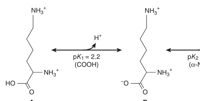
Numerically, it is the pH midway between the pKa values of the ionisations on either side of the isoelectric species.
For an amino acid with only two ionisable groups the calculation is unambiguous. For alanine, the α-carboxyl has pKa 2.35 and the α-amino group pKa 9.69, so the isoelectric point is the average of the two — about 6.02. For an amino acid with a third ionisable group in its side chain, you average the two pKa values that flank the neutral form, which is why acidic amino acids have a low pI and basic ones a high pI.
| Amino acid | pKa₁ (–COOH) | pKa₂ (–NH₃⁺) | pI |
|---|---|---|---|
| Alanine | 2.35 | 9.69 | ≈ 6.02 — average of the two |
| Aspartate (acidic) | — | — | Low — average of the two carboxyl pKa values |
| Lysine (basic) | — | — | High — average of the two amino pKa values |
Because a molecule stops moving in an electric field at its pI, this is the whole basis of electrophoresis and isoelectric focusing — the techniques used to separate serum proteins, to detect abnormal haemoglobins, and to identify the monoclonal band of myeloma on serum protein electrophoresis.
At a pH above its pI a protein is net negative and migrates to the anode; below its pI it is net positive and migrates to the cathode. That single sentence lets you predict the direction of migration in any exam question.
- Define the isoelectric point? → The pH at which a molecule has no net charge and does not migrate in an electric field
- How is it calculated? → The pH midway between the pKa values flanking the isoelectric species
- pI of alanine, and from what? → About 6.0 — the average of pKa 2.35 and 9.69
- Which way does a protein migrate above its pI? → It is net negative, so toward the anode
Absorbance at 280 nm
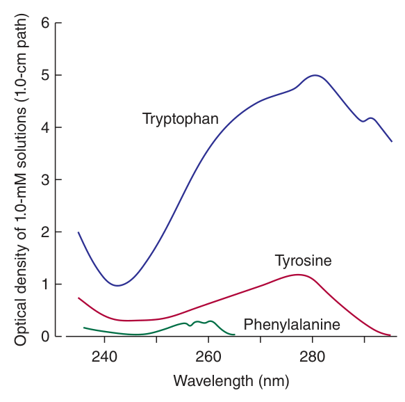
One small physical fact earns marks out of proportion to its size. The aromatic amino acids — tryptophan, tyrosine and phenylalanine — absorb ultraviolet light, and tryptophan absorbs most strongly. Harper's notes that tryptophan makes the major contribution to the ability of most proteins to absorb light around 280 nm.
Because almost all proteins contain tryptophan and tyrosine, reading the absorbance of a solution at 280 nm gives a fast estimate of protein concentration with no reagents and no destruction of the sample. It is the default measurement on every spectrophotometer and nanodrop in every biochemistry lab — including the ones in your own practicals.
The caveat is also anatomical, in a sense: a protein unusually poor in aromatic residues will read falsely low, which is why colorimetric assays such as the BCA method in your Experiment 2 are used when accuracy matters.
- Which amino acids absorb UV at 280 nm? → Tryptophan, tyrosine and phenylalanine
- Which contributes most? → Tryptophan
- What is this used for? → Rapid estimation of protein concentration by spectrophotometry
The peptide bond ★★★
Join the carboxyl group of one amino acid to the amino group of the next, losing a molecule of water, and you have a peptide bond. Repeat it a few hundred times and you have a protein. But the bond has one property that shapes everything above it, and it is the reason protein structure is possible at all.
The amide linkage formed between the α-carboxyl group of one amino acid and the α-amino group of the next, with the elimination of a molecule of water.
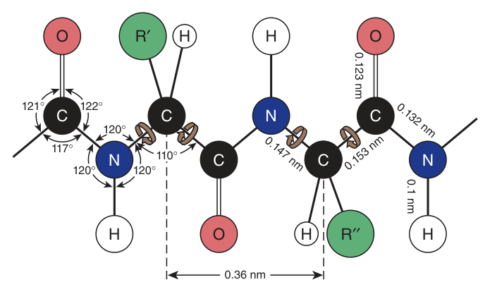
It has partial double-bond character, so the six atoms of the peptide unit are held rigidly in one plane and rotation about the C–N bond is restricted.
The lone pair on the peptide nitrogen delocalises onto the carbonyl, so the C–N bond behaves as though it were partly a double bond. Two consequences follow, and both are examinable:
1. The peptide unit is planar and rigid, and almost always in the trans configuration.
2. Rotation is therefore possible only about the two other backbone bonds — the N–Cα bond (angle φ, phi) and the Cα–C bond (angle ψ, psi).
That restriction is not a limitation, it is the whole point: a backbone free to rotate anywhere would never fold reproducibly. Because only φ and ψ can turn, the chain has a limited, predictable set of conformations — which is exactly what the α-helix and β-sheet of Unit 3 are.
Two conventions to state when you write about peptides. They are always numbered and read from the N-terminus to the C-terminus — and protein synthesis itself proceeds in that direction, which is a favourite MCQ. And the residue names change ending: in a chain, glycine becomes glycyl, alanine alanyl, with only the C-terminal residue keeping its full name.
- How is a peptide bond formed? → Between the α-carboxyl of one amino acid and the α-amino of the next, losing water
- What is its key physical property? → Partial double-bond character — planar, rigid, usually trans
- Which two backbone bonds can rotate? → N–Cα (phi) and Cα–C (psi)
- In which direction is a peptide written and synthesised? → N-terminus → C-terminus
Revision layer
Everything above was to make it make sense. What follows is the cram layer — and note that four of these are Section I definition terms, worth three marks each.
Definitions from this unit — Section I material
| Term | Definition |
|---|---|
| Zwitterion | A molecule containing an equal number of positively and negatively charged groups, therefore bearing no net charge |
| Isoelectric point (pI) | The pH at which a molecule has no net charge and does not migrate in a direct-current electrical field; the pH midway between the pKa values flanking the isoelectric species |
| Peptide bond | The amide linkage between the α-carboxyl of one amino acid and the α-amino of the next, formed with loss of water; has partial double-bond character |
| Essential amino acid | An amino acid that humans cannot synthesise in amounts adequate for growth and health, and which must therefore be supplied in the diet — ten in number |
The twenty, by side-chain class
| Class | Members |
|---|---|
| Non-polar aliphatic | Gly · Ala · Val · Leu · Ile · Pro · Met |
| Aromatic | Phe · Tyr · Trp (UV absorbance at 280 nm) |
| Polar uncharged | Ser · Thr · Cys · Asn · Gln |
| Acidic | Asp · Glu (low pI) |
| Basic | Lys · Arg · His (high pI) |
The special cases examiners like
| Amino acid | Why it is special |
|---|---|
| Glycine | Smallest; R group is H, so not chiral; fits where nothing else can, found at sharp bends |
| Proline | Side chain bonds back to the α-nitrogen — a secondary amine; rigid ring breaks α-helices |
| Cysteine | –SH group forms disulfide bonds, the only covalent cross-link in tertiary structure |
| Histidine | pKa near 6 — the only side chain that buffers at physiological pH |
| Tryptophan | Strongest 280 nm absorber |
| Selenocysteine | The 21st protein amino acid; selenium replaces sulfur |
- Define zwitterion, isoelectric point and peptide bond in exam wording
- Classify all twenty amino acids by side chain
- Explain why glycine is achiral and why proline breaks helices
- Explain what partial double-bond character does to the backbone, and name φ and ψ
- Explain why 280 nm absorbance measures protein, and when it fails